DOI: http://dx.doi.org/10.5007/1980-0037.2014v16n4p390
original article
Licence Creative Commom
CC
RBCDH
Analysis of localized muscle fatigue in
elderly women with and without a history
of falls using electromyographic signal
frequency parameters
Análise da fadiga muscular localizada em idosas
caidoras e não caidoras através de parâmetros de
frequência do sinal eletromiográfico
Lissara Ellen Silva1
Cristiane Rodrigues Pedroni1
Marcelo Tavella Navega1,2
Marcos Eduardo Scheicher1,2
Abstract– One of the problems that most concerns the elderly population is the falling, which can be caused by muscle fatigue. The objective of this study was to compare median electromyographic frequency values for the vastus lateralis muscle and the lateral portion of the gastrocnemius muscle (as parameters of electromyographic fatigue) in elderly people with and without a history of falls. A total of 20 healthy, physically active, elderly women who had or had not suffered falls were enrolled on the study. Median electromyographic signal frequencies were analyzed for the vastus lateralis muscle and the lateral portion of the gastrocnemius muscle in isometric contraction. ANOVA and Student’s t test were used for comparisons, to a cutoff of p ≤ 0.05. Median frequencies for the gastrocnemius and vastus lateralis muscles were both higher in the group with a history of falling, but without statistical significance. It was also observed that the rate of decline in median frequency was greater in the group with a history of falling. It can be concluded that elderly people with a history of falling have a greater tendency to fatigue of the gastrocnemius and vastus lateralis muscles than their peers with no history of falling.
Key words: Electromyography; Elderly; Balance; Muscle fatigue; Postural equilibrium; Seniors.
Resumo – Um dos problemas que mais preocupam a população idosa são as quedas, que podem ser causadas pela fadiga muscular. O objetivo do estudo foi comparar os valores de frequência mediana, como um parâmetro de fadiga eletromiográfica, dos músculos vasto lateral e porção lateral do gastrocnêmio em idosos com e sem história de quedas. Participaram do estudo 20 idosas saudáveis, ativas fisicamente (que sofreram e não sofreram quedas). Analisou-se a frequência mediana do sinal eletromiográfico dos músculos vasto lateral e porção lateral do gastrocnêmio em isometria. As comparações foram feitas pela ANOVA e teste t de Student, considerando p ≤ 0,05. O músculo grastrocnêmio apresentou maiores Fmed no grupo caidor do que no não caidor, o mesmo acontecendo com o vasto lateral, sem significância estatística. Também foi possível observar que a taxa de queda dos valores da frequência foi maior no grupo caidor do que no não caidor. Pode-se concluir que há maior tendência de fadiga dos músculos gastrocnêmio e vasto lateral de idosos caidores.
1 Universidade Estadual Paulista. Faculdade de Filosofia e Ciências. Departamento de Fisioterapia e Terapia Ocupacional. Marília, SP. Brasil.
2 Universidade Estadual Paulista. Instituto de Biociências. Programa Desenvolvimento Humano e Tec-nologias. Rio Claro, SP. Brasil.
The number of people over the age of 60 is predicted to double over the next 20 years. Demographic data indicate that there will be 33 million Brazilians over the age of 60 in 2025, making Brazil’s elderly population the sixth-largest in the world1.
One of the complications of aging is the emergence of chronic degen-erative conditions. It can be observed that functional capacity goes into decline during aging and that this is associated with a significant reduction in physical activity, meaning that elderly people are ever more limited in terms of their daily activities, resulting in a compromised musculoskeletal system, problems with balance, reduced resistance and frequent falls2-5.
It has been observed that muscle strength is reduced by 20% at 60 years of age, which is caused by changes in the number and size of muscle fib-ers, loss of motor units and changes to motor end plates6. This reduction
in strength increases the probability of falls, which are the most serious consequences of disorders of postural control. Around 29% of the elderly fall at least once a year and 13% fall repeatedly7.
One factor that is associated with falls is muscle fatigue, which can be defined as the point at which a given muscle or muscle group is no longer able to maintain a given level of force8,9. Muscle fatigue involves disturbances
in the neuromuscular system involving changes in muscle strength and control, altering amplitude and velocity of movements8. Muscle fatigue
af-fects both the proprioceptive and peripheral systems, and also processing of sensory information10, leading to attenuation of capacities and of precision,
reducing motor control, impacting negatively on balance and predisposing to falls11, which in turn cause physical, psychological and social problems2.
There is a lack in the literature of studies that have compared elec-tromyographic fatigue among elderly people with and without a history of falling. The hypothesis raised here is that elderly people with a history of falling could be differentiated from elderly people without a history of falling on the basis of electromyographic median frequency values, which would therefore be an indicator of differences in myoelectric conduction during the tasks assessed.
The objective of this study was therefore to compare median frequency values as a parameter of electromyographic fatigue of the vastus lateralis muscle and the lateral portion of the gastrocnemius muscle in elderly people with and without a history of falling.
METHODOLOGICAL PROCEDURES
Sample
Falling among elderly women: analysis of muscle fatigue Silva et al.
All of the participants were informed about the study objectives and signed free and informed consent forms. The study was approved by the local Research Ethics Committee under protocol number 0047/2011.
A questionnaire was used to acquire data for inclusion in the sample and personal details were then recorded. In order to meet the inclusion criteria, individuals had to be 60 or older, be defined as active according to the Brazilian Society for Sports Medicine criterion (SBME - Sociedade Brasileira de Medicina do Esporte), which requires activity at least three times per week with a minimum duration of 30 minutes13, be able to walk
unaided and to understand the verbal commands used during the proce-dures, this last assessed against a cutoff of ≥ 18 on the Mini Mental State Examination, duly adjusted for educational level14.
Individuals were excluded if they had clinical conditions that interfered with motor performance and/or balance, such as sequelae of neuromusculo-skeletal diseases or labyrinthine disorders, uncorrected visual problems or pos-tural hypotension on continuous sedative, antidepressant or hypnotic drugs. Occurrence of falls was measured using the responses to a dedicated questionnaire, with participants who reported suffering two or more falls during the previous year defined as having a history of falling15. The same
questionnaire also investigated fear of falling using a verbal response item.
Instruments
Participants’ vital signs (arterial blood pressure, heart rate and respiratory rate) were recorded at the start and end of procedures by the researchers. Electrical muscle activity was detected using a portable 8-channel Myo-system Br1_P84® Electromyograph (Datahominis, Uberlândia, Brazil), with analog band-pass filters for cutoff frequencies of 10-1000Hz, digitization to a sample frequency of 4KHz, 12-bit resolution and simultaneous signal sampling. Simple differential active surface electrodes by Datahominis Ltda. (model DHT-easd) were used. These were made of two parallel pure silver bars, were 1mm thick and 10mm long, with a 10 mm gap between elec-trodes, and had input impedance of 10 GW, 130 dB CMRR and 100 times gain. Electromyographic signals were viewed using the Myosystem-Br1 software, version 3.5, which was also used for digital processing. Signals were analyzed in terms of their Median Frequencies (Fmed) and a But-terworth 15-500Hz band-pass filter was used.
Procedures
The reference electrode was smeared with gel and attached at the lateral malleolus and active differential electrodes were placed over the muscle bellies after procedures to reduce the impedance of skin, trichotomy and cleaning of the skin with 70% alcohol16. All equipment was running on
batteries during data collection.
of Muscles17,18. The gastrocnemius muscle (lateral part) was monitored in
the proximal third between the head of the fibula and the ankle and the vastus lateralis muscle was monitored in the distal third along a line joining the anterior superior iliac spine to the superior lateral edge of the patella.
Electrical activity of the gastrocnemius muscle was recorded with the participant in a standing position performing a closed kinetic chain plantar flexion until standing on tiptoes, starting from a position at which neutral isometric contraction was registered.
For the vastus lateralis muscle, the electromyographic signal was re-corded while the participant performed a squat to 40 degrees of knee flexion. The participant was requested to stand on a squat platform and perform a wall slide exercise with the lumbar region supported by a Swiss ball against the wall, with feet shoulder-width apart and the lower extremities in semi-flexion and away from the wall. Participants were also requested to keep their heads erect and cross each arm across their chest in the direction of the contralateral shoulder, while maintaining light pressure between back and Swiss ball throughout the squat, in order to allow the greatest possible proportion of body weight to load the lower extremities bilaterally19.
The isometric contraction was held for 30 seconds, since this is the minimum duration suggested by the International Society of Electrophysi-ology and KinesiElectrophysi-ology - ISEK for fatigue analyses17.
Statistical analysis
The Kolmogorov-Smirnov test was used to confirm that the distributions of median frequency values were normal for both groups. ANOVA was used to conduct intragroup comparisons of median frequency values for 3-second time windows. Each time window was compared between groups using Student’s t test for unpaired observations. GraphPad InStat 3® software
was used for data analysis. The cutoff for significance was set at p ≤ 0.05. The minimum sample size (interactions method with stabilization at n=9 individuals)20, was estimated at n
minimum= 10 individuals, for a significance
level of 5% (a=0.05) and a type II error of 20% (1 – b= 80%). The values for vastus lateralis muscle activity (which resulted in the largest nminimum) used to calculate nminimum were obtained from the results of a pilot study in which the means of median frequencies for the three windows were 60.0 Hz (SD=11.1 Hz) for the group with no history of falling and 75.0 Hz (SD=7.6 Hz) for the group with a history of falling.
All procedures used for recording and analysis of electromyographic signals were in accordance with the Standards for Reporting EMG Data. 17
Falling among elderly women: analysis of muscle fatigue Silva et al.
RESULTS
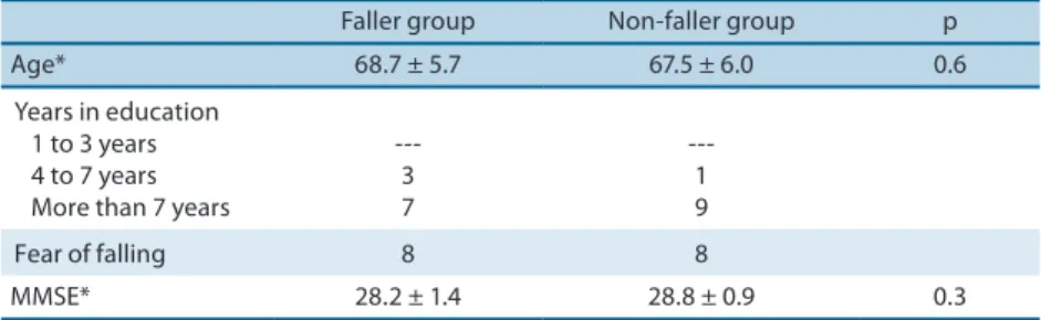
Twenty elderly women were enrolled who had or had not suffered falls during the 12 months preceding the tests and with mean ages of 68.7 ± 5.7 and 67.5 ± 6.0, respectively. Eighty-eight percent of the volunteers had studied for 7 years or more and 20% had spent from 4 to 7 years in educa-tion. Eighty-eight percent of the study population reported fear of falling (Table 1). The sample was selected by convenience, according to the inclu-sion and excluinclu-sion criteria.
Table 1. Characteristics of the sample: age, educational level, fear of falling and cognitive status.
Faller group Non-faller group p
Age* 68.7 ± 5.7 67.5 ± 6.0 0.6
Years in education 1 to 3 years 4 to 7 years More than 7 years
---3 7
---1 9
Fear of falling 8 8
MMSE* 28.2 ± 1.4 28.8 ± 0.9 0.3
*Figures shown are Mean ± Standard Deviation, MMSE: Mini Mental State Examination.
Tables 2 and 3 list means for MedFqW1 and MedFqW3 for the groups of elderly people with and without history of falling and their respective results after statistical analysis of the data. It was found that only the falling group had significantly lower results for MedFqW1 than for MedFqW3 for both muscles assessed.
Table 2. Comparison of means of normalized values for median frequencies for the gastrocnemius muscle for first and third analysis windows.
Moments Groups
Faller group Non-faller group p
MedFqW1 1.043 1.038 0.70
MedFqW3 0.953 0,987 0.42
Rate of loss 8.60% 4.90%
p 0.02 0.08
MedFqW1 - means of median frequencies for window 1, MedFqW3 - means of median frequencies for window 3.
Table 3. Comparison of means of normalized values for median frequencies for the vastus lateralis muscle for first and third analysis windows.
Moments Groups
Faller group Non-faller group p
MedFqW1 1.02 1.01 0.87
MedFqW3 0.97 0.99 0.31
Rate of loss 4.5% 1.8%
p 0.02 0.66
MedFqW1 - means of median frequencies for window 1, MedFqW3 - means of median frequencies for window 3
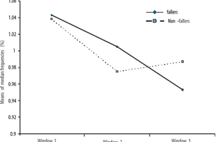
Figures 1 and 2 illustrate the behavior of Fmed during the data capture win-dows, for the groups with and without history of falling. The reduction in Fmed, which indicates fatigue, was greater among elderly people with a history of falling.
Figure 1. Means of normalized values of median frequency for the vastus lateralis muscle for fallers (n = 10) and non-fallers (n = 10).
Figure 2. Means of normalized values of median frequency for the gastrocnemius muscle for fallers (n = 10) and non-fallers (n = 10).
DISCUSSION
If falls are to be avoided after loss of postural stability, the central nervous system and the musculoskeletal system must both react accordingly21,22.
Falling among elderly women: analysis of muscle fatigue Silva et al.
In this study we opted to use a submaximal contraction lasting 30 sec-onds17 to test for muscle fatigue. With regard to this protocol, we observed
that holding the position for 30 seconds was sufficient to induce localized muscle fatigue in the muscles being tested. This claim is supported by the observation that initial and finalFmed values reduced in both groups. This behavior is widely reported in the literature as an indication that a muscle is working in a fatigued state23,24.
Muscle fatigue can be defined as a reduction in the neuromuscular system’s capacity to generate force during a sustained effort, leading to reduced muscle performance25. In electromyographic fatigue, the median
frequency can be observed to reduce, indicating a lower frequency of muscle motor unit firing. During the tasks undertaken in this study, the group with a history of falling had lower median frequencies than the group without a history of falling in both muscles tested, indicating a lower motor unit firing rate, which translates into a greater tendency to fatigue, and this de-cline could be a primary factor in the incidence of falls among the elderly26.
Muscle fatigue causes changes in the neuromuscular system, resulting in reduced muscle performance. Mian et al.27 claim that an increase in
co-contraction of the musculature needed to maintain balance increases metabolic costs for elderly people, which may explain the higher levels of fatigue among elderly people with a history of falling. The elderly develop adaptive muscle mechanisms to provide increased stability when walking, with or without obstacles in their path, which generates higher levels of muscle activity than are observed in young adults28,29, increasing the
likeli-hood of fatigue and, consequently, of falling.
When the rate of loss of median frequency was compared across groups the history of falling group had a higher rate of loss, indicating greater fa-tigue and greater risk of falling. There are two physiological consequences related to fatigue: peripheral disorders of muscle activation and failures of the central nervous system to adequately activate motor neurons25. These
disturbances involve changes in muscle strength and neuromuscular con-trol, impacting on amplitude and velocity of movements8. Postural control
is therefore inevitably affected, increasing the risk of falling.
One limitation to this study is the number of participants in each group. This was the result of the difficulties we encountered in trying to identify elderly people with a diagnosis of falling, since falls are not seen as diagno-ses, but as the causes of, for example, fractures. Another possible limitation is the fact that both groups were considered physically active, which may have contributed to the absence of statistical differences between them.
CONCLUSIONS
Report Series, 779). Available from: <http://whqlibdoc.who.int/trs/WHO_TRS_779. pdf> [2012 may 18].
2. Spirduso WW. Dimensões físicas do envelhecimento. Barueri: Manole; 2005.
3. Novaes RD, Santos EC, Miranda AS, Lopes KT, Riul TR. Causas e conseqüências de quedas em idosos como indicadores para implementação de programas de exercício físico. Lecturas en Educación Física y Deportes 2009;14(131).
4. Zinni JVS, Pussi FA. O papel da fisioterapia na prevenção da instabilidade e quedas em idosos. Available from: http://www.wgate.com.br/conteudo/medicinaesaude/ fisioterapia/traumato/instabilidade_postural_idoso.htm [2012 oct 16].
5. Fabrício SCC, Rodrigues RAP, Costa Junior ML. Causas e conseqüências de quedas de idosos atendidos em hospital público. Rev Saúde Públ 2004; 38(1):93-9.
6. Ferreira EAG, Marques AP. Postura e envelhecimento. In: Perracini, MR, Fló CM, editores. Funcionalidade e envelhecimento. 2a ed. Rio de Janeiro: Guanabara Koogan; 2011. p. 153-165.
7. Perracini MR. Prevenção e manejo de quedas. In: Ramos LR, coordenação. Guia de geriatria e gerontologia. Barueri: Manole; 2005. p.193-208.
8. Enoka RM, Stuart DG. Neurobiology of muscle fatigue. J Appl Physiol 1992; 72(5):1631-48.
9. Mademli L, Arampatzis A, Karamanidis K. Dynamic stability control in forward falls: postural corrections after muscle fatigue in young and older adults. Eur J Appl Physiol 2008;103(3):295-306.
10. Taylor JL, Butler JE, Gandevia SC. Changes in muscle afferents, motoneurons and motor drive during muscle fatigue. Eur J Appl Physiol 2000; 83(2-3):106-15.
11. Sharpe MH, Miles TS. Position sense at the elbow after fatiguing contractions. Exp Brain Res 1993;94(1):179-82.
12. Boyas S, Guevel A. Neuromuscular fatigue in healthy muscle: underlying factors and adaptation mechanisms. Ann Phys Rehabil Med 2011;54(2):88-108.
13. Carvalho T, Nóbrega ACL, Lazzoli JK. Posição oficial da Sociedade Brasileira de Medicina do Esporte: atividade física e saúde. Rev Bras Med Esporte 1996;2:1-3.
14. Brucki SMD, Nitrini R, Caramelli P, Bertolucci PHF, Okamoto IH. Sugestões para o uso do Mini-Exame do Estado Mental no Brasil. Arq Neuro-Psiquiat 2003;61(3B):777-81.
15. Sai AJ, Gallagher JC, Smith LM, Logsdon S. Fall predictors in the community dwelling elderly: a cross sectional and prospective cohort study. J Musculoskelet Neuronal Interact 2010;10(2):142-50.
16. Hermens HJ, Freriks B, Disselhorst-Klug C, Rau G. Development of recommenda-tions for SEMG sensors and sensor placement procedures. J Electromyogr Kinesiol 2000;10(5):361-74.
17. Merletti R. The standards for reporting EMG data. J Electromyogr Kinesiol 1999;9(1):3-4.
18. Hermens HJ, Merletti R, Rix H, Freriks B, editors. The state of the art on signal processing methods for surface electromyography: deliverable of the SENIAM pro-ject: SENIAM 7. Eschende (NE): Roessingh Research and Development; 1999. v. 7.
19. Earl JE, Schmitz RJ, Arnold BL. Activation of VMO and VL during dynamic mini-squat exercises with and without isometric hip adduction. J Electromyogr Kinesiol 2001;11(6):381-6.
20. Callegari-Jacques S. Bioestatística princípios e aplicações. Porto Alegre: Artmed; 2003.
21. Fasano A, Plotnik M, Bove F, Berardelli A. The neurobiology of falls. Neurol Sci 2012;33(6):1215-23.
Falling among elderly women: analysis of muscle fatigue Silva et al.
Corresponding author
Marcos Eduardo Scheicher Av. Hygino Muzzi Filho, 737, CEP 17525-900 - Marília, SP, Brasil. E-mail: [email protected]
23. Merletti R. Electromyography: physiology, engineering, and noninvasive aplica-tions. Italy: Philip Paker, 2004.
24. Maisetti O, Guevel A, Legros P, Hogrel J. SEMG power spectrum changes during a sustained 50% maximum voluntary isometric torque do not depend upon the prior knowledge of the exercise duration. J Electromyogr Kinesiol 2002;12:102-9.
25. Gandevia SC. Spinal and supraspinal factors in human muscle fatigue. Phys Rev 2001;81(4):1725-89.
26. Bellew JW, Fenter PC. Control of balance differs after knee or ankle fatigue in older women. Arch Phys Med Rehab 2006;87(11):1486-9.
27. Mian OS, Thom JM, Ardigò LP, Narici MV, Minetti AE. Metabolic cost, mechani-cal work, and efficiency during walking in young and older men. Acta Physiol 2006;186(2):127-39.
28. Brown LA, Gage WH, Polych MA, Sleik RJ, Winder TR. Central set influences on gait: age-dependent effects of postural threat. Exp Brain Res 2002;145(3):286-96.