www.jcol.org.br
Journal of
Coloproctology
Original article
Anatomical and physiological changes in pelvic
diaphragm in patients with chagasic megacolon
submitted to Duhamel surgery
Hélio Moreira Júnior, José P. T. Moreira, Raniere R. Isaac,
Arminda C. de Almeida, Hélio Moreira, Wilmar A. Klug
Hospital das Clínicas da Universidade Federal de Goiás (UFGO), Goiânia, GO, Brazil
a r t i c l e i n f o
Article history:
Received 12 February 2013 Accepted 20 April 2013
Keywords: Chagas disease Megacolon/surgery Intestinal constipation Anal canal
Surgical procedures of the digestive system
Perioperative care Evaluation
a b s t r a c t
Original contribution: understand the reasons why Duhamel surgery results in clinical im-provement of constipation in patients with Chagasic colopathy.
Background: Duhamel surgery is one of the most widespread techniques for the treatment of Chagasic megacolon, with low rates of recurrence of constipation.
Objective: evaluate the anatomical and physiological changes in the pelvic diaphragm of pa-tients with chagasic colopathy, as well as changes occurring after undergoing Duhamel surgery. Design: clinical data and results of cinedefecography, electromanometry and anorectal ul-trasound of the anal canal were evaluated in patients with Chagasic colopathy, before and after Duhamel surgery.
Location: Service of Coloproctology – Departament of Surgery, Faculdade de Medicina da Universidade Federal de Goiás. Patients: patients with positive serology for Chagas Disease, with constipation and radiological megacolon, who presented consecutively to the Chagas outpatient clinic and freely agreed to participate in this study, were prospectively included. Results: a total of 20 patients were included, with a mean age of 53.2 years, of which 16 were women. The following parameters were observed in the postoperative period: change in bowel frequency, of, on average, one evacuation every ten days to daily bowel movement; 16 patients used laxatives preoperatively and only one did, intermittently, in postopera-tive period. Electromanometry showed, postoperapostopera-tively, a decrease in anal resting pressure (60.88 to 37.2 mmHg p < 0.001) and anal squeeze pressures (244.3 mL to 161.25 p = 0.01), whereas ultrasound showed that 75% of the patients had abnormalities of the internal anal sphincter in the posterior anal canal juxtaposed to the pulled-through colon. Postoperative rectal emptying observed in cinedefecographic tests occurred more quickly and with less effort when compared with the preoperative i ndings. There was a change in the anorectal angle postoperatively, which became more obtuse, both during rest, straining and during evacuation.
Conclusions: the anatomical and functional changes in the pelvic diaphragm are signii cant after Duhamel surgery and together, they result in clinical improvement of patients.
© 2013 Elsevier Editora Ltda. All rights reserved.
* Corresponding author.
E-mail: [email protected] (H. Moreira-Júnior)
Palavras-chave: Doença de Chagas Megacolon/cirurgia Constipação intestinal Canal anal
Procedimentos cirúrgicos do sistema digestório Cuidados peri-operatórios Avaliação
r e s u m o
Avaliação das alterações anatômicas e funcionais do diafragma pélvico de pacientes portadores de colopatia chagásica submetidos à cirurgia de Duhamel
Contribuição original: compreender os motivos pelos quais a cirurgia de Duhamel resulta na melhora clinica da obstipação intestinal de pacientes com colopatia chagásica.
Antecedentes: a cirurgia de Duhamel é uma das técnicas mais difundidas para o tratamento do megacólon chagásico, com baixos índices de recidiva dos sintomas de obstipação intestinal. Objetivo: avaliar as alterações anatômicas e i siológicas do diafragma pélvico de pacientes portadores de colopatia chagásica e as mudanças ocorridas após serem submetidos à cirur-gia de Duhamel.
Desenho do estudo: foram avaliados os dados clínicos e os resultados de exames de cinedefe-cograi a, eletromanometria anorretal e o ultrassom do canal anal de pacientes portadores de colopatia chagásica, no pré e pós-operatório da cirurgia de Duhamel.
Localização: Serviço de Coloproctologia – Departamento de Cirurgia Faculdade de Medicina da Universidade Federal de Goiás.
Pacientes: foram inclusos, prospectivamente, pacientes com sorologia positiva para Doença de Chagas, com obstipação intestinal e megacólon radiológico, que se apresentaram conse-cutivamente ao ambulatório de Chagas e que livremente aceitaram participar desse estudo. Resultados: foram incluídos 20 pacientes, com média de idade de 53,2 anos, sendo 16 mulhe-res. Observou-se, no pós-operatório, uma mudança do ritmo intestinal de, em média, uma evacuação a cada dez dias para uma evacuação diária; 16 pacientes faziam uso de laxantes no pré-operatório e somente um o fazia, intermitentemente, no pós-operatório. A eletro-manometria evidenciou, no pós-operatório, uma diminuição das pressões anais de repouso (60,88 para 37,2 mmHg com p < 0,001), e da capacidade retal (244,3 para 161,25 mL, p = 0,01) e o ultrassom revelou que em 75% dos pacientes haviam alterações anatômicas do esfínc-ter inesfínc-terno na porção posesfínc-terior do canal anal, justaposto ao local de abaixamento do cólon. O esvaziamento da ampola retal, observado durante a cinedefecograi a pós-operatória, se processou mais rapidamente e com menor esforço quando comparado com os achados pré--operatórios. Houve mudança do ângulo anorretal no pós-operatório, que se tornou mais obtuso, tanto durante o repouso como durante o esforço evacuatório.
Conclusões: as alterações anatômicas e funcionais do diafragma pélvico são signii cativas após a cirurgia de Duhamel e que ela determina, em conjunto, a melhoria clínica dos pacientes.
© 2013 Elsevier Editora Ltda. Todos os direitos reservados.
Introduction
It is estimated that there are between 70 and 90 million
peo-ple worldwide at risk of being infected by Chagas disease.1
Recently, there was a signii cant increase in the number of
individuals infected by Trypanosoma cruzi in developed
coun-tries such as United States (USA), Japan, Canada and Spain.2
There are approximately 300,000 individuals with Chagas
disease in the United States,3 therefore, it constitutes a
new challenge to be faced by health authorities in these countries.
Surgical treatment of chagasic megacolon is indicated when there is progression and worsening of symptoms of constipation or progression to complicated forms of the disease, such as sigmoid volvulus or fecal impaction. A wide variety of surgical techniques has been described for the treatment of chagasic megacolon; many of them were based on prevalent physiopathological concepts of the dis-ease whenever they were described and others have been abandoned because they had high rates of of recurrence
of constipation or high rates of postoperative complica-tions.4-13
Bernardes et al. (1965) employed for the i rst time the Du-hamel technique in the treatment of chagasic megacolon,14
us-ing the proposed method made by this french surgeon for the treatment of congenital megacolon.15 This technique was latter
modii ed by Haddad et al.16
The Service of Coloproctology, of Universidade Federal de Goiás, has used Duhamel surgery as a routine technique for the treatment of chagasic megacolon since 1966, with some of the various modii cations that have been incorporated over the years.17-22 Despite of good surgical results, to date there has been
no detailed study that explained what anatomo-physiological changes result from this operation, which would be, in the end, responsible for the functional improvement of the patients.
Aim
Cha-gas colopathy after undergoing Duhamel surgery, using cinedefecography, electromanometry and anal ultrasound for the evaluation.
Materials and methods
Type and location of study
The study was developed at the Service of Coloproctology of the Department of Surgery, Hospital das Clínicas da Uni-versidade Federal de Goiás.
Ethical aspects
The study was performed in accordance with the Declaration of Helsinki and local regulations. The project was approved by the Research Ethics Committee (Hospital das Clínicas, Universidade Federal de Goiás). All patients enrolled in this study were informed on the purpose of the tests performed and signed a free and informed consent form.
Patients and Eligibility
A total of 20 patients with the following characteristics were prospectively selected from May 2006 to June 2008:
• positive serology for Chagas disease in at least one se-rological test;
• chronic constipation, dei ned as frequency of bowel movements of less than twice a week; and
• barium enema showing megacolon and/or anorectal electromanometry demonstrating achalasia of the in-ternal anal sphincter.
All patients underwent Duhamel-Haddad surgery and were submitted to cinedefecography, electromanometry and anal canal ultrasound preoperatively and at the third month after surgery. Patients also answered a specii c questionnaire to assess demographic data, the presence and severity of constipation, association with other clini-cal forms of Chagas disease functional status at these two distinct moments. The continence was assessed using Jorge
and Wexner incontinence scoore,23 ranging from 0 to 20,
with 0 being perfect continence. The classii cation of cha-gasic megaesophagus described in the study is based on radiological i ndings of these segment, with patients being assigned to groups I, II, III and IV, in which I is the least
advanced.28 The classii cation of sigmoidocele and/or
en-terocele used in our material was described by Jorge et al.,29
and only grade 3 cases were considered clinically signii -cant, i.e., the ones that reaches below the ischiococcygeal line.
Statistical analysis
Consensual statistic analysis show that a homogeneous sample of 20 patients with Chagas colopathy evaluated at two different times, by the same tests, in which control and post-treatment cases corresponded to the same group of
patients are adequate to evaluate anatomic and physiolgic changes after Duhamel surgery.
The comparison of qualitative variables observed in the pre- and postoperative periods was performed by McNemar test. Quantitative variables were assessed using Student’s t-test for paired samples or Wilcoxon t-test. The signii cance level was set at 5%.
Results
A total of 20 patients with Chagas colopathy were included in the study. The mean age of patients was 53.2 ± 11.2 years. Seventy-i ve percent of cases were females. The mean time of symptom onset was 16 years. The mean time without a bow-el movement was 10.3 days. Sixteen patients routinbow-ely used laxatives. Fourteen patients complained of abdominal pain and distension, 17 of sensation of incomplete evacuation, and seven had a history of fecal impaction. One case had a sig-moid volvulus and was treated by endoscopic distortion. Half of patients had Chagasic heart disease and seven patients presented with chagasic megaesophagus (i ve in group II and two in group III). Five patients had all three clinical forms of the disease (colopathy, esophagopathy and Chagas cardiopa-thy). Three patients had symptoms of anal incontinence and, in two cases, incontinence was related to gas, at a rate of one episode every 15 days (incontinence score = 2). Another pa-tient complained involuntary lost of liquid stool (speccially after using enemas) and gas, every 20 days and solid stools every 30 days (incontinence score = 5). Symptoms did not in-terfere with patient daily activities in any of these cases.
The postoperative bowel movement was a median of one daily bowel movement, ranging from three times a day to once every four days. Only one patient used laxatives, inter-mittently (once every 15 days on average). Two patients had persistent abdominal pain and distension, but with less in-tensity and frequency. Seven patients reported incomplete evacuation, being necessary to evacuate at least twice, the second almost immediately after the i rst evacuation.
There were no cases of fecal impaction or sigmoid volvu-lus postoperatively. Among the seven patients with chagasic megaesophagus, four reported dysphagia improvement after surgical treatment of megacolon. Four patients had symp-toms of anal incontinence after surgery, and three of these patients already had symptoms preoperatively.
There was a worsening of anal incontinence in two of these patients, who reported incontinence to gas almost daily and rarely for liquid stools (incontinence score = 5). The other patient reported that incontinence to gas became a weekly event (incontinence score = 3). The third patient, who had a preoperative incontinence score of 5, reported symptom im-provement after discontinuing the use of enemas and also reported having involuntary escape of gas daily, with no loss of liquid or formed stools (incontinence score = 4). No patient reported that symptoms of anal incontinence interfered with their usual daily activities.
showed abnormalities in 75% of patients. Three patients had complete injuries, with or without i brotic tissue surrounding the internal sphincter at the posterior hemicircumference of the anal canal, juxtaposed to the pulled-through colon (Fig. 1A) and another 12 patients had partial lesions of the sphinc-ter with or without scar tissue, at the possphinc-terior hemicircum-ference of the anal canal (Fig. 1B). Only i ve patients showed an intact sphincter anatomy. There were no signii cant changes at the perineal body thickness of female patients (mean 13.04 preoperatively to 12.6 mm postoperatively).
Cinedefecography showed, preoperatively, prolonged opening of the anal canal despite of an adequate relaxation of the puborectalis during evacuation in 95% of patients (Table 1). Eight female patients had rectocele. However, only two were considered clinically signii cant (greater than 3 cm and partially emptied after evacuation). Four patients had sig-moidocele (grades 1 and 2), but these i ndings did not cause obstruction of rectal contents during evacuation. One patient had partial relaxation of the puborectalis.
Static perineal descent was diagnosed in 35% of patients; 10% had associated static and dynamic perineal descent and 15% had dynamic descent, exclusively. Eighty-i ve percent of the patients had, preoperatively, a sensation of incomplete evacuation despite of a prolonged straining (Table 2). Objec-tive data with their respecObjec-tive means and standard deviations
observed at the cinedefecography preoperatively are summa-rized in Table 3.
Rectal emptying in postoperative cinedefecography ocurred faster and with less effort when compared with pre-operative i ndings (Table 1). The funnel-shaped image in the proximal portion of the anal canal during straining persist-ed in only two patients; however it did not determinpersist-ed any symptoms (both had daily stool frequency postoperatively, without effort).
Two out of eight patients who had preoperative rectocele were not diagnosed with the same problem postoperatively. Rectocele remained clinically signii cant postoperatively in the same two patients diagnosed before (Fig. 2). Both persist-ed with symptoms of incomplete evacuation, one of which eventually used digital maneuvers to have a successful bowel movement, both pre and postoperatively.
Four patients had sigmoidocele, three grade 1 and one grade 2, but these findings did not cause outlet obstruc-tion. Postoperative mean static and dynamic perineal de-scent decreased, when compared with values observed preoperatively. However, the difference between these two moments was only statistically significant for the static descent (Table 3).
Four patients postoperatively showed incomplete evacua-tion, two of them with signii cant rectocele. One patient had
A
B
Fig. 1 – Partial lesion (A) and thinning (B) of the internal anal sphincter at the posterior hemicircumference of the anal canal, juxtaposed to the pulled-through colon in different patients who underwent Duhamel surgery.
Table 1 – Observation of the prolonged opening of the anal canal in patients with Chagas colopathy, evaluated before and after Duhamel surgery.
Prolonged opening of the anal canal in the postoperative
period
Total
Yes No
Prolonged opening of the anal canal in the preoperative period
Yes n (%) 3 (15) 16 (80) 19 (95) No n (%) 0 (0) 1 (5) 1 (5)
Total n (%) 3 (15) 17 (85) 20 (100) p < 0.001
Table 2 – Observation of the sensation of incomplete evacuation in patients with Chagas colopathy, evaluated before and after Duhamel surgery.
Sensation of incomplete evacuation in the postoperative period
Total
Yes No
Sensation of incomplete evacuation in the preoperative period
Yes n (%) 3 (15) 14 (70) 17 (85) No n (%) 2 (10) 1 (5) 3 (15)
partial relaxation of the puborectalis muscle during straining. Another patient, despite the presence of residual contrast in the rectal stump, did not complain of a sensation of incom-plete evacuation.
There was a signii cant change in the anorectal angle post-operatively which became more obtuse, both during resting and evacuation (Table 3). The same was not observed when we compared the anorectal angle preoperatively with the angle formed between the pulled-through colon and the anal canal.
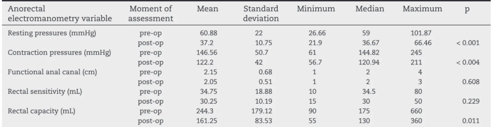
Postoperatively, the anorectal electromanometry showed a decrease in the mean resting and squeeze pressure of the anal canal of 60.88 mmHg to 37.2 mmHg (p < 0.01), and 146.56 mmHg to 122.2 mmHg (p < 0.004). The mean length of the functional anal canal did not change when compared pre- and postoperatively (2.15 to 2.05 cm, respectively, p = 0.608). The postoperative mean rectal capacity decreased from 244 mL to 161 mL (p = 0.01) (Table 4).
Discussion
Chronic constipation is the main clinical manifestation of Cha-gasic colopathy, which worsens progressively and become re-sistant to conservative medical treatment. The disease patho-genesis were identii ed by Habr-Gama et al.,26,27 as two basic
functional changes that could explain the onset of constipa-tion, both resulting from the destruction of Meissner and Auer-bach intestinal plexus: achalasia of the internal anal sphincter and colonic motor incoordination of the colon.
Cavenaghi et al.,28 in a series of 39 patients with Chagasic
megacolon, described that the rectoanal inhibitory rel ex (RAIR) is not always evident in patients with Chagas disease due to the use of insufi cient volumes of air insufl ation in the rectum during eletromanometric test. The authors observed that the RAIR was present in 43% of studied cases, with a mean volume of 196 mL. Other authors have shown that, in normal subjects, intrarectal balloon inl ation with up to two thirds of the rectal
threshold volume was enough to demonstrate RAIR.29,30 In our
study, we decreased the possibility of false negative RAIR con-sidering that rectal sensory threshold might be equivocal as it relies on patient’s information. The mean volume used to evaluate the rel ex was 80 mL. These values are below those re-ported by Cavenaghi et al.,28 and may be due to different
meth-odological approach used in our service. We think that eliciting RAIR with very high intrarectal volume does not consubstanti-ate a normal physiological stconsubstanti-ate, and may, as reported by Fang et al.,31 be an electromanometric evidence of the opening of the
anal canal caused by signii cant increase of intrarectal pres-sure other than a normal RAIR.
The reason why a large number of surgical techniques has been proposed for the treatment of Chagasic megacolon may
Fig. 2 – Radiographs of patients with Chagas colopathy, after Duhamel surgery, during evacuation straining and after evacuation, respectively. Note the presence of an incomplete emptied bulky anterior rectocele at the two distinct moments, with a partial emptied rectocele.
Table 3 – Anorectal and coloanal angles, static and dynamic perineal descents observed on cinedefecography in the pre and postoperative period of patients with chagasic colopathy who underwent Duhamel surgery.
Variable Moment of
assessment
Mean Standard
deviation
Minimum Median Maximum p
Anorectal angle at rest pre-op post-op
103.3 136.6
20.5 12.1
58 110
103 137.5
135
160 < 0.001 Coloanal angle at rest post-op 103.3 20.1 62 100 143 0.975 Anorectal angle at squeeze pre-op
post-op
81 120
21.5 11.4
42 95
76.5 121
120
145 < 0.001 Coloanal angle at squeeze post-op 84.7 19.7 51 80 135 0.411 Anorectal angle at evacuation pre-op
post-op
119 141
16.1 15.9
94 100
119 140.5
145
170 < 0.001 Coloanal angle at evacuation post-op 125.7 22.1 90 125 165 0.091 Static perineal descent pre-op
post-op
6.7 4.4
4.9 2.3
1.8 2
5.7 3.5
19.5
11 0.031 Dynamic perineal descent pre-op
post-op
2.8 2.3
2.1 2.2
0 0
3 1.75
6
be due to the lack of better knowledgement on the functional effects of surgery on these patient. The most appropriate sur-gery would be the one that, at least in part, addresses the de-scribed physiopathological changes for chagasic megacolon, with acceptable of morbidity and mortality rates.
The postoperative functional results of Duhamel surgery in the middle and long term are quite satisfactory, with ex-ceptional risk of recurrence of constipation.32,33,34 Even in
patients who complain of postoperative constipation, the severity of symptoms is much lower when compared to the preoperative state. However, there is a risk with post-opera-tive anal incontinence. In most cases, these symptoms are more common in the immediate postoperative period and improve over time.
Nevertheless, a minority of patients may have refractory symptoms of anal incontinence, which might impact their quality of life. In the present study, the functional results were satisfactory with signii cant improvement of constipation in all studied cases. The surgery resulted in the onset of subtile postoperative anal incontinence in only three cases, without interfering on their social or work activities. However, it should be noted that these results were postoperatively evaluated af-ter a short period of time.
The mechanisms for such good functional outcame after Duhamel surgery were, until now, partially understood. Sev-eral studies have shown that achalasia of the internal anal
sphincter persists after Duhamel surgery. Moreira35 observed
that motor incoordination between the pulled-through colon and rectal stump is restaured to a similar normal pattern as the contraction of the pulled-through colon is followed by rec-tal relaxation. It is speculated that this return to normal motor patterns between the colon and rectum would occur due to a side-to-side other than an end-to-end anastomosis of these two segments.
However, other anatomofunctional aspects resulting from the surgery could inl uence postoperative results. Our re-sults demonstrate that patients with chagasic megacolon, after undergoing Duhamel surgery, presented a decrease rest-ing pressure of the anal canal and rectal capacity, as well as an improvement in rectal sensory threshold. These electro-manometry i ndings are similar to those observed by Moreira JPT.36 Ultrasound of the anal canal provides an explanation for
these functional changes, as approximately 60% of patients af-ter surgery, showed reduced thickness of the inaf-ternal sphinc-ter and 15% had complete lesion of this muscle in the
poste-rior circumference of the anal canal; only 25% had an intact sphincter anatomy.
Fang et al.31 evaluated 29 patients with advanced
Chagas-ic megacolon through cinedefecography and detected a pro-longed time for evacuation when compared with asymptom-atic individuals.
These cinedefecography i ndings were similar to those ob-served in our study. Despite of an adequate relaxation of the puborectalis muscle during straining, the anal canal was not adequately opened, obstructing rectal emptying. Only after a considerable increase of intra-abdominal pressure that rectal contrast was eliminated, coni rming Fang et al.31 theory that
evacuation only occurs due to a difference of intrarectal pres-sure vs. resting pressure of the anal canal. At maximum stage of pushing, a funnel shape image was noted in the upper por-tion of the anorectal ring, which represents the dynamic dis-play of the internal anal sphincter achalasia.
Furthermore, we observed, in the pre-operative period of female patients other mechanisms that corroborated with constipation, especially larger than 4 cm rectoceles which did not emptied after straining. These bulky rectoceles determined postoperative sensation of incomplete evacuation, and need for digital maneuvers to successfully achieve a complete emp-ty rectum.
It is noteworthy in this study, the change in the anorectal displacement that occurred after Duhamel surgery. The pulled-through colon on the retrorectal space shifted foward the rectal stump axis, originating an obtuser anorectal angle. It is known that after this surgery, bowel transit includes the rectal stump as the last segment to store the fecal content.37 Therefore an
obtuser anorectal angle between the anal canal and the rectal stump becomes a facilitating mechanism for evacuation in the postoperative period of patients undergoing Duhamel surgery. We can therefore summarize that the anatomophysiologi-cal changes determined by Duhamel surgery adresses the physiopathological changes of Chagasic colopathy, includes:
1) Low latero-lateral anastomosis between pulled-through
colon and rectal stump. Study published by Moreira H.35
showed that after Duhamel surgery, the pulled-through colon and rectal stump start once again to show propul-sive motor coordination, i.e., the contraction of the colon wall is accompanied by rectal wall relaxation. This phe-nomenon facilitates the progression of colonic contents into the rectal stump.
Table 4 – Electromanometric assessments, performed pre- and postoperatively in patients with Chagas colopathy submitted to Duhamel surgery.
Anorectal electromanometry variable Moment of assessment Mean Standard deviation
Minimum Median Maximum p
Resting pressures (mmHg) pre-op post-op 60.88 37.2 22 10.75 26.66 21.9 59 36.67 101.87
66.46 < 0.001 Contraction pressures (mmHg) pre-op
post-op 146.56 122.2 50.7 42 61 56.7 144.82 120.94 245
211 < 0.004 Functional anal canal (cm) pre-op
post-op 2.15 2.05 0.68 0.51 1 1 2 2 4 3 0.608 Rectal sensitivity (mL) pre-op
post-op 34.75 30.25 18.88 10.19 10 15 34.5 30 80 50 0.229 Rectal capacity (mL) pre-op
2) The resting pressure of the anal canal decreases after sur-gery as a consequence of partial damage or thinning of the internal anal sphincter, associated with i brotic process, at the posterior hemicircumference of the anal canal, whe-re the colon was pulled-through. This electromanometric i nding has important implications for rectal empting as the intrarectal pressure needed to overcome internal anal sphincter achalasia will be reduced.
3) Postoperative cinedefecographic assessments showed that as the rectal stump is laid up with contrast, the anastomo-tic angle between the rectal stump and the pulled-through colon moves posteriorly and works as a valvular mecha-nism that precludes retrograde transit during the effort for evacuation, enhancing intrarectal pressure. Therefore, an effortless evacuation is achieved as a more efi cient rise of intrarectal pressure is expected. For a few other patients, the rectal stump and pulled-through colonic contrast were emptied at the same moment.
4) A retrorectal, endoanal space is created to pull-through the colon. Consequently, the axis of the remaining rectal stump is shifted anteriorly, determining an obtuser ano-rectal angle. During straining, there is an almost complete rectii cation (close to 180°) between the axis of the anal ca-nal and the remaining rectal stump, facilitating the emp-tying of the rectum.
This study provides a better understanding of why there are good functional results in the postoperative period of pa-tients with Chagasic megacolon who underwent Duhamel surgery. New knowledge can determine other technical varia-tions for different subgroups of patients with megacolon, in order to further improve postoperative results. We can, for in-stance, re-evaluate the use of this technique in patients who already have deteriorated sphincter function preoperatively, those with a large megarectum, or even in patients who have other conditions causing outlet obstruction in association to chagasic megacolon, such as rectocele and anismus.
Conclusions
Anal sphincter pressure in the pre-operative time measured by eletromanometry is within normal limits; there is a sig-nii cant decrease in resting and squeeze pressures of the anal canal after Duhamel surgery, as well as a decreased capacity and increased rectal sensitivity.
Ultrasound of the anal canal, postoperatively, detected partial or total lesion of the internal anal sphincter muscle, such as perimuscular i brosis, in 75% of cases, always at the posterior hemicircumference of the anal canal, where the co-lon was pulled-through.
Postoperative cinedefecography coni rmed a signii cant im-provement in rectal emptying during straining. A signii cant change in the anorectal angle was also observed at rest and during straining (which became, postoperatively, more obtuse). Secondary factors in the genesis of constipation may be present in some patients with Chagasic colopathy (rectocele, perineal descent and anismus).
Signii cant anatomical and functional changes were identii ed by anorectal eletromanometry, anal ultrasound
and cinedefecography in patients who underwent Duhamel surgery for chagasic megacolon, which ultimately resulted in clinical impovement.
Confl ict of interest
The authors declare no conl icts of interest.
R E F E R E N C E S
1. Coura JR, Dias JCP. Epidemiology, control and surveillance of Chagas disease: 100 years after its discovery. Memórias do Instituto Oswaldo Cruz 2009; 104: 31-40.
2. Parker ER, Sethi A. Chagas disease: coming to a place near you. Dermatol Clin, v. 29, n. 1, p. 53-62, Jan 2011.
3. Bern C, Montgomery SP. An estimate of the burden of Chagas disease in the United States. Clin Infect Dis 2009;49:e52–4. 4. Almeida AD. Sigmoidectomia abdominal no tratamento do
megacolo. Rev Paul Med 1963; 62: 349-60.
5. Capelhuchnik P, Silva Prado W. Hemicolectomia esquerda no tratamento do megacólon-resultado em 57 casos. In: Congresso da International Society of University Colon and Rectal Surgeons [Anais] São Paulo; 1970.
6. Cardoso AA. Modii cação na técnica de anastomose cólon-retal na cirurgia do megacólon chagásico. Rev Bras Cir 1963; 15:16-22. 7. Celso NM. Tratamento do megacolo adquirido pela
anorretomiectomia. Rev Assoc Med Minas Gerais 1962;13:139-46. 8. Cutait DE. Megacolo. Nova técnica de retossigmoidectomia
abdômino- perineal sem colostomia. In: 1º Congresso Latino-Americano, 11º Internacional e 10º Brasileiro de Proctologia [Anais]. São Paulo. 1960; 2:831-46.
9. Ferreira-Santos R, Carril CF. Megacólon chagásico – Análise de 36 casos tratados cirurgicamente. 1º Congresso Latino Americano, 2º Internacional e 10º Brasileiro de Proctologia [Anais] São Paulo. 1960; 2:863-5.
10. Paula Pinto E. Tratamento cirúrgico do megacólon adquirido. 1º Congresso Latino Americano, 2º Internacional e 10º Brasileiro de Proctologia. [Anais] São Paulo 1960; 1:294-5.
11. Simonsen O, Habr-Gama A, Gazal P. Retossigmoidectomia endoanal com ressecção da mucosa retal. Rev Paul Med 1960; 57:116-7.
12. Vasconcelos E. Nova técnica de abaixamento do cólon sem suturas, no megacólon. Rev Hosp Clin Fac Med São Paulo 1961; 16:355-62.
13. Vasconcelos E. Colectomia sub-total e anastomose ceco-retal no tratamento do megacólon do adulto. Rev Hosp Clin Fac Med São Paulo 1964; 19:332-7.
14. Bernardes de Oliveira A, Oliveira C, Goldenberg S. A anastomose ano-reto-cólica no tratamento cirúrgico do megacólon (operação de Duhamel). An Paul Med Cir 1965; 89:45-61; 83-94; 103-18.
15. Duhamel B. Une nouvelle operation pour le megacólon congenital: L’abaissement retro-rectal et trans-anal du cólon et son aplicattion possible au traitment de quelques autres malformations. Presse Med 1956; 64:2249-50.
16. Haddad J, Raia A, Corrêa-Neto A. Abaixamento retro-retal do cólon com colostomia perineal no tratamento do megacólon adquirido. Rev Assoc Med Bras 1965; 11:83-8.
19. Moreira H. Megacólon chagásico. In: Speranzini M, Ramos M. Manual do residente de cirurgia. 2ª. ed. Rio de Janeiro: Guanabara-Koogan; 1981. p.286-92.
20. Moreira H. Tratamento cirúrgico do megacolo chagásico pela técnica de Duhamel-Haddad. Aspectos técnicos. In. Pinotti HW. Atualização cirúrgica. São Paulo: Robe; 1982. v. 7, p.161-82. 21. Lins Neto MAF. Operação de Duhamel modii cada com
anastomose colorretal imediata para o tratamento do megacólon chagásico. [Tese - Mestrado]. São Paulo: Faculdade de Medicina da Universidade de São Paulo; 1997.
22. Moreira H, Moreira JPT, Moreira Jr H. Avaliação crítica do tratamento cirúrgico de megacólon chagásico. Castro LP, Savassi-Rocha PR, Lacerda Filho A, Conceição AS, (Eds) Tópicos em Gastroenterologia 11 - Avanços em coloproctologia. Rio de Janeiro: Medsi; 2001. p.403-13.
23. Jorge JMN, Wexner SD. Etiology and management of fecal incontinence. Disease of Colon and Rectum 1993; 36: 77-97. 24. Rezende JM, Lauar KM, Oliveira AR. Aspectos clínicos e
radiológicos da aperistalsis do esôfago. Rev Bras Gastroenterol 1960;12:2473/462.
25. Jorge JMN, Yang YK, Wexner SD. Incidence and clinical signii cance of sigmoidoceles as determined by a new classii cation system. Dis Colon Rectum 1994; 37:1112-7. 26. Habr-Gama A. Motilidade do cólon sigmóide e do reto.
Contribuição à i siopatologia do megacólon chagásico. [Tese - Doutorado]. São Paulo: Faculdade de Medicina da Universidade de São Paulo; 1966.
27. Habr-Gama A, Costa Curta L, Raia A. Anatomia e i siologia do esfíncter interno do ânus. Rev Soc Bras Proctol. 1970; 3:21-30. 28. Cavenaghi S, Felicio OC, Ronchi LS, Cunrath GS, Melo MM,
Netinho JG. Prevalence of rectoanal inhibitory rel ex in chagasic megacolon. Arq Gastroenterol.. 2008; 45:128-31.
29. Coller JA. Clinical application of anorrectal manometry. Gastroenterol Clin North Am. 1987; 16:17-33.
30. Stein BL, Roberts PL. Manometry and the rectoanal inhibitory rel ex. In: Wexner SD, Bartolo DCC. (Eds). Constipation: Etiology, evaluation and management. Oxford: Butterworth Heineman; 1995. p 63-76.
31. Fang CB, Klug WA, Aguida HASC, Ortiz JÁ, Capelhunik P. Avaliação das pressões anais em doentes com o megacólon chagásico. Rev Bras Colo-Proctol. 1998; 18:173-7.
32. Moreira H, Rezende JM, Sebba F, Azevedo IF, Leite ACA, Soares EP. Chagasic megacólon. Coloproctology. 1985; 5:260-6. 33. Moreira H, Rezende JM. Megacólon chagásico:
clínica, diagnóstico e tratamento. In: Moreira H. (Ed.) Coloproctologia: conceitos. Goiânia: Escaleno; 1993. p.15-60. 34. Moreira H. Megacólon chagásico. In: Cordeiro F, Meneghelli U,
Resende JM (Eds). Federação Brasileira de Gastroenterologia. A gastroenterologia no Brasil: subsídios para sua história até o ano 2000. Rio de Janeiro: Revinter; 2001. p.377-407.
35. Moreira H. Estudo eletromanométrico da atividade motora do coto retal e do cólon descendente em pacientes chagásicos submetidos às cirurgias de Hartmann e de Duhamel. [Tese - Mestrado]. Goiânia: Faculdade de Medicina da Universidade Federal de Goiás; 1972.
36. Moreira JPT. Avaliação eletromanométrica no pré e no pós operatório de pacientes portadores de megacolon chagásico submetidos á cirurgia de Duhamel. [Tese - Mestrado] Goiânia: Instituto de Patologia Tropical -Universidade Federal de Goiás; 2001.