Introduction
Congenital bicuspid aortic valve and advanced age are leading risk factors for calcific aortic valve stenosis (CAVS), the third leading cause of heart disease in adults [1-3]. However, epidemi-ological databases have helped to define other clinical risk factors, including smoking, male sex, hypertension, hypercholesterolemia, excess body mass index, and diabetes [4, 5].
Genetic predisposition has also been suggested as a mechanism for CAVS development [6], al-though few studies have elucidated the genes and genetic variants involved. Probst et al [7] demonstrated a familial aggregation for CAVS in western France. Polymorphisms in the vitamin D receptor are more common in patients with
CAVS [8], and loss of function through mutation of a polymorphism in the NOTCH1 receptor ac-celerates calcification of the valve in patients with CAVS and other congenital heart abnor-malities [9, 10].
Two studies have correlated genetic lipoprotein abnormalities in patients predisposed to CAVS development [11, 12] and Nordstrom et al [13] demonstrated that Pvull polymorphisms in the estrogen receptor alpha gene are related to presence of aortic stenosis (AS) in postmeno-pausal women and to lipid levels in adolescent females, suggesting that this polymorphism may influence CAVS risk by affecting lipid levels. The importance of oxidative modification of low-density lipoprotein (LDL) in the pathogenesis of
Original Article
Relationship of PON1 192 and 55 gene polymorphisms to
calcific valvular aortic stenosis
Luis M Moura1,6, Susana Faria2, Miguel Brito3, Fausto J Pinto4, Steen D Kristensen5, Isabel M Barros6,
Nalini Rajamannan7, Francisco Rocha-Gonçalves1
1Department of Medicine, Oporto School of Medicine, University of Oporto, Portugal; 2CMAT; Mathematical Research
Centre, Department of Mathematics and Applications, University of Minho, Campus de Azurém, Portugal; 3
Depart-ment of Biology, Technological School of Health, Lisbon Polytechnic Institute, Portugal; 4Department of Cardiology,
Lisbon School of Medicine, University of Lisbon, Portugal; 5Department of Cardiology, Aarhus University Hospital,
Skejby, Denmark; 6Pedro Hispano Hospital, Matosinhos, Portugal; 7Northwestern School of Medicine, Chicago, IL,
USA
Received March 8, 2012; accepted April 22, 2012; Epub May 15, 2012; Published June 15, 2012
Abstract: Introduction and Objectives: Paraoxonases may exert anti-atherogenic action by reducing lipid peroxidation. Previous studies examined associations between polymorphisms in the paraoxonase 1 (PON1) gene and develop-ment of coronary artery disease (CAD), with inconsistent results. Given the similarities in clinical and pathophysiologi-cal risk factors of CAD and pathophysiologi-calcific aortic valve stenosis (CAVS), we postulated a link between PON1 alleles and CAVS progression. Methods: We investigated the association between PON1 55 and 192 single nucleotide polymorphisms (SNPs), their enzyme activity, and CAVS progression assessed by aortic valve area and transvalvular peak velocity in 67 consecutive patients with moderate CAVS and 251 healthy controls. Results: PON1 paraoxonase activity was higher in CAVS patients (P<0.001). The PON1 genotype Q192R SNP (P=0.03) and variant allele (R192) (P=0.01) frequencies differed between CAVS patients and controls. Significant association existed between PON1 enzyme activity, phenotypic effects of PON1 192 genotype polymorphisms, and CAVS progression, but not between PON1 55 and high-density lipoprotein (P=0.44) or low-density lipoprotein cholesterol (P=0.12), between 192 genotype and high -density lipoprotein (P=0.24) or low-density lipoprotein cholesterol (P=0.52). Conclusion: The PON1 genotype Q192R SNP has an important effect on CAVS disease progression. This study helps outline a genotype-phenotype relation-ship for PON1 in this unique population.
atherosclerosis is well recognized [14, 15]. As an anti-atherogenic mediator, high-density lipo-protein (HDL), besides its key role in reverse cholesterol transport [16], protects itself and LDL against oxidation and reduces lipoprotein-associated peroxides[17-19]. The antioxidant property of HDL has been attributed, in part, to the paraoxonase 1 (PON1) enzyme [15, 18-20]. PON1 is an aryldialkylphosphatase synthesized in the liver and transported in systemic circula-tion exclusively in associacircula-tion with an HDL sub fraction containing apolipoproteins (apo) A-I and apoJ [21-23]. Serum PON1 activity is inversely related to coronary artery disease (CAD), hyper-cholesterolemia, and diabetes [23, 24]. Previous studies [23] reported two protein poly-morphisms in PON1 Q192R and L55M that ex-ert significant effects on PON1 activity. We typed these two polymorphisms in all subjects and determined allele frequencies. Individually, both polymorphisms exert significant effects on all substrate activities. The previous studies showed that QQ individuals had lower PON1 activity compared to RR, whereas LL individuals had higher activity for all substrates compared to MM. The M55 variant is associated with lower PON1 activity levels, probably as a result of a large imbalance with the Q192 PON1 vari-ants [24, 25]. The variability of PON1 activity towards paraoxon is partly determined by poly-morphisms in the paraoxonase gene located on chromosome 7 between q21.3 and 22.1. Of these, the codon 192 polymorphism produces two alloenzymes, with low and high activity, re-spectively [26, 27]. In the high-activity alloen-zyme, glutamine (Q allele) is replaced by argin-ine (R allele) at codon 192. In several but not all studies, the R192 allele was associated with CAD risk [28-33] and adverse lipoprotein profile [28, 32, 34-36]. PON1 has other common poly-morphisms at codon 55 [Leu (L)/Met (M)] that correlate with enzyme activity [26, 37]. In-creased enzyme activity has been associated with higher HDL levels in some populations, but not in all studies [38]. The PON 192 and 55 polymorphisms also have been associated with risk of ischemic stroke [39].
In the past decade, several studies have investi-gated the association between the single nu-cleotide polymorphisms (SNPs) of the PON gene cluster and CAD susceptibility, but have yielded apparently conflicting results. The PON1 geno-type Gln192Arg (Q192R) SNP was significantly associated with stroke in CARE trial patients
[25] and PON1 activity is lower in patients with CAVS and inversely correlated with CAVS sever-ity [26]. Inconsistent results could be due to insufficient power, the small effect of the poly-morphism on CHD risk, and/or false-positive results. A recent meta-analysis suggested a weak association, overall, between Q192R SNP and CHD risk [27]. Altogether, these clinical studies focused on Q192R PON1 SNP suggest that a certain genetic background may be re-lated to higher risk of developing CAVS. We chose PON1 as a candidate marker to test the hypothesis that common PON1 genetic variants could influence aortic valve calcification and CAVS progression. The study aimed to evaluate (1) the allele frequency of Q192R and L55M SNPs, (2) the effects of this polymorphism on serum lipoprotein variables, and (3) the relation-ship between Q192R and L55M PON1 gene SNPs and PON1 enzyme activity in CAVS pro-gression in our patient population.
Methods Research ethics
The study complied with the Helsinki declara-tion and the Pedro Hispano Hospital Institu-tional Review Board (Matosinhos, Oporto, Portu-gal) approved the protocol (IRB-22352). All par-ticipants were fully informed and provided signed consent before enrollment.
Study population
Participants in the prospective Rosuvastatin Affecting Aortic Valve Endothelium (RAAVE) study were asymptomatic patients with moder-ate AS, defined as an aortic valvular area (AVA) of 1.0 to 1.5 cm2. We selected 255 consecutive,
statin-naïve, new AS referrals to our inpatient and outpatient cardiology clinic [28] and ex-cluded those with history of CAD (myocardial infarction and/or angiographically demon-strated coronary artery stenosis), aortic valve surgery, congenital cardiac disease (bicuspid aortic valve), statin therapy, active or chronic liver disease, or currently taking an angiotensin-converting enzyme inhibitor. No other medica-tions were contraindicated, including other anti-hypertensives, oral hypoglycemics, or insulin. Other exclusion criteria were echocardiographic evidence of rheumatic mitral valve disease, aor-tic regurgitation, subaoraor-tic obstruction, and creatinine concentration ≥2.0 mg/dL (to mini-mize the risk of hypercalcemia as a potential
confounding factor).
Of the remaining 135 eligible participants, 14 were unsuitable due to technical problems with initial echocardiography or difficulties in obtain-ing medical history. Sufficient DNA was avail-able for genetic analysis of 67 of the 121 as-ymptomatic AS patients finally included, before randomization to treatment. To estimate 10-year cardiovascular risk of death, we included a control group of 251 healthy volunteers (age >45 years, without CAD or AS, with low cardio-vascular risk based on sex, age, smoking status, blood pressure, and total cholesterol). Data ob-tained for all participants included age, sex, smoking history, and presence of hypercholes-terolemia, arterial hypertension (>140/90 Hg), and diabetes. At inclusion, no participant had detectable evidence of inflammatory, neoplas-tic, metabolic, or vascular disease in an initial history, examination, or routine tests. Patients received standard clinical care throughout the study.
Biochemistry
Blood was drawn after 12-hour fasting for rou-tine clinical biochemistry in the Pedro Hispano Hospital laboratory, following standard institu-tional protocols. In-hospital audit had previously demonstrated sample result variability between 5% and 15% for these assays (data not shown). PON1 activity
Serum PON1 paraoxonase (PON1-para) activity was determined using paraoxon (Aldrich Chemi-cal Co, St. Louis, MO) as previously described [29]. Briefly, the assay buffer was prepared from 0.132 M Tris–HCl, pH 8.5, and 1.32 mM CaCl2. Each set of assays used 6 mM freshly prepared paraoxon substrate solution (120 mM paraoxon in acetone diluted with 0.132 mM Tris –HCl). The assay tube contained 152 µl Tris buffer, 8 µl serum (1:2 diluted with water), and 40 µl 6-mM paraoxon. The reaction was initi-ated at 37.8°C by adding the substrate solu-tion; absorbance was continuously monitored at 405 nm. A molar extinction coefficient of 18.05 x 10³ was obtained and used to calculate
activ-ity, and units were expressed as U/L. PON1 genotyping
Genetic samples were taken following 10 min-utes of supine rest and immediately prior to
echocardiography. Whole blood samples were collected, stored, and frozen at -80°C. Extrac-tion was done using phenol-chloroform purifica-tion, and DNA was eluted in 50 μl TE. Polymor-phisms were genotyped using a TaqMan allelic discrimination assay. Q192R (rs662) and L55M (rs854580) PON1 polymorphisms were typed using primers and probes from the Applied Bio-systems Predesigned Drug Metabolism Geno-typing Assays program (Applied Biosystems, Foster City, CA). Genotyping was done with the ABI Prism 7900HT Sequence Detection System and typing errors were reduced using Simwalk2 (Sobel and Lange, 1996). The Q192R genotype was 97% concordant with previous assess-ments based on PON1 activity status [30] and, in a subset of unrelated individuals, both poly-morphisms were in Hardy-Weiberg equilibrium. Echocardiography
Comprehensive transthoracic echocardiograms were performed and reported in a single central echocardiography laboratory (Acuson Sequoia C512, Siemens Healthcare, Erlangen, Germany) by either of two experienced cardiologists spe-cialized in echocardiography (LM and IB). Imme-diate inter-observer review determined which studies should be repeated to maintain quality control. Everyone involved in performing and interpreting echocardiograms was blinded to patient treatment status. Hemodynamic pro-gression was assessed using serial echocardio-graphic studies at 6-month intervals.
Standard Doppler recordings were made of the left ventricular outflow tract and aortic valve from multiple views to obtain peak transvalvular jet velocity (Vmax), mean and peak gradients,
and AVA in accordance with international guide-lines [31, 32]. Left atrial volume relative to body surface area was estimated from end-systolic measurements [33-37]. Reproducibility of the echocardiography observations by the two cardi-ologists was assessed in a 30-patient subset. All echocardiography parameters examined showed intra-class coefficient correlation (ICC) between 0.962 and 0.989 (intra-observer) and between 0.955 and 0.992 (inter-observer). Con-ventional Doppler and tissue Doppler imaging (TDI) parameters [38, 39] had good intra- and inter-observer coefficients of reproducibility (1.56-9.02). Coefficients of variation and repro-ducibility were, for intra-observer variability, 1.88% and 0.16 m/sec for Vmax and 3.89% and
and 2.01% and 0.14 m/sec for Vmax, and 3.25% and 0.08 cm2 for AVA, respectively, for
observer 2, and for inter-observer variability, 2.0% and 0.14 m/sec for Vmax and 2.91% and
0.04 cm2 for AVA, respectively.
Statistical analysis
Patient characteristics are presented as mean±standard deviation (SD) for normally dis-tributed continuous variables; categorical vari-ables are expressed as frequencies (percentages) and non-normally distributed con-tinuous variables as the median (1st-3rd
inter-quartile ranges). Continuous variables were as-sessed usingKolmogorov-Smirnov test to verify the assumption of normality.
Reproducibility was assessed with the Bland and Altman method [40, 41] and results ex-pressed as a coefficient of reproducibility (twice the SD of the differences). Differences in base-line characteristics between CAVS patients and controls were compared with independent t-tests for normally distributed continuous vari-ables, Mann-whitney U tests for non-normally distributed continuous variables, and chi-square for categorical variables.
Differences between PON1 genotypes groups and lipid profile were compared using one-way ANOVA. The relationship between enzyme activ-ity and alleles was studied using one-way ANOVA followed by Tukey HSD multiple compari-son test. Differences between AS patients and controls in the distribution of genotype and al-lele frequencies and estimates of Hardy-Weinberg equilibrium were tested using chi-squared. Logistic regression modeling deter-mined the association between the independ-ent effect of the Q192R SNP and the effect of interaction between genotype, PON1 activity, and other covariates on lipids and CAVS pro-gression. All statistical analysis was performed using SPSS, version 17.0.1 for Windows SPSS Inc, Chicago, IL). Significance level was .05 for all analyses.
Results
Population characteristics
Patients and controls were chosen from con-secutive referrals to our inpatient and outpa-tient cardiology clinic. Social and clinical
differ-ences between the two groups have been pub-lished in detail elsewhere [28]; a summary is provided in Table 1. In addition to having similar serum lipid levels, the groups did not differ with respect to sex (male: AS group 62.7% vs. con-trols 49.0%, P=0.320) or other established car-diovascular risk factors. The AS group was more likely to have elevated total cholesterol (227.0±52.0 md/dL vs. 189.1±44.1 mg/dL in controls; P=0.004), elevated HDL-cholesterol (53.4±10.7 md/dL vs. 43.7±11.4. mg/dL, re-spectively; P=0.001), and diabetes mellitus (44.8% vs. 11.8%, respectively; P=0.001). Baseline cardiac function
Both groups had similar normal left ventricular systolic function (P=0.067), with ejection frac-tion 54.3±2.1% in AS patients and 55.6±4.4% in controls (Table 1). End-diastolic long axis di-ameter was 51.4±5.7 mm in the AS group vs. 52.3±4.2 mm in controls (P=0.311). End-systolic long-axis diameter was 33.4±4.8 mm in the AS group vs. 34.6±3.9 mm in controls (P=0.165). There was no difference between groups in left ventricular mass (P=0.091) or left atrial volume (P=0.625).
Paraoxonase1 55 and 192 polymorphism and the presence of CAVS
The PON1 55 and 192 polymorphisms had al-lele frequencies of 51.8% for L and 83.2% for R in AS patients, and 42% and 72.9%, respec-tively, in controls (Table 2). Analysis of Q192R distributions in the different PON1 genotypes showed significant prevalence in the frequency of R alleles in the AS patients (P=0.01); in addi-tion, 62.7% were carriers of RR variant allele vs. 45.0% of controls (P=0.03). With respect to PON1 L55M, there were no significant differ-ences in allelic frequency and allelic variants (LL, MM and LM) between groups (P>0.05) (Table 2). For both PON1 55 and 192 polymor-phisms, good agreement was found between the observed and expected genotype frequen-cies according to the Hardy-Weinberg equilib-rium (data not shown).
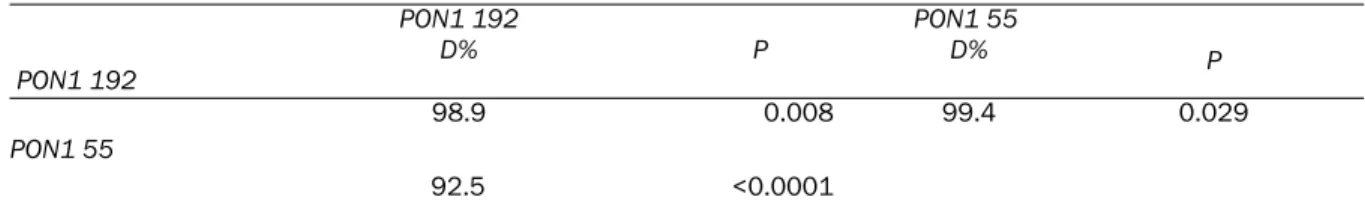
Linkage disequilibrium
The analysis of linkage disequilibrium between PON1 polymorphisms is presented in Table 3. We observed, as have other studies, a strong (D>90%) genetic linkage disequilibrium
between PON1 polymorphisms at the promoter and coding regions.
Paraoxonase1 55 and 192 polymorphisms and lipid profile
Mean levels of serum lipoproteins for Q192R and L55M are presented in Table 4. After ad-justing for age, sex, and blood pressure, the Q and R alleles were not associated with signifi-cantly increased HDL cholesterol (P=0.44); the Table 1. Baseline Clinical Characteristics
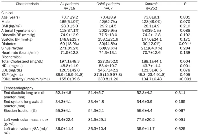
Characteristic All patients
n=318 CAVS patients n=67 Controls n=251 P
Clinical Age (years) 73.7 ±9.2 73.4±8.9 73.8±9.1 0.831 Male 165(51.9%) 42(62.7%) 123(49.0%) 0.070 BMI (kg/m2) 28.3 ±5.0 29.2 ±4.5 28.1±4.9 0.210 Arterial hypertension 118(37.1%) 20(29.9%) 98(39.1 %) 0.088 Diastolic BP (mmHg) 74.9±12.9 77.5±13.0 74.2±12.8 0.192 Systolic BP(mmHg) 148.8±23.7 153.1±21.9 147.6±24.1 0.171 Diabetes 60 (18.9%) 30(44.8%) 30(12.0%) 0.001* Sinus rhythm 271(85.2%) 60(89.6%) 211(84.0 %) 0.284
Heart rate (beats/min) 71.5±12.8 74.3±13.1 70.7±12.6 0.138
Biochemical
Total Cholesterol (mg/dL) 197.1±48.3 227.0±52.0 189.1±44.1 0.004 HDL (mg/dL) 45.8±11.9 53.4±10.7 43.7±11.4 0.001 LDL (mg/dL) 126.5±42.0 145.7±42.2 121.3±40.5 0.078 BNP (pg/mL) 39.9 (15.9-91.8) 37.9 (15.9-87.3) 45.3 (23.4-91.8) 0.405
PON1 activity (µmol/min/mL) 155.0±39.6 230.8±1.20 134.7±6.48 <0.001
Echocardiography
End-diastolic long-axis di-ameter (mm)
52.1±4.6 51.4±5.7 52.3±4.2 0.311 End-systolic long-axis
di-ameter (mm)
34.3±4.1 33.4±4.8 34.6±3.9 0.165 Ejection fraction (%) 55.3±4.1 54.3±2.1 55.6±4.4 0.067 Left ventricular mass index
(g/m2)
78.4±22.4 81.9±29.1 77.5±20.2 0.091 Left atrial volume/SA (mL/
m2)
36.0±11.4 36.3±10.4 35.9±11.7 0.625
Values are means±standard deviations for normally distributed continuous variables, medians (interquartile range) for non-normally distributed continuous variables, and frequencies (percentages) for categorical variables. Abbrevia-tions: CAVS, calcific aortic valvular stenosis; BMI, body mass index; BP, blood pressure; HDL, high-density lipoprotein cholesterol; LDL, low-density lipoprotein cholesterol; BNP, brain natriuretic peptide; PON1, Paraoxonase 1; SA,
sur-face area.
Table 2. Frequency of Paraoxonase 1 (PON1) 55 and 192 Polymorphisms
Polymorphism
(frequencies and percentages)
Genotype CAVS patients n=67 % P PON1: Q192R 0.03 RR 42 62.7 113 45.0 QR 20 29.9 98 39.1 QQ 5 7.5 40 15.9 Allele Frequency 0.01 R 124 83.2 344 72.9 Q 25 16.8 128 27.1 PON1: L55M >0.05 LL 20 29.9 54 21.6 MM 20 29.9 94 37.3 LM 27 40.3 103 41.1 Allele Frequency >0.05 L 67 50.8 211 42.0 M 65 49.2 291 58.0 Controls n=251 %
same was true for L and M alleles (P=0.24). Neither Q nor R allele genotypes were associ-ated with serum levels of total cholesterol (P=0.25 and P=0.34, respectively) or LDL cho-lesterol (P=0.12 and P=0.52, respectively). Paraoxonase 1 55 and 192 polymorphisms and PON1 enzyme activity
The carriers of RR variant allele demonstrated a significantly higher AVA reduction than the
carri-ers of QQ variant allele (one-way ANOVA, Tukey’s HSD test p=0.04) and QR variant allele (one-way ANOVA, Tukey’s HSD test p=0.02). Moreover the carriers of QR variant allele have showed a significantly lower Vmax increase than
QQ allele (one-way ANOVA, Tukey’s HSD test p=0.04) and RR variant allele (one-way ANOVA, Tukey’s HSD test p=0.04) (Table 5).
Moreover, individually, both polymorphisms ex-erted significant effects on all substrate activi-Table 3. Linkage disequilibrium among Paraoxonase 1(PON1) polymorphisms
PON1 192 PON1 192 D% P PON1 55 D% P 98.9 0.008 99.4 0.029 PON1 55 92.5 <0.0001 D%=percentage of linkage disequilibrium. P values from chi-square analysis.
Table 4. Relationship between paraoxonase 1(PON1) 55 and 192 genotype and lipid serum levels
Lipid Profile RR QQ QR P Total Cholesterol (mg/dL) 210.9±52.3 240.0±49.2 226.6±52.8 0.25 HDL Cholesterol 51.8±11.4 45.3±8.0 51.2±124 0.44 LDL Cholesterol 134.7±42.4 161.8±42.5 152.7±41.1 0.12 LL MM LM P Total Cholesterol (mg/dL) 244.1±61.3 260.2±79.2 246.7±75.6 0.34 HDL Cholesterol 79.2±9.8 55.8±7.8 51.2±159 0.24 LDL Cholesterol 144.9±53.4 161.8±42.5 172.3±63.3 0.52 Values are means±standard deviations. Statistically significant (P <0.05) between compared groups. Abbreviations: HDL, high-density lipoprotein; LDL, low-density lipoprotein.
Table 5. Correlation of enzyme activity with PON1 192 and 55 genotype and CAVS progression
Q192R Activity (U/L) RR QQ QR P 168.74±4.16 103.52±2.13 145.45±3.17 <0.001 L55 M Activity (U/L) LL MM LM P 136.56±2.45 116.93±7.23 129.77±2.92 0.12 Echo Parameters RR QQ QR P AVA Reduction (cm2) -0.14±0.06 -0.07±0.06a -0.04±0.07a 0.029 Vmax Increase (m/s) 0.19±0.16b 0.17±0.16b 0.05±0.16 0.041 LL MM LM P AVA Reduction (cm2) -0.17±0.06 -0.14±0.08 -0.12±0.10 0.62 Vmax Increase (m/s) 0.17±0.19 0.19±0.13 0.15±0.11 0.84
Values are means±standard deviations . Statistically significant (P <0.05) between compared groups. PON activity in U/L=micromole of substrate hydrolysed/min. Statistically significant (P <0.05) between compared groups. (a) signifi-cant differences compared to RR allele, (b) signifisignifi-cant differences compared to QR allele Abbreviations: AVA: aortic valvular area e VMAX:transvalvular jet velocity
ties. In addition, RR individuals had higher activ-ity than QQ or QR individuals, and LL individuals, compared to MM and LM, had even lower PON1 substrate activities.
Paraoxonase 1 55 and 192 polymorphisms and CAVS progression
To test the genotype effect on CAVS progression we evaluated the independent association be-tween PON1 activity and genotypes as markers of disease progression using a multiple logistic regression analysis, having adjusted the model for predictors such as baseline AVA, sex, age, hypertension, diabetes mellitus, LDL choles-terol, degree of valve calcification, LV hypertro-phy, MR, LV end-diastolic volume, statin use, and baseline CAVS severity. Disease progres-sion was considered a variable of independent outcome and was classified according to the criteria presented above. Results of the step-wise multiple logistic regression are shown in Table 6. The odds ratio and associated 95% confidence intervals were estimated to assess the magnitude of the association between the various factors and disease progression. PON1 activity was strongly associated with CAVS dis-ease progression. In comparison to Q192 allele, the presence of R192 allele was associated with a 23% greater increase in CAVS disease progression.
Discussion
The present study demonstrates that (1) the relative allele frequency of the Q192R SNP of the PON1 gene differs markedly between CAVS patients and controls, (2) genotype has no ef-fect on lipid profile, and (3) the variation of PON1 activity could explain the variation in phe-notypic results, particularly CAVS progression. PON1 is considered anti-atherogenic because of its ability to destroy the inflammatory lipid
per-oxides formed by LDL oxidation [15, 42], which promotes many of the steps in CAVS progres-sion. Because PON1 protects the aortic valve against degenerative processes by inhibiting LDL oxidation, decreased PON1 activity will in-crease oxidative stress and inflammation, while excessive inflammation will accelerate valve degeneration and stenosis. To date, however, no specific genetic marker has been identified that predicts CAVS development. Whether or not there is a genetic contribution, in addition to multiple clinical atherosclerotic risk factors, to the development and progression of degenera-tive CAVS remains controversial: some in vitro studies have shown increased protection of the Q 192 allele against LDL oxidation compared to the R192 allele [19, 42], while other studies have found equal [43] or decreased protection [44]. Because it has been suggested that poly-morphisms negatively influence PON1 transcrip-tion or activity (increasing the rate of atheroscle-rosis), we analyzed the influence of Q192R and L55M SNPs in patients with CAVS.
Previous studies associated the R192R variant with increased levels of serum LDL cholesterol, HDL cholesterol, and triglycerides [45, 46]; re-cent studies have found either no adverse geno-typic effect on lipoprotein variables [47-49] or a positive association between the Q192 allele and levels of HDL cholesterol and apo-I [50]. In addition, the physical relationship of PON1 with HDL and the existence of cholesterol regulatory elements at the PON1 locus suggest a further relationship of PON1 with lipoproteins, which may contribute to its role in vascular disease [51]. The biological basis for these divergent observations, including ours, is not clear. Our study suggests that Q192 PON1 bearers were better protected from oxidative damage caused by serum LDL cholesterol than were bearers of R192 PON1 and confirms recent in vitro find-ings [35]. In our study, PON1 genotype Q192R SNP and its activity were also significantly corre-lated with CAVS progression evaluated by AVA and Vmax. These results are consistent with
ear-lier findings on CAD progression and stroke [25, 52].
The main limitation of this study is the small number of patients. Although our patient popu-lation was homogeneous in terms of demo-graphic characteristics, our results could have been influenced at least in part, as in many ob-servational studies, by differences other than Table 6. Predictors of CAVS progression by
step-wise multiple logistic regression
PON1 activity OR 95% CI P
1.35 1.1-1.7 0.003
R192 allele
1.23 1.1-14 0.002
Statistically significant (P <0.05) between compared groups. Abbreviations CI: confidence interval; OR: odds ratio
genetics. As a genetic association study, there are certain limitations in that it cannot provide evidence of causative relationship between the Q192R genotype and outcome phenotypes. Al-though the observed associations are consis-tent with biological plausibility, it is likely that the genotypic effect may be due to linkage dis-equilibrium with another closely linked func-tional mutation in the PON1 gene or a nearby gene in the chromosome. Further, because this is a non-randomized and observational study, our results are necessarily limited by the popu-lation frequencies of individual alleles, and gene -gene and/or gene-environment interaction might have uniquely influenced the outcome measures in this study cohort. For these rea-sons additional studies with a larger sample size are needed to confirm our observations. In conclusion, we show that the Q192R polymor-phism of the PON1 gene and PON1 activity influ-ence the risk of CAVS progression. However, knowledge of a simple aspect of the PON1 phe-notype may not provide a complete picture of the role PON1 may play in metabolism and even in an individual’s susceptibility to cardiovascular disease. PON1 is controlled primarily by varia-tion at the structural gene, but also by variavaria-tion at several other unidentified genes. Neverthe-less, the approach we used holds potential for future experimental and clinical trials designed to shed light on the role of polygenic factors in the CAVS progression.
Acknowledgements
We thank E Lilly, PhD, for improving the use of English in the manuscript.
Address correspondence to: Prof. Dr. Luis M Moura, Serviço de Medicina A, Faculdade de Medicina da Universidade do Porto, Alameda Prof. Hernani Monteiro; 4200-319, Porto, Portugal. Tel: +351 225513659; Fax: +351226092318; E-mail: [email protected]
References
[1] Freeman RV, Crittenden G and Otto C. Acquired aortic stenosis. Expert Rev Cardiovasc Ther 2004; 2: 107-116.
[2] Sverdlov AL, Ngo DT, Chapman MJ, Ali OA, Chirkov YY and Horowitz JD. Pathogenesis of aortic stenosis: not just a matter of wear and tear. Am J Cardiovasc Dis 2011; 1: 185-199. [3] Sverdlov AL, Ngo DT and Horowitz JD.
Patho-genesis of aortic sclerosis: association with low
BMI, tissue nitric oxide resistance, but not sys-temic inflammatory activation. Am J Cardiovasc Dis 2012; 2: 43-49.
[4] Aronow WS, Ahn C, Kronzon I and Goldman ME. Association of coronary risk factors and use of statins with progression of mild valvular aortic stenosis in older persons. Am J Cardiol 2001; 88: 693-695.
[5] Stewart BF, Siscovick D, Lind BK, Gardin JM, Gottdiener JS, Smith VE, Kitzman DW and Otto CM. Clinical factors associated with calcific aortic valve disease. Cardiovascular Health Study. J Am Coll Cardiol 1997; 29: 630-634. [6] Rajamannan NM. Calcific aortic stenosis:
les-sons learned from experimental and clinical studies. Arterioscler Thromb Vasc Biol 2009; 29: 162-168.
[7] Probst V, Le Scouarnec S, Legendre A, Jous-seaume V, Jaafar P, Nguyen JM, Chaventre A, Le Marec H and Schott JJ. Familial aggregation of calcific aortic valve stenosis in the western part of France. Circulation 2006; 113: 856-860.
[8] Ortlepp JR, Hoffmann R, Ohme F, Lauscher J, Bleckmann F and Hanrath P. The vitamin D receptor genotype predisposes to the develop-ment of calcific aortic valve stenosis. Heart 2001; 85: 635-638.
[9] Garg V, Muth AN, Ransom JF, Schluterman MK, Barnes R, King IN, Grossfeld PD and Srivastava D. Mutations in NOTCH1 cause aortic valve disease. Nature 2005; 437: 270-274.
[10] Garg V. Molecular genetics of aortic valve dis-ease. Curr Opin Cardiol 2006; 21: 180-184. [11] Avakian SD, Annicchino-Bizzacchi JM, Grinberg
M, Ramires JA and Mansura AP. Apolipopro-teins AI, B, and E polymorphisms in severe aor-tic valve stenosis. Clin Genet 2001; 60: 381-384.
[12] Novaro GM, Sachar R, Pearce GL, Sprecher DL and Griffin BP. Association between apolipopro-tein E alleles and calcific valvular heart dis-ease. Circulation 2003; 108: 1804-1808. [13] Nordstrom P, Glader CA, Dahlen G, Birgander
LS, Lorentzon R, Waldenstrom A and Lorentzon M. Oestrogen receptor alpha gene polymor-phism is related to aortic valve sclerosis in postmenopausal women. J Intern Med 2003; 254: 140-146.
[14] Steinberg D, Parthasarathy S, Carew TE, Khoo JC and Witztum JL. Beyond cholesterol. Modifi-cations of low-density lipoprotein that increase its atherogenicity. N Engl J Med 1989; 320: 915-924.
[15] Navab M, Berliner JA, Watson AD, Hama SY, Territo MC, Lusis AJ, Shih DM, Van Lenten BJ, Frank JS, Demer LL, Edwards PA and Fogelman AM. The Yin and Yang of oxidation in the devel-opment of the fatty streak. A review based on the 1994 George Lyman Duff Memorial Lec-ture. Arterioscler Thromb Vasc Biol 1996; 16: 831-842.
[16] Fielding CJ and Fielding PE. Molecular physiol-ogy of reverse cholesterol transport. J Lipid Res 1995; 36: 211-228.
[17] Mackness MI, Arrol S, Abbott C and Durrington PN. Protection of low-density lipoprotein against oxidative modification by high-density lipopro-tein associated paraoxonase. Atherosclerosis 1993; 104: 129-135.
[18] Watson AD, Berliner JA, Hama SY, La Du BN, Faull KF, Fogelman AM and Navab M. Protec-tive effect of high density lipoprotein associated paraoxonase. Inhibition of the biological activity of minimally oxidized low density lipoprotein. J Clin Invest 1995; 96: 2882-2891.
[19] Aviram M, Rosenblat M, Bisgaier CL, Newton RS, Primo-Parmo SL and La Du BN. Paraoxonase inhibits high-density lipoprotein oxidation and preserves its functions. A possi-ble peroxidative role for paraoxonase. J Clin Invest 1998; 101: 1581-1590.
[20] Mackness B, Davies GK, Turkie W, Lee E, Rob-erts DH, Hill E, RobRob-erts C, Durrington PN and Mackness MI. Paraoxonase status in coronary heart disease: are activity and concentration more important than genotype? Arterioscler Thromb Vasc Biol 2001; 21: 1451-1457. [21] Blatter MC, James RW, Messmer S, Barja F and
Pometta D. Identification of a distinct human high-density lipoprotein subspecies defined by a lipoprotein-associated protein, K-45. Identity of K-45 with paraoxonase. Eur J Biochem 1993; 211: 871-879.
[22] Kelso GJ, Stuart WD, Richter RJ, Furlong CE, Jordan-Starck TC and Harmony JA. Apolipopro-tein J is associated with paraoxonase in human plasma. Biochemistry 1994; 33: 832-839. [23] McElveen J, Mackness MI, Colley CM, Peard T,
Warner S and Walker CH. Distribution of paraoxon hydrolytic activity in the serum of patients after myocardial infarction. Clin Chem 1986; 32: 671-673.
[24] Mackness MI, Harty D, Bhatnagar D, Winocour PH, Arrol S, Ishola M and Durrington PN. Serum paraoxonase activity in familial hypercholester-olaemia and insulin-dependent diabetes melli-tus. Atherosclerosis 1991; 86: 193-199. [25] Ranade K, Kirchgessner TG, Iakoubova OA,
Devlin JJ, DelMonte T, Vishnupad P, Hui L, Tsu-chihashi Z, Sacks FM, Sabatine MS, Braunwald E, White TJ, Shaw PM and Dracopoli NC. Evaluation of the paraoxonases as candidate genes for stroke: Gln192Arg polymorphism in the paraoxonase 1 gene is associated with increased risk of stroke. Stroke 2005; 36: 2346-2350.
[26] Maganti K, Rigolin VH, Sarano ME and Bonow RO. Valvular heart disease: diagnosis and man-agement. Mayo Clinic proceedings. Mayo Clinic 2010; 85: 483-500.
[27] Wang M, Lang X, Zou L, Huang S and Xu Z. Four genetic polymorphisms of paraoxonase gene and risk of coronary heart disease: a
meta-analysis based on 88 case-control studies. Atherosclerosis 2011; 214: 377-385.
[28] Moura LM, Ramos SF, Zamorano JL, Barros IM, Azevedo LF, Rocha-Goncalves F and Rajaman-nan NM. Rosuvastatin affecting aortic valve endothelium to slow the progression of aortic stenosis. J Am Coll Cardiol 2007; 49: 554-561. [29] Rainwater DL, Mahaney MC, Wang XL, Rogers J,
Cox LA and Vandeberg JL. Determinants of variation in serum paraoxonase enzyme activity in baboons. J Lipid Res 2005; 46: 1450-1456. [30] Richter RJ, Jampsa RL, Jarvik GP, Costa LG and
Furlong CE. Determination of paraoxonase 1 status and genotypes at specific polymorphic sites. Curr Protoc Toxicol 2004; Chapter 4: Unit4 12.
[31] Bonow RO, Carabello BA, Chatterjee K, de Leon AC, Faxon DP, Freed MD, Gaasch WH, Lytle BW, Nishimura RA, O'Gara PT, O'Rourke RA, Otto CM, Shah PM, Shanewise JS, Smith SC, Jacobs AK, Adams CD, Anderson JL, Antman EM, Fuster V, Halperin JL, Hiratzka LF, Hunt SA, Lytle BW, Nishimura R, Page RL and Riegel B. ACC/AHA 2006 guidelines for the management of patients with valvular heart disease: a report of the American College of Cardiology/American Heart Association Task Force on Practice Guidelines (writing Committee to Revise the 1998 guidelines for the manage. Journal of the American College of Cardiology 2006; 48: e1-148.
[32] Galderisi M, Henein MY, D'Hooge J, Sicari R, Badano LP, Zamorano JL and Roelandt JR. Rec-ommendations of the European Association of Echocardiography: how to use echo-Doppler in clinical trials: different modalities for different purposes. Eur J Echocardiogr 2011; 12: 339-353.
[33] Rodriguez L, Garcia M, Ares M, Griffin BP, Naka-tani S and Thomas JD. Assessment of mitral annular dynamics during diastole by Doppler tissue imaging: comparison with mitral Doppler inflow in subjects without heart disease and in patients with left ventricular hypertrophy. Am Heart J 1996; 131: 982-987.
[34] Nishimura RA and Tajik AJ. Evaluation of dia-stolic filling of left ventricle in health and dis-ease: Doppler echocardiography is the clini-cian's Rosetta Stone. J Am Coll Cardiol 1997; 30: 8-18.
[35] Nagueh SF, Middleton KJ, Kopelen Ha, Zoghbi Wa and Quiñones Ma. Doppler tissue imaging: a noninvasive technique for evaluation of left ventricular relaxation and estimation of filling pressures. Journal of the American College of Cardiology 1997; 30: 1527-1533.
[36] Jassal DS, Tam JW, Dumesnil JG, Giannoccaro PJ, Jue J, Pandey AS, Joyner CD, Teo KK and Chan KL. Clinical usefulness of tissue Doppler imaging in patients with mild to moderate aortic stenosis: a substudy of the aortic stenosis pro-gression observation measuring effects of
rosu-vastatin study. J Am Soc Echocardiogr 2008; 21: 1023-1027.
[37] Steine K, Rossebo AB, Stugaard M and Peder-sen TR. Left ventricular systolic and diastolic function in asymptomatic patients with moder-ate aortic stenosis. Am J Cardiol 2008; 102: 897-901.
[38] Lund O, Flo C, Jensen FT, Emmertsen K, Niel-sen TT, RasmusNiel-sen BS, HanNiel-sen OK, Pilegaard HK and Kristensen LH. Left ventricular systolic and diastolic function in aortic stenosis. Prog-nostic value after valve replacement and under-lying mechanisms. Eur Heart J 1997; 18: 1977-1987.
[39] Weber M, Arnold R, Rau M, Elsaesser A, Brandt R, Mitrovic V and Hamm C. Relation of N-terminal pro B-type natriuretic peptide to pro-gression of aortic valve disease. Eur Heart J 2005; 26: 1023-1030.
[40] Bland JM and Altman DG. Statistical methods for assessing agreement between two methods of clinical measurement. Lancet 1986; 1: 307-310.
[41] Bland JM and Altman DJ. Regression analysis. Lancet 1986; 1: 908-909.
[42] Durrington PN, Mackness B and Mackness MI. Paraoxonase and atherosclerosis. Arterioscler Thromb Vasc Biol 2001; 21: 473-480.
[43] Cao H, Girard-Globa A, Berthezene F and Moulin P. Paraoxonase protection of LDL against peroxidation is independent of its es-terase activity towards paraoxon and is unaf-fected by the Q-->R genetic polymorphism. J Lipid Res 1999; 40: 133-139.
[44] Kuremoto K, Watanabe Y, Ohmura H, Shimada K, Mokuno H and Daida H. R/R genotype of human paraoxonase (PON1) is more protective against lipoprotein oxidation and coronary ar-tery disease in Japanese subjects. J Atheroscler Thromb 2003; 10: 85-92.
[45] Saha N, Roy AC, Teo SH, Tay JS and Ratnam SS. Influence of serum paraoxonase polymor-phism on serum lipids and apolipoproteins. Clin Genet 1991; 40: 277-282.
[46] Hegele RA, Brunt JH and Connelly PW. Multiple genetic determinants of variation of plasma lipoproteins in Alberta Hutterites. Arterioscler Thromb Vasc Biol 1995; 15: 861-871.
[47] Antikainen M, Murtomaki S, Syvanne M, Pahlman R, Tahvanainen E, Jauhiainen M, Frick MH and Ehnholm C. The Gln-Arg191 polymor-phism of the human paraoxonase gene (HUMPONA) is not associated with the risk of coronary artery disease in Finns. J Clin Invest 1996; 98: 883-885.
[48] Herrmann SM, Blanc H, Poirier O, Arveiler D, Luc G, Evans A, Marques-Vidal P, Bard JM and Cambien F. The Gln/Arg polymorphism of hu-man paraoxonase (PON 192) is not related to myocardial infarction in the ECTIM Study. Atherosclerosis 1996; 126: 299-303.
[49] Mackness B, Mackness MI, Arrol S, Turkie W, Julier K, Abuasha B, Miller JE, Boulton AJ and Durrington PN. Serum paraoxonase (PON1) 55 and 192 polymorphism and paraoxonase activ-ity and concentration in non-insulin dependent diabetes mellitus. Atherosclerosis 1998; 139: 341-349.
[50] Ruiz J, Blanche H, James RW, Garin MC, Vaisse C, Charpentier G, Cohen N, Morabia A, Passa P and Froguel P. Gln-Arg192 polymorphism of paraoxonase and coronary heart disease in type 2 diabetes. Lancet 1995; 346: 869-872. [51] Deakin S, Leviev I, Guernier S and James RW.
Simvastatin modulates expression of the PON1 gene and increases serum paraoxonase: a role for sterol regulatory element-binding protein-2. Arterioscler Thromb Vasc Biol 2003; 23: 2083-2089.
[52] Wheeler JG, Keavney BD, Watkins H, Collins R and Danesh J. Four paraoxonase gene polymor-phisms in 11212 cases of coronary heart dis-ease and 12786 controls: meta-analysis of 43 studies. Lancet 2004; 363: 689-695.