ORIGINAL ARTICLE
Analysis of synaptic proteins in the cerebrospinal fluid
as a new tool in the study of inborn errors
of neurotransmission
Sofia T. Duarte&Carlos Ortez&Ana Pérez&Rafael Artuch&Angels García-Cazorla
Received: 26 September 2010 / Revised: 22 November 2010 / Accepted: 25 November 2010 / Published online: 13 January 2011 # SSIEM and Springer 2010
Abstract In a few rare diseases, specialised studies in cerebrospinal fluid (CSF) are required to identify the underlying metabolic disorder. We aimed to explore the possibility of detecting key synaptic proteins in the CSF, in particular dopaminergic and gabaergic, as new procedures that could be useful for both pathophysiological and diagnostic purposes in investigation of inherited disorders of neurotransmission. Dopamine receptor type 2 (D2R), dopamine transporter (DAT) and vesicular monoamine transporter type 2 (VMAT2) were analysed in CSF samples
from 30 healthy controls (11 days to 17 years) by western blot analysis. Because VMAT2 was the only protein with intracellular localisation, and in order to compare results, GABA vesicular transporter, which is another intracellular protein, was also studied. Spearman’s correlation and Student’s t tests were applied to compare optical density signals between different proteins. All these synaptic proteins could be easily detected and quantified in the CSF. DAT, D2R and GABA VT expression decrease with age, particularly in the first months of life, reflecting the expected intense synaptic activity and neuronal circuitry formation. A statistically significant relationship was found between D2R and DAT expression, reinforcing the previous evidence of DAT regulation by D2R. To our knowledge, there are no previous studies on human CSF reporting a reliable analysis of these proteins. These kinds of studies could help elucidate new causes of disturbed dopaminergic and gabaergic transmission as well as understanding different responses to L-dopa in inherited disorders affecting dopamine metabolism. Moreover, this approach to synaptic activity in vivo can be extended to different groups of proteins and diseases.
Introduction
Specialised investigations in cerebrospinal fluid can be crucial to identify some inborn errors of metabolism. Defects of biogenic amines (dopamine and serotonine), GABA and glycine make up the group of diseases that are currently considered to be inborn errors of neurotransmitters. In these disorders, quantification of some particular CSF metabolites (biogenic amines, GABA or glycine) is necessary in the diagnostic process. However, although growing evidence suggests that genetic or pathologic alterations of
Communicated by: Sedel Frederic Competing interests: None declared.
S. T. Duarte
:
C. Ortez:
A. Pérez:
A. García-CazorlaDepartment of Neurology, Hospital Sant Joan de Déu, Barcelona, and CIBER-ER (Biomedical Network Research Centre
on Rare Diseases, Instituto de Salud Carlos III, Madrid, Spain
S. T. Duarte
Neuropaediatric Department, Hospital D. Estefânia, CHLC, EPE and CEDOC, Faculdade de Ciências Médicas da Universidade Nova de Lisboa,
Lisboa, Portugal R. Artuch
Department of Biochemistry, Hospital Sant Joan de Déu, Barcelona, and CIBER-ER (Biomedical Network Research Centre on Rare Diseases, Instituto de Salud Carlos III,
Madrid, Spain
A. García-Cazorla (*)
Neurology Department, Hospital Sant Joan de Deu, Passeig Sant Joan de Deu, 2,
08950 Esplugues, Barcelona, Spain e-mail: [email protected]
proteins involved in synaptic transmission may underlie a number of neurological and psychiatric disorders (Südhof and Malenka 2008; Südhof 2008; Witzmann et al. 2005; Corradini et al.2009; Kauer and Malenka 2007), the study of these proteins is not normally included in the array of investigations carried out in disorders of this kind. In vivo measurement of synaptic activity is difficult to carry out in humans. CSF samples are easily accessible by standard lumbar puncture techniques. Additionally, biochemical changes in CSF may reflect pathological alteration of CNS physiology, as previously demonstrated (Ormazabal et al. 2005). Accordingly, CSF receives a wide variety of molecules released by different CNS cell populations (Thouvenot et al. 2008). Several authors, including some from our group, have shown that there is a strong variability of CSF biogenic amines with age in children not suffering neurologic disease (Ormazabal et al. 2005), which suggests that the same variability may be expected when analysing synaptic proteins related to classical neurotransmitter function.
Synaptic transmission depends on neurotransmitter pools stored within vesicles that undergo regulated exocytosis. In the case of dopaminergic transmission, the vesicular monoamine transporter-2 (VMAT2) is responsible for the loading of dopamine (DA) and other monoamines into synaptic vesicles (Cartier et al.2010). The DA transporter (DAT) carries DA across the plasmalemmal membrane from the synaptic cleft into the cytoplasm (Volz et al.2009). The central actions of DA are mediated by five distinct receptors that belong to the G-protein receptor family. They are classified as D1-like (D1 and D5) and D2-like (D2, D3 and D4), and their interaction with dopamine translates into activation/inhibition of specific neurons and circuitries (De Mei et al. 2009). Those suffering D2 receptor (D2 R) genetic mutations exhibit postural abnormalities, bradykinesia with delayed initiation of movements, impaired coordination and prolonged periods of immobility (Glickstein and Schmauss 2001). These symptoms are also characteristic of patients suffering from dopaminergic neurotransmission disorders.
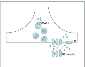
In spite of the critical role of these proteins in dopaminergic transmission (Fig. 1), information about procedures for studying them in CSF is very scarce, to our knowledge. However, such knowledge would be useful to increase understanding of the mechanisms of disturbed dopaminergic transmission in primary (inherited disorders of dopamine) and secondary deficiencies.
We aimed to analyse, in the CSF of a control population, the synaptic proteins VMAT2, DAT and D2 R. Because VMAT2 was the only protein with intracel-lular localisation, GABA vesicular transporter (GABA VT), which is another intracellular protein, was also included in the study. We evaluated the correlation of these proteins with each other and with the age of controls. Additionally, because total CSF protein
concen-tration is known to decrease with age, we introduced this variable in the age correlation studies.
Methods CSF samples
The study was performed in 30 subjects (age range: 11 days–17 years; average: 1.8 years) whose CSF samples were submitted to Hospital San Joan de Déu laboratory for analysis under suspicion of viral or bacterial meningitis or encephalitis. Lumbar puncture was performed in the emergency room, and the first ten drops were used for routine cytochemical/microbiological studies. Inflammatory markers (C-reactive protein and procalcitonin) were also analysed. Exclusion criteria were diagnosis of viral or bacterial meningitis, a chronic neurological condition, and hematic or xanthochromic CSF (blood contamination). Samples from patients were obtained in accordance with the Helsinki Declaration of 1964, as revised in 2000. The ethical committee of the Hospital Sant Joan de Déu approved the study. After collection, samples were stored at−80°C up to the moment of the analysis.
Western blot
Western blot analysis was performed for each protein (DAT, D2 R, VMAT2, GABA VT). A sample of 20 μL of CSF was loaded into the gel and proteins were separated on 10% sodium dodecyl sulphate-polyacrylamide gels and trans-ferred to polyvinylidene difluoride (PVDF) membranes (Amersham™ Hybond™–ECL; GE Healthcare). The
mem-VMAT 2
DAT
D2 receptor
Fig. 1 Schematic representation of the dopaminergic synapse, illustrating the link between VMAT2 (vesicular monoamine transporter 2), DAT (dopamine transporter ) and D2 R (D2 receptor)
branes were incubated in TBST buffer (0.02 M Tris-base, pH7.6, 0.8% NaCl, 0.1% Tween 20) supplemented with 5% dried skimmed milk for 60 min to block non-specific binding. Anti-DAT extracellular loop (1:1,000; Sigma®), anti-D2 R (1:1,000; Millipore®), anti-VMAT 2 (1:1,000; Santa Cruz Biotechnology®), and anti-GABA VT (1:500; Millipore®) antibodies were added, and the preparations were incubated at 4°C overnight. The membranes were washed three times with TBST buffer and then incubated with appropriate anti-rabbit (1:3,000; Promega®) or anti-mouse (1:5,000; Promega®) IgG secondary antibodies at room temperature for 1 h. The blot was then washed six times with TBST and prepared with ECL (Pierce® ECL Western Blotting Substract; Thermo Scientific) for developing. Relative levels of each protein were quantified by measuring optical densities (OD) of the corresponding bands with Quantity One® V 4.3.1. software.
CSF total protein concentration, procalcitonin and C-reactive protein determination
CSF total protein and C-Reactive Protein concentrations were measured by standard automated procedures in an Architect ci8200 analyser (Abbott, USA). Procalcitonin was measured by fluorimetric detection, with Brahams-Kryptor-Atom® analyser.
Statistical analysis
Statistical analysis was performed with the SPSS 17.0 program. Spearman’s correlation test was applied to search for associations among the OD signals of the different proteins and with the age of controls. Multivariate linear regression analysis was used to adjust the results by total CSF protein concentration. Student’s t test was applied to compare OD signals of the different proteins according to gender. Statistical significance was considered as p<0.05.
Results
DAT, D2R, VMAT2 and GABA vesicular transporter were clearly detectable at different ages using conventional western blot analysis in all the CSF samples studied (Fig.2).
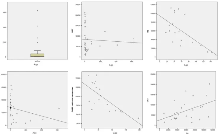
VMAT2 expression measured as OD showed a statistically significant negative correlation with age (Table1.). Correlation between DAT expression and age showed a strong tendency to significance (p value=0.05). D2R expression and GABA vesicular transporter expression did not show statistical association with age when all the CSF samples were included in the statistical study. However, since our sample population was predominantly younger than 200 days (for age distribution, see Fig.3), we searched for a correlation between D2R and
GABA VT expression with age in this group. A significant negative correlation was found for D2R and GABA VT. In order to rule out interference of the expected decrease in total CSF protein concentration with age, we adjusted these results by total CSF protein concentration (range, 7–77 g/l; mean, 34.7; median, 31; standard deviation, 17.2. All values were within normal limits according to different age ranges (Biou et al. 2000)). Except for VMAT2, the relation between age and expression of these synaptic proteins remained statistically significant (Table1).
No differences were observed when we compared DAT, D2, VMAT2 and GABA VT protein expression according to gender.
Concerning association studies among the different proteins, OD signal of CSF, D2R, and DAT showed a statistically significant positive correlation (Spearman’s rho correlation test r=0.016; p=0.443), while no correlation was found between D2 R and VMAT, or between DAT and VMAT.
Because VMAT2 is also involved in adrenaline and noradrenaline transport, and its release can be triggered by acutely stressful situations, we also tried to correlate VMAT2 expression with acute phase reactants (C-reactive protein and procalcitonin), in order to rule out the possible influence of fever or acute disease in the expression of this protein, since our control population was selected from a group of children under suspicion of an active infectious process. C-reactive protein and procalcitonin levels were
Fig. 2 D2, DAT, VMAT and GABAVT detection in the CSF by western blot in different control samples (a), and a representation of them according to age ranges (b)
not related to total CSF protein concentration, nor with VMAT 2 expression.
Discussion
We aimed to explore the possibility of detecting key dopaminergic and gabaergic synaptic proteins in a control
population of children. To our knowledge, this is the first report of detection of these proteins in CSF in a paediatric population.
In spite of their low abundance compared to global CSF proteome (Thouvenot et al. 2008), the intensity and definition of the different bands obtained with the present procedure (Fig.1) lends strong support to the applicability of this analysis in neurochemical research.
Fig. 3 Upper left Population age distribution. Upper middle, upper right, lower left, lower middle Correlation between age and synaptic proteins. Lower right Correlation between DAT and D2
Table 1 Statistical data of the results for the synaptic proteins studied
D2 R DAT VMAT2 GABA VT
Mean (OD measure) 580,755.5 825,805.8 654,848.24 945,490.3
Standard deviation (OD measure) 302,541.9 598,210.0 385,135.9 356,870.5
Upper Limit (OD measure) 112,6336 211,5196 50,255 1,582,609
Lower Limit (OD measure) 99,224.4 87,053.87 1,574,680 395,287.83
Spearman’s Rho correlation test between age and OD measure
r=−0.591; p=0.01 (if age under 200 days)
r=−0.329; p=0.05 r=−0.532; p=0.002 r=−0.681; p=0.002
(if age under 200 days) r=−0.482; p=0.015
(if age under 200 days) Coefficients of a multivariate linear
regression model adjusted by total protein concentration
β=−6139.738; p=0.007 (if age under 200 days)
β=16.294; p=0.834 β=−28.736; p=0.358 β=−5,886.624; p=0.001
(if age under 200 days) β=−8,119.042; p=0.016
(if age under 200 days) Optic densities are expressed in arbitrary units
D2R D2 receptor, DAT dopamine transporter, VMAT2 vesicular monoamine transporter 2, GABA VT GABA vesicular transporter, OD optic density
The presence of transmembrane synaptic proteins in CSF can be explained in several ways. It has been suggested that membrane protein detection is the consequence of extensive protease actions that cleave the fragments from proteins that are embedded in membranes, and that these fragments enter the CSF (Egaña et al.2009; Zougman et al 2008). However, our study demonstrates that these proteins are detected at the expected molecular weight, indicating that there is no fragmentation, or at least that they are intact in part. Harrington et al. (2009) identified the presence of CSF membranous nanostructures, thought to play a physiologically active role, and not merely the result of blebbing, apocrine secretion, or apoptosis events, or cellular debris. These structures can provide an appropriate environment for transmembrane proteins which are hydrophobic in nature. Their morphology is similar to that of synaptic vesicles and exosomes; their structure resembles that of nanotubules, cell-to-cell inter-actions that facilitate the selective transfer of membrane vesicles and organelles but which seem to impede the flow of small molecules (Rustom et al. 2004). In the first months of life, a period of intense synaptogenesis and neuronal circuitry formation, cell-to-cell exchange of synaptic machinery is thought to be carried out by small dense core vesicles (∼80 nm) (Sorra et al. 2006). However, extracellular localisation of these structures is not well documented.
Based on our previous experience with the quantification of biogenic amines in a population of children without neurological disease (Ormazabal et al.2005) who exhibited a decrease from the first days of life until reaching adult age, we aimed to learn whether the same tendency was observed with the synaptic proteins included in the study. For DAT, D2R and GABA VT, it was possible to establish a statistically significant relationship, particularly in the group of children under 6 months of age from whom most of our sample was drawn. Age-related changes in DAT functionally active protein have been described in rats (Volz et al.2009), showing a decrease between young adolescent rat levels and adult levels. Interestingly, in the same report, no difference between VMAT 2 immunore-activity in the two groups was found, although conflicting data had previously been published (Volz et al. 2006). Furthermore, because VMAT2 is involved in fast neuro-transmission, its regulation could appear to be related to rapid communication needs and not to age. Another result that deserves discussion is the relationship discovered between D2 and DAT expression. Numerous studies have supported the notion that DAT is subjected to dynamic regulation in the plasma membrane, and this regulation has been extensively reviewed by Eriksen et al. (2010). There is strong evidence that D2 R causes an increase in dopamine uptake through an increase in DAT surface expression.
Additionally, loss of D2R/DAT co-immunoprecipitation has been described in schizophrenia, suggesting a role for the loss of the interaction in the disease process (Bolan et al. 2007).
Currently, investigations are underway of CSF in relation to disorders of suspected neurometaolic origin; these include examination of cytochemistry (cells, glucose, proteins), lactate, amino acids, biogenic amine metabolites, pterins and 5-methyltetrahydrofolate. However, only a few disorders can be detected by means of these analyses (García-Cazorla et al.2010). Furthermore, the diagnosis of monogenic defects of dopaminergic neurotransmission is almost exclusively based on the quantitative determination of their metabolites in CSF (Marín-Valencia et al. 2008). Secondary deficiencies of dopamine are found in about 10% of patients who undergo a lumbar puncture in the diagnostic work-up of a suspected neurometabolic disorder (García-Cazorla et al.2007; Van Der Heyden et al. 2003). Disorders such as mitochondrial diseases (García-Cazorla et al.2008) and Lesch-Nyhan disease (Serrano et al.2008) are amongst the main known causes of secondary deficiencies. The analysis of these and other synaptic proteins is expected to be useful to both increased understanding of the mecha-nisms of disturbed neurotransmission and identification of new causes of dopaminergic defects. Concerning primary deficiencies such as tyrosine hydroxylase deficiency, this approach could be useful to improving the understanding of the variable response that patients have to L-dopa therapy (from normalisation to almost absent effect) (Willemsen et al. 2010), but we need further data to document this hypothesis.
To conclude, we report the detection of transmembrane synaptic proteins in the CSF of a population of children without neurological disorders. This approach was made as a means of monitoring neurotransmitter genetic disease outcome and response to treatment, but its applications can be extended to the investigation of a growing group of neurological and psychiatric disorders related to neuro-transmission. The proteins included in this study were chosen because of their relevance for dopaminergic and gabaergic transmission, but there are many other proteins of interest that can be selected for analysis with this methodology.
The findings reported here deserve further investigation, especially in the field of CSF-neuron and inter-cellular interactions, in order to better understanding of their physiological implications in the developing brain and their pathophysiological role in disease states. Significant correlation with age was found with D2R, DAT and GABA VT expression, as expected from the previously reported decrease of biogenic amines in a paediatric population. Coordinated regulation of DAT by stimulation of D2 R is further reinforced by our results.
Acknowledgements We greatly appreciate the technical assistance of Nuria Valmanzo and Belén Ramos (Mental Health laboratory, Fundació Sant Joan de Déu, Barcelona). Statistical studies were done with the collaboration of Raquel Iniesta (Fundació Sant Joan de Déu, Barcelona). CIBERER is an initiative of the ISCIII (MICINN, Spain). This study was funded by the grant FIS PS09/01132. C.O. is supported by a grant from Caja Navarra.
References
Biou D, Benoist JF, Nguyen-Thi Xuan Huong C, Morel P, Marchand M (2000) Cerebrospinal fluid protein concentrations in children: age-related values in patients without disorders of the central
nervous system. Clin Chem 2000 46(3):399–403
Bolan EA, Kivell B, Jaligam V et al (2007) D2 receptors regulate dopamine transporter function via an extracellular signal-regulated kinases 1 and 2-dependent and phosphoinositide 3
kinase-independent mechanism. Mol Pharmacol 71(5):1222–1232
Cartier EA, Parra LA, Baust TB et al (2010) A biochemical and functional protein complex involving dopamine synthesis and transport into synaptic vesicles. J Biol Chem 285(3):1957–1966 Corradini I, Verderio C, Sala M et al (2009) SNAP-25 in neuropsychiatric
disorders. Ann NY Acad Sci 1152:93–99
De Mei C, Ramos M, Iitaka C et al (2009) Getting specialized: presynaptic and postsynaptic dopamine D2 receptors. Curr Opin
Pharmacol 9(1):53–58
Egaña LA, Cuevas RA, Baust TB et al (2009) Physical and functional interaction between the dopamine transporter and the synaptic
vesicle protein synaptogyrin-3. J Neurosci 29(14):4592–4604
Eriksen J, Jørgensen TN, Gether U (2010) Regulation of dopamine transporter function by protein-protein interactions: new discoveries
and methodological challenges. J Neurochem 113(1):27–41
García-Cazorla A, Serrano M, Pérez-Dueñas B et al (2007) Secondary abnormalities of neurotransmitters in infants with neurological
disorders. Dev Med Child Neurol 49:740–744
García-Cazorla A, Duarte S, Serrano M (2008) Mitochondrial diseases mimicking neurotransmitter defects. Mitochondrion 8(3):273–278 García-Cazorla A, Wolf NI, Hoffmann GF (2010) Neurological
disease. In: Hoffmann GF et al (eds) Inherited metabolic diseases: a clinical approach. Springer, Berlin, pp 127–159 Glickstein SB, Schmauss C (2001) Dopamine receptor functions: lessons
from knockout mice [corrected]. Pharmacol Ther 91(1):63–83 Harrington MG, Fonteh AN, Oborina E et al (2009) The morphology
and biochemistry of nanostructures provide evidence for synthesis
and signaling functions in human cerebrospinal fluid. Cerebrospinal Fluid Res 6:10
Kauer JA, Malenka RC (2007) Synaptic plasticity and addiction. Nat
Rev Neurosci 8(11):844–858
Marín-Valencia I, Serrano M, Ormazabal A et al (2008) Biochemical diagnosis of dopaminergic disturbances in paediatric patients: analysis of cerebrospinal fluid homovanillic acid and other biogenic amines. Clin Biochem 41(16–17):1306–1315
Ormazabal A, García-Cazorla A, Fernández Y et al (2005) HPLC with electrochemical and fluorescence detection procedures for the diagnosis of inborn errors of biogenic amines and pterins. J
Neurosci Methods 142(1):153–158
Rustom A, Saffrich R, Markovic I et al (2004) Nanotubular highways for
intercellular organelle transport. Science 303(5660):1007–1010
Serrano M, Pérez-Dueñas B, Ormazábal A et al (2008) Levodopa therapy in a Lesch-Nyhan disease patient: pathological, biochemical,
neuroimaging, and therapeutic remarks. Mov Disord 23(9):1297–
1300
Sorra KE, Mishra A, Kirov SA, Harris KM (2006) Dense core vesicles resemble active-zone transport vesicles and are diminished following synaptogenesis in mature hippocampal slices. Neuroscience 141 (4):2097–2106
Südhof TC (2008) Neuroligins and neurexins link synaptic function to cognitive disease. Nature 455(7215):903–911
Südhof TC, Malenka RC (2008) Understanding synapses: past, present, and future. Neuron 60(3):469–476
Thouvenot E, Urbach S, Dantec C et al (2008) Enhanced detection of CNS cell secretome in plasma protein-depleted cerebrospinal
fluid. J Proteome Res 7(10):4409–4421
Van Der Heyden JC, Rotteveel JJ, Wevers RA (2003) Eur J Paediatr
Neurol 7(1):31–37
Volz TJ, Hanson GR, Fleckenstein AE (2006) Kinetic analysis of developmental changes in vesicular monoamine transporter-2
function. Synapse 60(6):474–477
Volz TJ, Farnsworth SJ, Rowley SD (2009) Age-dependent differences in dopamine transporter and vesicular monoamine transporter-2 function and their implications for methamphetamine neurotoxicity.
Synapse 63(2):147–151
Willemsen MA, Verbeek MM, Kamsteeg EJ et al (2010) Tyrosine hydroxylase deficiency: a treatable disorder of brain catecholamine biosynthesis. Brain 133(6):1810–1822
Witzmann FA, Arnold RJ, Bai F et al (2005) A proteomic survey of rat cerebral cortical synaptosomes. Proteomics 5(8):2177–2201 Zougman A, Pilch B, Podtelejnikov A et al (2008) Integrated analysis
of the cerebrospinal fluid peptidome and proteome. J Proteome