UNIVERSIDADE DE LISBOA
FACULDADE DE CIÊNCIAS
DEPARTAMENTO DE BIOLOGIA VEGETAL
The Tat pathway in
Listeria monocytogenes
Henrique Ramalho Machado
Mestrado em Microbiologia Aplicada
UNIVERSIDADE DE LISBOA
FACULDADE DE CIÊNCIAS
DEPARTAMENTO DE BIOLOGIA VEGETAL
The Tat pathway in
Listeria monocytogenes
Dissertação orientada por Professora Luísa Brito (ISA/UTL)
e Doutora Sandra Chaves (FCUL-BioFIG)
Henrique Ramalho Machado
Mestrado em Microbiologia Aplicada
The Tat pathway in
Listeria monocytogenes
Henrique Ramalho Machado
Master Thesis
2011
This thesis was performed at the Microbiology Lab of the Instituto Superior de
Agronomia (CBAA), at the Group of Molecular Microbiology of the Institute for
Molecular and Cell Biology (IBMC) and at the Department of Biochemistry and
Molecular Microbiology of the Southern Denmark University (BMB-SDU).
i
Acknowlegments
Ao Instituto Superior de Agronomia, por me ter acolhido na realização deste trabalho. À Professora Luísa um enorme OBRIGADO por ter aceite orientar-me, já sabendo que não seria fácil, e por toda a paciência, compreensão e apoio que teve para comigo.
À Doutora Sandra Chaves por me co-orientar neste trabalho e pela disponibilidade que demonstrou para me receber ao longo deste ano.
Ao Professor Rogério Tenreiro pelo excelente trabalho realizado como coordenador de mestrado e por ser sempre tão prático e disponível.
To Dr. Didier Cabanes for having me in his lab, allowing me to learn so many things, and for his words of wisdom when I left.
To Prof. Dr. Birgitte Kallipolitis a special thanks, not only for having me in her lab as an Erasmus student, but most of all for everything I learned there and for the great time I had in Denmark, loved every second I spent in the lab, it made me grow both as an individual and as a scientist.
Aos Micro Team (Catarina, Elisa, Espiga, Né, Sofia, Sara, Rui, Zé e Mariana) que se encontram por todo o mundo, por estarem sempre presentes, por todo o apoio e carinho, a todos vocês um “Up and down, up and down, up and down, Esgota!”.
A todos os meus amigos da Benedita e Truquel (sim, Tru) (em especial, Anita, Rafael, Sarinha, Carol, Briii, Piga e Rasko) por não me abandonarem apesar das longas estadas ausente e por estarem sempre quando mais preciso.
A todo o grupo de Listeria (e não só) do ISA (António, Carla, Elsa, Ana [Fifi], Paula, Mara, André, Suse e Rute) por todos os conhecimentos transmitidos, apoio e amizade. To Mahesh for all the wisdom sentences he came up with, when I was felling down, and for being such a good hearted person. À D. Manuela e à D. Lena por tudo o que fazem e pela boa companhia que são. Aos restantes membros do laboratório de Microbiologia, e aos membros dos laboratórios de Fisiologia e de Genética pelo tanto que me ensinaram e por toda a ajuda que me deram.
Aos membros do grupo de Microbiologia Molecular do IBMC (Filipe, Ana Filipa, Teresa, Olga, Ana, Elsa e Rita) por me terem recebido e integrado tão bem, e por me terem ensinado tanta coisa.
À Lúcia e à Inês por me receberem como só elas sabem na Residência das Relvas, no Porto. =)
ii À Mariana Ayala por sempre acreditar que eu tinha uma estrelinha, espero ainda vir a comprová-lo.
To everyone in Birgitte’s Lab for including me and teaching me so much, I really enjoyed. Thank you for making my stay abroad so enjoyable. A special thanks to Eva for being so patient with me and teaching me everything.
To all the friends I made in Denmark (Simon, Susana, Debbie or “Deburah”, Gonçalo, Annika, Simone, Fabian, Katrien, Nina, Marc, Oscar, Claudia, Pedro, Carolina, Nicola, Loïc, Rougec, and so on…), it was such a great and fun time with you guys! You made my first experience abroad unbelievable! It was, wait for it… AWESOME!
A very special thanks to my schnücki Katharina for all the love, support, patience and advice.
Finalmente, e mais importante que tudo, à minha Família! Aos meus pais por tudo o que são e por tudo o que me proporcionam, principalmente o apoio incondicional. À minha madrinha Berta por tudo, tudo tudo, és incrível! Aos Carochos pelos bons fins-de-semana passados em casa da avó e por todo o apoio e carinho, demonstrado à sua particular maneira! =P Aos Ramalhos por sempre se preocuparem e quererem saber como ia o trabalho.
E a todos os que me esqueci e que de alguma forma contribuiram para este trabalho ou para a minha formação como pessoa, muito obrigado!
And to everyone else I forgot and that somehow contributed to this work or to my formation as a human being.
iii
Abstract
Listeria monocytogenes, a foodborne pathogenic bacterium, remains a serious public health
concern due to its frequent occurrence in food products coupled with a high mortality rate, specially among immunocompromised hosts. Bacterial pathogenicity depends greatly on the ability to secrete virulence factors to or beyond the bacterial cell surface. Thus, the study of secreted proteins is of crucial importance to further develop defensive strategies.
The Tat pathway, one of the secretion systems present in L. monocytogenes, was until now only investigated in silico. Therefore, a better understanding of the Tat pathway was needed. In L. monocytogenes, strain EGDe, two proteins constitute the Tat pathway and are encoded in the genes tatC (lmo0361) and tatA (lmo0362).
In the present study, a L. monocytogenes mutant strain lacking the genes coding for the Tat pathway was successfully constructed (EGDe ΔtatAC). The mutant showed the ability to grow at the same growth rate as the parent strain, proving that the Tat pathway is not essential for L. monocytogenes survival. Moreover this study showed that both genes are transcribed in a bicistronic and growth-phase dependent manner.
The deletion mutant for the Tat pathway showed no differences in the in vitro virulence potential, but significant differences (p < 0.05) were found when in vivo virulence potential was assessed, being the tat mutant more virulent than the wild-type strain.
Regulation of tatAC was also investigated, and a deletion mutant for lmo0364, a gene coding for a transcription regulator, localized close to the tat genes, was constructed. Our results show that Lmo0364 is not related to the other genes in the locus, as it is not involved in the transcription regulation of any of the genes analyzed.
This is, to our knowledge, the first experimental study on the Tat pathway of L.
monocytogenes. Here we show that this pathway is not essential for the bacterium and that it
might be impairing its virulence ability.
Keywords:
iv
Resumo
Listeria monocytogenes é actualmente considerada uma bactéria patogénica de risco para a
Indústria Alimentar, afectando essencialmente grávidas, crianças, idosos e indivíduos imunocomprometidos. Este risco torna-se cada vez mais importante, à medida que aumentam os casos de doentes imunocomprometidos, quer por infecções por VIH ou por tratamentos como quimioterapia e radioterapia, entre outros.
L. monocytogenes é uma bactéria gram-positiva em forma de bastonete com dimensões de
0,4 µm por 1 a 1,5 µm. Esta bactéria caracteriza-se pelo seu baixo teor em G+C, por ser anaeróbia facultativa, catalase positiva e oxidase negativa. A sua principal característica é a sua versatilidade no que diz respeito às condições de crescimento, nomeadamente, temperatura (1 a 45 ˚C), pH (4,4 a 9,6), cloreto de sódio (10 a 20% (m/v)), sais biliares (10 a 40% (m/v)), actividade da água (aw) (≥0,92) e a tolerância a alguns metais, geralmente
tóxicos para outras bactérias, como o lítio, o tálio e o telúrio.
Listeriose é o nome dado à doença provocada por L. monocytogenes e encontra-se associada uma elevada taxa de mortalidade (20 a 30%). Na Europa, o número de casos confirmados aumentou 19% em 2009, comparativamente com 2008. L. monocytogenes é um parasita intracelular facultativo. Após ingestão de alimentos contaminados, a bactéria tem a capacidade de atravessar o epitélio intestinal e disseminar-se através dos vasos linfáticos ou sanguíneos, para tecidos mais profundos. O fígado e o baço são órgãos-alvo primários para a sucessiva multiplicação bacteriana, com a possível formação de abcessos. Uma das principais características fisiopatológicas de L. monocytogenes é a capacidade de atravessar barreiras epiteliais, como a barreira hemato-encefálica e a barreira placentária, levando a meningo-encefalite ou infecção do feto, podendo eventualmente causar aborto e morte ou meningite neonatal.
A patogenicidade bacteriana encontra-se bastante dependente da capacidade das bactérias segregarem factores de virulência, que são expostos à superfície da célula, segregados para o ambiente extracelular ou, até mesmo, injectados directamente nas células hospedeiras. Enquanto que em bactérias gram-negativas, para que haja secreção de proteínas para o exterior da célula têm de ser transpostas duas membranas biológicas, em bactérias gram-positivas, os mecanismos de transposição da membrana citoplasmática permitem a segregação eficaz de proteínas para o exterior da célula.
Em bactérias gram-positivas, são, actualmente, reconhecidos sete sistemas de secreção: Sec (Secretion), Tat (Twin-arginine translocation), FPE (Fimbrilin – Protein Exporter), transportadores ABC (ATP-binding cassette), FEA (Flagella Export Apparatus), Holinas e Sistema Wss.
v Relativamente a L. monocytogenes, embora a secreção de proteínas seja de extrema importância, quer no processo de colonização de ambientes bióticos e abióticos, quer na sua virulência, ainda muito pouco é conhecido. Assim, a partir dos genomas sequenciados e com recurso à bioinformática, foram indentificados, em L. monocytogenes, todos os sistemas descritos para gram-positivos.
O sistema Tat é responsável pela secreção de proteínas na sua conformação final, tendo as proteínas secretadas por este sistema um motivo N-terminal [(S/T)TRRXFLK] de consenso. Este sistema de secreção foi primeiramente identificado em bactérias gram-negativas, sendo essencial para a sua funcionalidade três proteínas membranares: TatA, TatB e TatC. A maioria das bactérias gram-positivas possui um sistema Tat “minimalista” pois apenas as proteínas TatA e TatC estão presentes, sendo TatA bifuncional, colmatando a inexistência de TatB.
Este sistema de secreção está presente em vários microrganismos patogénicos, para os quais foi demonstrada a sua importância para os estilos de vida, saprófito ou patogénico. A secreção de proteínas por este sistema demonstrou ser importante para a mobilidade em
Escherichia coli O157:H7, para a infecção e para a aquisição de ferro em Pseudomonas aeruginosa, para o crescimento, divisão e formação de biofilme em Legionella pneumophila
e para a capacidade infecciosa in vivo de Staphylococcus aureus.
Em L. monocytogenes apenas uma cópia dos genes tatA e tatC foi identificada, sendo que a proteína considerada como secretada por este sistema se encontra codificada num operão muito próximo deste locus.
O trabalho que aqui se apresenta teve como objectivo o estudo do sistema Tat em L.
monocytogenes.
Para avaliar o papel deste sistema de secreção, construiu-se um mutante de delecção da estirpe EGDe nos genes codificantes para o sistema Tat. O mutante foi construído usando um sistema de dois passos: um primeiro passo de integração e segundo passo de desintegração do plasmídeo no genoma, através de regiões homólogas às regiões anterior e posterior aos genes. Tanto quanto se sabe, trata-se do primeiro mutante de delecção descrito para o sistema Tat em L. monocytogenes. O sistema Tat não se mostrou essencial ao crescimento de L. monocytogenes em meio completo e em meio mínimo, apresentando o mutante e a estirpe selvagem taxas específicas de crescimento semelhantes.
Foram efectuados estudos transcriptionais relativos ao locus do sistema, nomeadamente análises Northern blot e estudos da actividade promotora, fazendo uso do gene codificante para a β-galactosidase, como gene repórter. A transcrição dos genes tatA e tatC revelou-se dependente da fase de crescimento, sendo a transcrição mais evidente na fase exponencial,
vi comparativamente com a fase estacionária. Demonstrou-se ainda que a transcrição dos genes codificantes para o sistema Tat é bicistrónica, apresentando o operão correspondente uma forte actividade promotora.
A existência de um regulador de transcrição (Lmo0364) codificado no locus estudado, levou à hipótese de este se encontrar relacionado com a regulação da transcrição do sistema Tat. Esta hipótese foi investigada através da construção de um mutante de delecção neste regulador de transcrição. No entanto, não foram registadas alterações, quer na transcrição, quer na actividade promotora dos genes estudados.
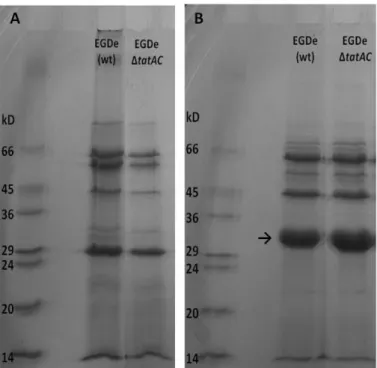
Estudos prévios realizados in silico, por outros autores, identificaram apenas uma proteína secretada através deste sistema. Esta proteína está codificada no operão lmo0365-67, sendo a última do operão (lmo0367). De forma a identificar esta e/ou outras proteínas secretadas por este sistema, procedeu-se a uma análise das proteínas secretadas por SDS-PAGE, não tendo sido no entanto possível detectar alterações entre o secretoma do mutante e o secretoma da estirpe selvagem, possivelmente devido às limitações desta metodologia (limites de resolução e detecção).
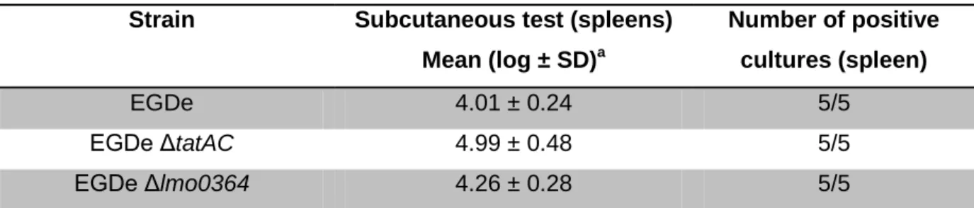
Os potenciais de virulência dos dois mutantes (EGDe ΔtatAC e EGDe Δlmo0364) foram também avaliados in vitro, através da infecção de células epiteliais humanas, e in vivo, através da contagem de bactérias no baço, após três dias de inoculação subcutânea em ratinhos. Apesar de não terem sido identificadas diferenças no potencial virulento in vitro, foram verificadas diferenças significativas (p < 0,05) in vivo, sendo que o mutante tatAC demonstrou um potencial virulento mais elevado do que a estirpe selvagem, demonstrando que a presença deste sistema de secreção nesta estirpe poderá dificultar o processo de infecção.
Ao contrário do que se verificou noutros microrganismos patogénicos, o sistema Tat não é necessário à sobrevivência de L. monocytogenes e muito menos à sua capacidade infecciosa. Tanto quanto se sabe, este trabalho constitui o primeiro trabalho experimental realizado em L. monocytogenes, com o objectivo de estudar o sistema de secreção Tat.
Palavras-chave:
vii
Contents
Acknowlegments ... i Abstract ... iii Resumo ... iv Figure Index ... ix Table Index ... x 1. Introduction ... 1 1.1. Listeria monocytogenes ... 1 1.1.1. Historical facts ... 1 1.1.2. Characteristics ... 1 1.1.3. Infectious cycle ... 21.2. Bacterial Secretion Systems ... 4
1.3. The Tat Pathway ... 7
2. Materials and Methods ... 9
2.1. Bacterial strains and plasmids ... 9
2.2. Growth conditions ...10
2.3. Genomic DNA extraction ...10
2.4. Construction of mutants ...11
2.4.1. Construction of double deletion mutant strain (L. monocytogenes EGDe Δlmo0361 Δlmo0362) ... 11
2.4.2. Construction of deletion mutant strain (L. monocytogenes EGDe Δlmo0364) ... 12
2.5. Preparation of total RNA ...12
2.6. Northern blot analysis ...13
2.7. Transcriptional fusions ...13
2.8. Beta-galactosidase assay ...14
2.9. Secreted protein analysis ...14
2.9.1. Bacterial cultures ... 14
2.9.2. Protein precipitation ... 15
2.9.3. Protein quantification... 15
viii
2.10. In vitro test of virulence ...15
2.10.1. Cell line and culture conditions ... 15
2.10.2. Plaque-forming assay (PFA) ... 16
2.11. In vivo test of virulence ...16
2.12. Expression of results and data analysis ...17
3. Results ...18
3.1. Deletion mutant for the Tat pathway ...18
3.2. Transcriptional analysis ...19
3.2.1. Northern blot ... 19
3.2.2. Promoter activity ... 21
3.3. Proteomic analysis ...24
3.3.1. SDS-PAGE analysis of secreted proteins ... 24
3.4. Phenotypical analysis ...25
3.4.1. In vitro virulence ... 25
3.4.2. In vivo virulence ... 26
4. Discussion ...27
4.1. The Tat pathway in Listeria monocytogenes ...27
4.2. The lmo0365-67 operon ...29
4.3. Transcriptional Regulator Lmo0364 ...30
5. References ...31
Appendix 1 – List of Primers ...37
Appendix 2 – Plasmid Maps ...38
ix
Figure Index
Figure 1 – Schematic representation and electron micrographs of the Listeria
monocytogenes life cycle. ... 3
Figure 2 – Schematic representation of the secretion systems and the number of proteins secreted by each system in Listeria monocytogenes. ... 5 Figure 3 – Schematic representation of Tat pathways (A) in negative and (B) gram-positive bacteria. ... 7 Figure 4 – Organization of the genes coding for the Tat pathway and the lmo0365-7 operon of Listeria monocytogenes EGDe. ... 8 Figure 5 – Schematic representation of primer annealing positions for primers used in the construction of the tatAC deletion mutant. ...12 Figure 6 – Polymerase chain reaction results confirming the construction of a tatAC deletion mutant from the parent strain L. monocytogenes EGDe. ...18 Figure 7 – Growth curves of L. monocytogenes EGDe and EGDe ΔtatAC mutant grown in complete medium (BHI) at 37 ˚C. ...19
Figure 8 – Northern blot analysis of lmo0361, lmo0362 and lmo0363 transcripts in wild-type and tatAC mutant strain ...20 Figure 9 - Northern blot analysis of lmo0361, lmo0362 and lmo0363 transcripts in wild-type,
tatAC and lmo0364 mutant strains ...21
Figure 10 – Determination of promoter activity by using lacZ fusion. ...21
Figure 11 – Comparison of lmo0362 promoter activity at different time points during growth (3, 4, 5, 7 and 24 hours in BHI, at 37 ˚C). ...22
Figure 12 – Determination of promoter activities in the EGDe (wt), ΔtatAC and Δlmo0364 mutant strais. (A) Promoter activity measured for the gene lmo0362. (B) Promoter activity measured for the operon lmo0365-67. ...23 Figure 13 – Determination of promoter activity measured for the gene lmo0364. ...24
Figure 14 - The secretome of L. monocytogenes strains grown in minimal medium and harvested at late exponential phase (A600 = 0.8). ...25
x
Table Index
Table 1 – Bacterial strains used in this study. ... 9
Table 2 – Plasmids used in this study. ...10
Table 3 – In vitro virulence potential of EGDe wild-type strain and ΔtatAC mutant. ...25
Introduction
1
1. Introduction
1.1.
Listeria monocytogenes
1.1.1. Historical factsIn 1924, after an outbreak in laboratory animals in Cambridge, Murray, Webb and Swann have isolated a gram-positive bacterium which they named Bacterium monocytogenes (1), due to the monocytosis disease observed in the infected animals. This was considered the official discovery of Listeria, which after the name of Bacterium monocytogenes had other names as Erysipelothrix, Listerella and finally, Listeria (2), in honor of Lord Lister (3).
Despite its official discovery date in 1924, there is evidence that this organism had probably been observed in histological sections many years before its official discovery and it had been cultivated and described by Hülphers in 1911 (4).
Although the clinical descriptions of infection by L. monocytogenes in animals and humans date from the twenties of the last century, only in 1952, in Germany, listeriosis was recognized as an important cause of neonatal meningitis and septicemia (5). In 1981, L.
monocytogenes was for the first time associated with the Food Industry and considered as a
foodborne pathogen, following an outbreak of listeriosis in Nova Scotia, Canada, with 41 cases and 18 deaths reported, mostly children and pregnant women, epidemiologically linked by the consumption of coleslaw contaminated with sheep feces containing L. monocytogenes (6).
1.1.2. Characteristics
The presence of L. monocytogenes in food products is currently considered a risk in the Food Industry. This pathogenic microorganism affects primarily pregnant women, children, the elderly and immunocompromised individuals. This risk becomes more important as the cases of immunocompromised patients, either by HIV infection or by treatments such as chemotherapy and radiotherapy, among others increases.
Taxonomically, L. monocytogenes belongs to the Bacteria domain, phylum Firmicutes, class
Bacilli, order Bacillales, family Listeriaceae and gender Listeria. Eight species have been
described for the genus Listeria: L. monocytogenes, L. ivanovii, L. seeligeri, L. innocua, L.
welshimeri, L. grayi, L. marthii (7) and L. rocourtiae (8). L. monocytogenes infects ruminants
and it has been considered the only one pathogenic for humans (9) (10), L. ivanovii was thought to infect only ruminants but recently a case of infection by this species in a man was
Introduction
2 reported, suggesting that the rarity of human listeriosis due to this species can be explained not only by the host tropism factors but also by the rare occurrence of this species in the environment, compared with L. monocytogenes (11).
Thirteen serovars (1/2a, 1/2b, 1/2c, 3a, 3b, 3c, 4a, 4ab, 4b, 4c, 4d, 4e and 7), from three different lineages (lineages I, II and III) have been described for L. monocytogenes (12).
L. monocytogenes is a gram-positive rod-shaped bacterium with dimensions of 0.4 µm for 1
to 1.5 µm. This bacterium has a low G+C content, is a facultative anaerobe, catalase positive and oxidase negative. It has peritrich flagella, which presence is very limited when grown at 37 ˚C (13). L. monocytogenes can grow in a range of temperatures from 1 to 45 ˚C (14) (15), although its optimal growth temperature ranges from 30 to 37 ˚C. The growth versatility of this bacterium is also observed with respect to pH, as it has the ability to grow at pH values between 4.4 and 9.6 (14), although the optimal pH for its growth is neutral or slightly alkaline.
L. monocytogenes is quite tolerant to sodium chloride (10 to 20% (w/v)) (16) (13) (17), bile
salts (10 to 40% (w/v)), and to some metals, which are usually toxic for other bacteria, such as lithium, thallium and tellurium. It has also the ability to grow in low water activity (aw)
values, equal or greater than 0.92 (18), but its optimal growth occurs at 0.97.
1.1.3. Infectious cycle
L. monocytogenes is responsible for listeriosis, a disease with a high mortality rate (20% to
30%). In the EU, the number of confirmed cases increased 19% in 2009 compared to 2008 (19). L. monocytogenes is a facultative intracellular bacterium. After ingestion of contaminated food it is able to cross the intestinal epithelium and disseminate via the lymph and blood-streams to deeper tissues. The liver and the spleen are primary target organs for further bacterial multiplication, resulting in possible abscess formation (20). One of the main characteristics of L. monocytogenes physiopathology is its capacity of crossing major epithelial barriers, the blood-brain barrier and the placental barrier, leading to meningo-encephalitis or fetus infection, ultimately it can eventually cause abortion, stillbirth or neonatal meningitis.
Briefly, after being ingested this bacterium has the ability of entering the intestine epithelial cells and escape from the phagosome, becoming free in the cell cytoplasm. By actin polymerization it propels through the cell cytoplasm and into the adjacent cell’s outer membrane which encapsulates this protuberance and acquires the bacteria that will become once again free in the cell cytoplasm (Figure 1).
Introduction
3 Figure 1 – Schematic representation and electron micrographs of the Listeria monocytogenes life cycle.
a - L. monocytogenes induces its entry into a non-professional phagocyte. b - Bacteria are internalized in a vacuole (also known as a phagosome). c,d - The membrane of the vacuole is disrupted by the secretion of two phospholipases, PlcA and PlcB, and the pore-forming toxin listeriolysin O. Bacteria are released into the cytoplasm, where they multiply and start to polymerize actin, as observed by the presence of the characteristic actin tails. e - Actin polymerization allows bacteria to pass into a neighbouring cell by forming protrusions in the plasma membrane. f - On entry into the neighbouring cell, bacteria are present in a double-membraned vacuole, from which they can escape to perpetuate the cycle. F-actin, filamentous actin (21).
Introduction
4
1.2.
Bacterial Secretion Systems
Bacterial protein secretion is extremely important because secreted proteins are the main tool used by bacteria to interact with their environment. Bacterial pathogenicity depends greatly on the ability of bacteria to secrete virulence factors which are displayed on the bacterial cell surface, secreted into the extracellular milieu or even injected directly into the host cell (22). Many nonpathogenic organisms also secrete proteins that are important for its growth, for example degradative enzymes such as cellulases, secreted by saprophytic bacteria.
Protein secretion systems have been extensively investigated in a wide range of gram-negative bacterial species and six secretion systems were identified (numbered from I to V, plus the chaperone pathway), although they are not all systematically present in a single bacterium and its presence and activity varies from one bacterium to another (23). In contrast, information about secretion systems in gram-positive bacteria is still essentially restricted to Bacillus subtilis, although other studies concerning gram-positive bacteria such as L. monocytogenes (24) (25) and Staphylococcus aureus (26) (27) are starting to be reported.
In gram-positive bacteria seven protein secretion systems are currently recognized: the Sec (Secretion) pathway; the Tat (Twin-arginine translocation) pathway; the FPE (Fimbrilin-Protein Exporter); some ABC (ATP-binding cassette) protein exporters; the FEA (Flagellum Export Apparatus); the holins (hole-forming) and the Wss (WXG100 secretion system) (25). As occurs in gram-negative bacteria, not all of the secretion pathways are systematically present in a single organism, and their respective contribution in protein transportation varies from one organism to another.
All the secretion pathways described for gram-positive bacteria were identified in Listeria
Introduction
5 Figure 2 – Schematic representation of the secretion systems and the number of proteins secreted by each system in Listeria monocytogenes.
SP, signal peptide; Sec, secretion; Tat, twin-arginine translocation; FPE, fimbrilin-protein exporter; ABC, ATP-binding cassette exporter; FEA, flagella export apparatus; holin, hole forming; Wss, WXG100 secretion system; Cyto, cytoplasm; CM, cytoplasmic membrane; CW, cell wall; EM, extracellular milieu. Adapted from Desvaux et al, 2010 (25).
The Sec pathway is the major secretion system in prokaryotes and eukaryotes, not only because of the number of proteins considered as exported by this pathway, but also because of their importance. Although the presence of the Sec pathway in L. monocytogenes has not been experimentally proved, its existence was inferred due to the high number of proteins carrying the putative translocation signal peptide and the presence of these proteins in the extracellular milieu or in the cell surface (28) (29). This system consists of a heterotrimeric SecYEG complex, which is the central component as it forms a channel in the cytoplasmic membrane. A cytosolic ATPase SecA is also essential for protein transport. It acts through cycles of adenosine triphosphate (ATP) binding and hydrolysis, thereby facilitating the binding of proteins to the system, leading to stepwise export of the proteins (24). In L.
monocytogenes a SecA paralogue named SecA2 was identified (30) and unlike SecA this is
not essential for cell viability but it is involved in the secretion of proteins that contribute to virulence (31), such as p60 hydrolase (30), NamA (N-acetilmuramidase A) (32) and FbpA (Fribonectin-binding protein A) (33).
Introduction
6 The proteins secreted by the Tat pathway have a N-terminal signal peptide and an essential twin arginine motif, (S/T)TRRXFLK, which straddles the N-domain and the hydrophobic H-domain (34). Unlike the Sec system, the Tat pathway is considered to secrete proteins in their final conformation (35) and somehow rejects the ones that are unfolded. In L.
monocytogenes EGDe (serovar 1/2a) only one copy of the genes coding for tatA and tatC
was identified while in the sequenced L. monocytogenes 4b serovars no genes coding for the Tat pathway were identified.
The Fimbrilin-Protein Exporter (FPE) system is responsible for the secretion and agglomeration of proteins similar to pilin, and its components are encoded in comG locus. Although the existence of this system has never been experimentally demonstrated in L.
monocytogenes, it is referred because of its similarity with the genetic locus comG of Bacillus subtilis.
ATP-binding cassette (ABC) protein exporters are transmembrane proteins that utilize the energy of ATP hydrolysis to carry out various biological processes, including translocation of substrates across membranes. Only recently this type of system was considered as responsible for the secretion of four bacteriocins in L. monocytogenes (25).
In gram-negative bacteria, the Flagellum Export Apparatus (FEA) is related with the type III secretion system, together with the Hrp (Hypersensitive response and pathogenicity) pilus export apparatus and the injectisome export apparatus (24). From the nine FEA components identified in gram-negative bacteria, seven were identified in Listeria, FlhA, FlhB, FliP, FliQ, FliR, FliI and FliH.
Holins are small membrane proteins that allow the transport across the cytoplasmic membrane of proteins lacking N-terminal signal sequences. These proteins, which derived from phages, are involved in the secretion and activation of proteins with murein hydrolyzing activity in early stages of cell lysis, which is very important for apoptosis. Holins are homo-oligomeric complexes that form pores in the cytoplasmic membrane, allowing an energy independent translocation of proteins (24).
The Wss system is formed by a protein with 100 amino acids, which has a coil-coil domain and a conserved WXG motif, called WXG100 and a membrane-bound ATPase, homologous to B. subtilis YukAB. In Listeria only one copy of the gene coding for the protein YukAB was identified, which is also present in the non-pathogenic species L. innocua (36).
Introduction
7
1.3.
The Tat Pathway
The twin-arginine translocation (Tat) pathway is responsible for the secretion of proteins in their folded conformation and, as referred before, targeted proteins to be secreted via Tat pathway have a N-terminal consensus motif [(S/T)TRRXFLK] (34). The energy required for protein translocation is exclusively acquired from the transmembrane proton electrochemical gradient (Δp) (37).
This secretion system was first characterized in plant chloroplasts and in 1998 a homologous bacterial Tat pathway was identified in Escherichia coli, encoded by the tat genes (tatA, tatB,
tatC and tatE) (38).
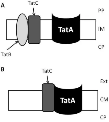
In gram-negative bacteria, where this pathway was first identified in prokaryotes, the Tat translocation requires three integral membrane proteins belonging to TatA, TatB and TatC families, and all of them have been demonstrated to be essential for protein translocation (Figure 3A) (34).
Most gram-positive bacteria and
Archaea possess a simpler and
minimalist Tat pathway as only tatA and tatC genes are present, lacking the gene for TatB component, which function is performed by a bifunctional TatA component (Figure 3B) (39). Although the TatB is missing in gram-positive bacteria, it has been proved its functionality in B.subtilis. This operon (tatAdCd, named d due to the existence of more than one copy of these genes in B. subtilis genome) was expressed in an E. coli tat null mutant and its ability to export several Tat substrates was proved (39).
Despite the differences between gram-positive and gram-negative bacteria, homologues of
E. coli tat genes have been identified in the genomes of many bacterial pathogens, including E. coli O:157, Agrobacterium tumefaciens, Pseudomonas aeruginosa, Vibrio cholerae,
Figure 3 – Schematic representation of Tat pathways (A) in negative and (B) gram-positive bacteria.
PP, periplasm; IM, inner membrane; CP, cytoplasm; Ext, extracellular milieu; CM, cytoplasmic membrane.
Introduction
8
Helicobacter pylori, Mycobacterium tuberculosis, L. monocytogenes, Salmonella enterica, Legionella pneumophila, Neisseria meningitidis, Haemophilus influenzae, Pasteurella multocida, Yersinia pestis, Xanthomonas campestris, X. axonopodis, Xyllela fastidiosa, S. aureus and Ralstonia solanacearum (38). For some of these pathogens the importance of
the Tat pathway was assessed and studied in detail. Those studies proved the involvement of the Tat pathway in different lifestyles of bacteria, from saprophytic to pathogenic. Translocation of proteins by this system showed to be important for E. coli O157:H7 motility, for infection and iron acquisition in P. aeruginosa and for cell growth, division and biofilm formation in L. pneumophila (38). In S. aureus, a bacterium taxonomically closer to L.
monocytogenes, there are evidences of the Tat pathway function in transporting an
iron-dependent peroxidase (FepB) and its need to successful in vivo infection was shown (26).
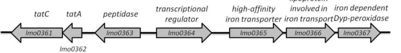
In L. monocytogenes EGDe, the two proteins which constitute the Tat pathway are encoded in the genes lmo0361 and lmo0362. The gene located four genes away of the tat genes corresponds to the gene coding for an iron-dependent Dyp-peoxidase (lmo0367), with 40 % similarity to FepB of S. aureus considered secreted by the Tat pathway. This indicates that probably Lmo0367 is also secreted by the Tat system. This protein is part of an operon formed by genes lmo0365, lmo0366 and lmo0367 (40), all of them related with iron. Between the two locus mentioned there is a gene coding for a peptidase and a transcriptional regulator, lmo0363 and lmo0364, respectively. A schematic representation of the locus coding for the Tat Pathway and the protein putatively secreted by this system is shown in Figure 4.
Figure 4 – Organization of the genes coding for the Tat pathway and the lmo0365-7 operon of Listeria monocytogenes EGDe.
Materials and Methods
9
2. Materials and Methods
2.1.
Bacterial strains and plasmids
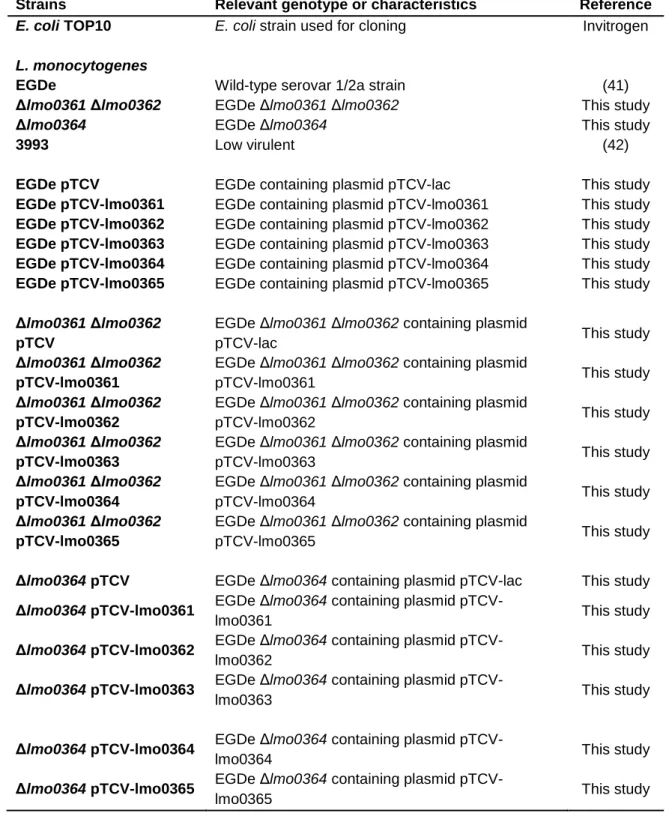
Bacterial strains and plasmids used in this study and its characteristics are described in Tables 1 and 2, respectively.
Table 1 – Bacterial strains used in this study.
Strains Relevant genotype or characteristics Reference
E. coli TOP10 E. coli strain used for cloning Invitrogen
L. monocytogenes
EGDe Wild-type serovar 1/2a strain (41)
Δlmo0361 Δlmo0362 EGDe Δlmo0361 Δlmo0362 This study
Δlmo0364 EGDe Δlmo0364 This study
3993 Low virulent (42)
EGDe pTCV EGDe containing plasmid pTCV-lac This study
EGDe pTCV-lmo0361 EGDe containing plasmid pTCV-lmo0361 This study
EGDe pTCV-lmo0362 EGDe containing plasmid pTCV-lmo0362 This study
EGDe pTCV-lmo0363 EGDe containing plasmid pTCV-lmo0363 This study
EGDe pTCV-lmo0364 EGDe containing plasmid pTCV-lmo0364 This study
EGDe pTCV-lmo0365 EGDe containing plasmid pTCV-lmo0365 This study
Δlmo0361 Δlmo0362 pTCV
EGDe Δlmo0361 Δlmo0362 containing plasmid
pTCV-lac This study
Δlmo0361 Δlmo0362 pTCV-lmo0361
EGDe Δlmo0361 Δlmo0362 containing plasmid
pTCV-lmo0361 This study
Δlmo0361 Δlmo0362 pTCV-lmo0362
EGDe Δlmo0361 Δlmo0362 containing plasmid
pTCV-lmo0362 This study
Δlmo0361 Δlmo0362 pTCV-lmo0363
EGDe Δlmo0361 Δlmo0362 containing plasmid
pTCV-lmo0363 This study
Δlmo0361 Δlmo0362 pTCV-lmo0364
EGDe Δlmo0361 Δlmo0362 containing plasmid
pTCV-lmo0364 This study
Δlmo0361 Δlmo0362 pTCV-lmo0365
EGDe Δlmo0361 Δlmo0362 containing plasmid
pTCV-lmo0365 This study
Δlmo0364 pTCV EGDe Δlmo0364 containing plasmid pTCV-lac This study
Δlmo0364 pTCV-lmo0361 EGDe Δlmo0364 containing plasmid
pTCV-lmo0361 This study
Δlmo0364 pTCV-lmo0362 EGDe Δlmo0364 containing plasmid
pTCV-lmo0362 This study
Δlmo0364 pTCV-lmo0363 EGDe Δlmo0364 containing plasmid
pTCV-lmo0363 This study
Δlmo0364 pTCV-lmo0364 EGDe Δlmo0364 containing plasmid
pTCV-lmo0364 This study
Δlmo0364 pTCV-lmo0365 EGDe Δlmo0364 containing plasmid
Materials and Methods
10 Table 2 – Plasmids used in this study.
Plasmids Relevant genotype or characteristics Reference
pAULA Ermr; Cloning plasmid for gene replacements in
gram-positive bacteria
(43)
pMAD Ampr; Ermr; Cloning plasmid for gene replacements
in gram-positive bacteria
(44)
pMAD – Δlmo0361 Δlmo0362
Ampr; Ermr; pMAD derivative containing
homologous regions upstream EGDe lmo0362 and downstream of EGDe lmo0361
This study
pAULA – Δlmo0364 Ermr; pAULa derivative containing homologous regions up- and downstream of EGDe lmo0364
This study
pTCV-lac Kanr; Transcriptional lacZ fusion vector; Low copy-number
(45)
pTCV-lmo0361 Kanr; Truncated lmo0361 region (-143 to +36) inserted in pTCV-lac upstream of lacZ
This study
pTCV-lmo0362 Kanr; Truncated lmo0362 region (-152 to +48) inserted in pTCV-lac upstream of lacZ
This study
pTCV-lmo0363 Kanr; Truncated lmo0363 region (-208 to +42) inserted in pTCV-lac upstream of lacZ
This study
pTCV-lmo0364 Kanr; Truncated lmo0364 region (-178 to +36) inserted in pTCV-lac upstream of lacZ
This study
pTCV-lmo0365 Kanr; Truncated lmo0365 region (-219 to +30) inserted in pTCV-lac upstream of lacZ
This study
2.2.
Growth conditions
Escherichia coli strains were grown at 37 ˚C with shaking in Luria-Bertani (LB) medium (1 %
tryptone, 0.5 % yeast extract, 0.5 % NaCl, pH 7.2) or on LA plates (LB supplemented with 1.5 % (w/v) agar). When required, ampicillin (Amp), erythromycin (Erm) or kanamycin (Kan) was added to a final concentration of 100 µg/mL, 150 µg/mL and 50 µg/mL, respectively.
L. monocytogenes strains were grown at 37 ˚C with shaking in brain heart infusion (BHI)
broth or on BHI plates (BHI supplemented with 1.5% (w/v) agar) and on minimal medium Modified Welshimer Broth (MWB) (46) or on Tryptone Soya Yeast Extract Agar (TSA-YE, 1.5% (w/v) agar). When required, erythromycin (Erm) or kanamycin (Kan) was added to a final concentration of 5 µg/mL and 50 µg/mL, respectively.
2.3.
Genomic DNA extraction
Overnight cultures of L. monocytogenes strains were centrifuged at 3500 g for 5 min. The pellets were resuspended in 100 µL 1xPBS (137 mM NaCl, 2.7 mM KCl, 4.3 mM Na2HPO4,
Materials and Methods
11 added, following 15 min incubation at 37 ˚C, with shaking. The DNA was extracted and purified using FastDNA kit and protocol from Bio101 (Bio101, USA).
The DNA quality and concentration was verified by agarose gel electrophoresis (1% agarose).
2.4.
Construction of mutants
2.4.1. Construction of double deletion mutant strain (L. monocytogenes EGDe Δlmo0361 Δlmo0362)
In order to generate the lmo0361 lmo0362 double deletion mutant, an approximately 1000 basepair (bp) large region upstream of lmo0362 and downstream of lmo0361 was generated from L. monocytogenes EGDe genomic DNA. To generate these regions, a standard polymerase chain reaction (PCR) was performed using DNA polymerase high-fidelity (Fermentas, Canada), primers Tat_A together with Tat_B, and primers Tat_C together with Tat_D, as listed in Appendix 1 (for schematic representation see Figure 5). The first fragment (AB) was digested with restriction enzymes SalI and MluI and cloned in the temperature sensitive plasmid pMAD (Appendix 2), resulting the pMAD-TatAB plasmid. The second fragment (CD) was digested with restriction enzymes MluI and BglII and cloned in the pMAD-TatAB plasmid, resulting the pMAD-TatAD plasmid which had two approximately 1000 bp region, each homologous to the up and down-stream of the genes of interest.
The pMAD-TatAD plasmid was electroporated into L. monocytogenes EGDe, according to Park et al. (47). To induce chromosomal integration of the pMAD-TatAD, the cells were grown at a non permissive temperature (42 ˚C) on BHI plates containing erythromycin (5 µg/mL). Colonies containing the integrated plasmid were grown for eleven days at a permissive temperature (30 ˚C) without antibiotics, allowing the plasmid excision from the chromosome and its loss from the cell, thus allowing excising the genes lmo0361 and
lmo0362 from the chromosome (schematic representation of gene deletion in Appendix 3).
Finally, the presence of lmo0361 and lmo0362 regions was investigated through amplification from the resulting mutants using the internal primers for both genes, TatA_fw with TatA_rv and TatC_fw with TatC_rv, respectively (for schematic representation see Figure 5).
Materials and Methods
12 Figure 5 – Schematic representation of primer annealing positions for primers used in the construction of the tatAC deletion mutant.
2.4.2. Construction of deletion mutant strain (L. monocytogenes EGDe Δlmo0364)
In order to generate the lmo0364 (transcription regulator) deletion mutant, an approximately 400 bp large region upstream and downstream of lmo0364 was generated from L.
monocytogenes EGDe genomic DNA. To generate these regions, a standard polymerase
chain reaction (PCR) was done using Phusion polymerase (Finnzymes, Finland), primers P1-lmo0364 and P2-P1-lmo0364, and primers P3-P1-lmo0364 together with P4-P1-lmo0364, as listed in Appendix 1. The resulting products were used to produce an approximately 800 bp fragment by splicing by overlap extension (SOE-ing) (48). After digestion of this fragment with EcoRI and BamHI it was cloned in the temperature sensitive plasmid pAULA (Appendix 2) resulting the pAULA–Δlmo0364, which construction was confirmed by sequencing with primers pAUL-1 and pAUL-2 (Appendix pAUL-1).
The pAULA–Δlmo0364 plasmid was electroporated into L. monocytogenes EGDe (47). To induce chromosomal integration of the pAULA–Δlmo0364, the cells were grown at a non permissive temperature (42 ˚C) on BHI plates containing erythromycin (5 µg/mL). Colonies containing the integrated plasmid were grown for eight days at a permissive temperature (30 ˚C) without antibiotics, allowing the plasmid excision from the chromosome and its loss from the cell, thus allowing the excision of gene lmo0364 from the chromosome (schematic representation of gene deletion in Appendix 3). Finally, to confirm that the gene was deleted from the chromosome, the lmo0364 region was amplified in wild-type and mutant strain DNA following sequencing using primers PC1_lmo0364 and PC2_lmo0364 (Appendix 1).
2.5.
Preparation of total RNA
Overnight cultures of L. monocytogenes strains were diluted to an A600 = 0.02 in BHI
(corresponding to time 0) and grown at 37 ˚C. At various time points (3, 4.5, 6 and 8 hours) 20 mL samples of the growing cultures were shortly cooled in liquid nitrogen (N2) and the
Materials and Methods
13 nitrogen and stored at -80 ˚C. The cells were resuspended in 900 µL TRI reagent (MRCGENE, USA) and lysed using a FastPrep instrument. RNA was subsequently extracted using chloroform and precipitated with ethanol and sodium acetate, overnight at -80 ˚C. The samples were centrifuged at 20000 g for 30 min at 4 ˚C, washed with 70% ethanol (-20 ˚C) and resuspended in 50 µL Milli-Q water.
The RNA quality and quantification was assessed on a NanoDrop 2000. The integrity and the predicted concentration of the RNA was verified by agarose gel electrophoresis (1% agarose).
2.6.
Northern blot analysis
For Northern blotting, loading buffer (95% formamide and 20 mM EDTA, 0.05% BPB and 0.05% xylene cyanol) was added to 10 µg of total RNA and samples were boiled at 95 ˚C for 3 min (to denaturate the RNA) and immediately cooled on ice. The total RNA was separated on a 4% polyacrylamide-7M urea gel and transferred to a Zeta probe nylon membrane (Bio-Rad, USA) by semi-dry electroblotting at 400 mA for 2 hours.
For detection of RNA, the membranes were pre-incubated for 30 min at 42 ˚C in PerfectHyb hybridization buffer (Sigma-Aldrich, USA) and hybridized with a specific 32P-labelled DNA probe, prepared using poly nucleotide kinase (PNK) (Fermentas, Canada) and γ-[32P]-ATP (PerkinElmer, USA). The membranes were then washed with a 2X saline-sodium citrate (SSC), 0.1% sodium dodecyl sulfate (SDS) solution for 5 min and a 0.5X SSC, 0.1% SDS solution for 20 min, both at 42 ˚C, and finally visualized by phosphor imaging using a Typhoon scanner.
2.7.
Transcriptional fusions
The promoter regions of lmo0361, lmo0362, lmo0363, lmo0364 and lmo0365-lmo0367, respectively (see Figure 4 in the Introdution, section 1.3.) were amplified from L.
monocytogenes EGDe chromosomal DNA under standard PCR conditions using Phusion
polymerase (Finnzymes, Finland) with primers listed in Appendix 2. The resulting products of approximately 200 bp were digested with EcoRI and BamHI and cloned in the low copy-number plasmid pTCV-lac (Appendix 2). The construction was confirmed by sequencing with primers vLac1 and vLac2 (Appendix 1).
The constructed plamids, pTCV-lmo0361, pTCV-lmo0362, pTCV-lmo0363, pTCV-lmo0364 and pTCV-lmo0365 were each one electroporated into L. monocytogenes EGDe, Δlmo0361
Materials and Methods
14 Δlmo0362 and Δlmo0364 (47) strains. Transformants were selected by kanamycin resistance.
2.8.
Beta-galactosidase assay
Strains for testing β-galactosidase activity were diluted from an overnight culture to an A600=0.02 in BHI. At different growth times (3, 4, 5, 7 and 24 hours) 1 mL of each sample
were collected by centrifugation at 14000 g for 4 min, thoroughly aspirated, frozen and stored at -80 ˚C until use. For the assay, the pelleted cells were thawn on ice and resuspended in 1 mL Z-buffer (60 mM Na2HPO4, 40 mM NaH2PO4, 10 mM KCl, 1 mM MgSO4, pH 7.0). Cells
were permeabilized by adding 0.5 % toluene and 4.5 % ethanol, vortexing after each addition. From the permeabilized cell suspension, 100 µL was mixed with 400 µL of Ortho-nitrophenyl-β-galactosidase (ONPG) solution (1 mg/mL ONPG, 0.075 % SDS dissolved in Z-buffer) and incubated at 37 ˚C until the samples present a yellow colour (A420 ≈ 0.6 - 0.9).
When this colour was reached, the reaction was stopped by addition of 500 µL of 1 M Na2CO3.
The β-galactosidase activity was determined as described by Miller (49), using the equation:
,
where t is the time of reaction in minutes, v is the volume of culture used in the assay (mL), A420 and A550 is the absorbance of the β –galactosidase sample at 420 nm and 550 nm,
respectively, and A600 is the absorbance of the cell culture when harvested.
Strains carrying an empty vector were also subjected to β-galactosidase measurements and this background activity was subtracted of the obtained data.
2.9.
Secreted protein analysis
2.9.1. Bacterial cultures
L. monocytogenes strains were cultured overnight at 37 ˚C on TSA-YE. A single isolated
colonie was suspended in 25 mL of minimal medium MWB (46) and incubated overnight at 37 ˚C with shaking. Overnight cultures were adjusted to an A600 = 0.05 using fresh MWB and
grown at the same conditions used before (37 ˚C with shaking) for about 20 hours. The cells were harvested at exponential phase (A600 ≈ 0.8) by centrifugation (3 000 g, 10 min, 4 ˚C).
The obtained supernatants were filtered with 0.22 µm Millipore Express Membranes and phenylmethylsulphonyl fluoride (PMSF) was added to a final concentration of 0.2 mM (29).
Materials and Methods
15
2.9.2. Protein precipitation
The proteins present in the supernatants were precipitated with 0.2 mg/mL sodium deoxycholate (DOC) for 30 min at 4 ˚C, followed by addition of 6 % (w/v) trichloroacetic acid (TCA) and overnight incubation at 4 ˚C. The precipitate was centrifuged (17 900 g, 15 min, 4 ˚C), washed with ice-cold acetone and resuspended in Milli-Q water.
2.9.3. Protein quantification
Protein concentration was determined by a modified Lowry method (50) using the bovine serum albumin (BSA) as standard.
2.9.4. SDS-PAGE
Samples containing 50 µg of total protein were dissolved in sample buffer: 80 mM Tris-HCl, pH 6.8, containing 2 % (w/v) SDS, 0.1 mM β-mercaptoethanol, 15 % glycerol and 0.01 % (w/v) m-cresol purple. Samples were then heated at 100 ˚C for 4 min and submitted to SDS-PAGE 15 % (w/v) in gels with 8 cm x 7.3 cm x 0.75 mm, on a Mini-PROTEAN 3 Cell (Bio-Rad, USA).
Separated proteins were visualized by colloidal Coomassie Brilliant Blue G-250 staining (51). Apparent masses were calculated based on the molecular masses of standard LPM (14 to 66 kDa) (Sigma-Aldrich, USA).
2.10. In vitro test of virulence
2.10.1. Cell line and culture conditions
The human adenocarcinoma cell line HT-29 (ECACC n.º 850611109) was used between passages 62 and 64. Cells were routinely grown in 75 cm2 flasks in high glucose Dulbecco’s modified Eagle’s medium (D-MEM) (Gibco/Invitrogen, UK), supplemented with sodium bicarbonate (3.7 g/L), fetal calf serum (10% (v/v)) and L-glutamine (2mM) (complete medium). Penicillin (100 IU/mL) and streptomycin (100 µg/mL) were always added to the culture medium except for the medium used 24 hours prior to the virulence assays. Cells were maintained in a humidified atmosphere using an incubator at 37 ˚C under 5% (v/v) CO2
Materials and Methods
16
2.10.2. Plaque-forming assay (PFA)
HT-29 cells were trypsinized from the 75 cm2 flasks and 3 x 10⁴ cells were deposited per well in a 96-well tissue culture plate. To obtain confluent monolayers, the plates were incubated for 3 days with antibiotics followed by incubation for 24 h without antibiotics.
The overnight grown Listeria strains (TSA-YE, 37 ˚C) were suspended in buffered saline to a concentration of 4 x 10⁸ cell mL-1 and serial dilutions were made in complete medium. HT-29 cell monolayers were infected with dilution series of 102 to 107 L. monocytogenes cells per well, and incubated for 2 h at 37 ˚C. The inocula concentration was assessed by duplicate plating of the appropriate dilutions onto TSA-YE and incubation at 37 ˚C for 24 h, before counting colonies.
After 2 h incubation with the bacterial cells, the suspensions were removed and cell monolayers were incubated for 1.5 h with complete D-MEM medium containing 100 µg gentamicin mL-1, and lacking penicillin and streptomycin. The wells were then covered with complete D-MEM medium supplemented with 10 µg gentamicin mL-1 and 2.5 % (m/v) agarose. Once the agar media was solidified, complete medium was added to the top of the agar media to prevent cell starvation. Culture plates were incubated for 24 to 48 h at 37 ˚C under 5 % (v/v) CO2 in air. Formed plaques were counted 24 h after bacteria deposition on
the HT-29 cell monolayer, and confirmed after 48 h of incubation. Enumeration of the plaques was done using an inverted microscope (52) (53).
2.11. In vivo test of virulence
To test the in vivo pathogenicity by subcutaneous injection (50 µL) of the wild-type and the mutants, seven-week-old immunocompetent Swiss female mice were used (54). They were maintained on sterilized wood shavings with free access to water and sterilized food. For each isolate, groups of five mice were inoculated subcutaneously into the left hind footpad. Bacterial strains were grown on BHI plates for 17 hours at 37 ˚C. The culture was standardized turbidimetrically and diluted appropriately in phosphate buffered saline (PBS, pH 7.3) to obtain 4 log CFU (colony forming unit) in 50 µL for subcutaneous infection. The inocula used were verified by determining viable counts on TSA (Trypic Soy Agar) plates. Mice were sacrificed by cervical dislocation three days after subcutaneous injection and spleens were aseptically removed. Homogenate samples were appropriately diluted in PBS and plated onto TSA plates. Viable numbers of bacteria were assessed after incubation at 37
Materials and Methods
17 ˚C for 48 hours. Animal handling and care conditions were consistent for all experimental runs.
2.12. Expression of results and data analysis
For SDS-PAGE gels, the images were analyzed and band quantification was done using the densitometric curves obtained in the Gel Compare II version 5.1 (Applied Maths, Belgium). For the galatosidase assay, the promoter activities were expressed as the mean of β-galactosidase units, from experiments done in duplicate, from three independent trials. The standard deviation was also assessed and it is shown in the graphic representations of promoter activities.
For the in vitro test, the pathogenic potential of the isolates was expressed as the mean log of the number of plaques formed (for 107 Listeria per well) (log PFA), in duplicate, from at least two independent trials. ANOVA of the log PFA values from the plaque-forming isolates was carried out using least significant differences (LSD) post hoc multiple comparison tests by running the program Statistica, version 6 (Statsoft).
For the mouse virulence assay, the virulence potential of the isolates was expressed as the mean log of the number of colony forming units (log CFU) per spleen, multiplied by the ratio of number of positive cultures in the spleen to inoculated mice in the subcutaneous infection test. The significant differences between strains were assessed using the log CFU mean and the standard deviations by applying a t-test.
Those values whose probability of occurrence was greater than 95 % (p < 0.05) were considered as significant values.
Results
18
3. Results
3.1.
Deletion mutant for the Tat pathway
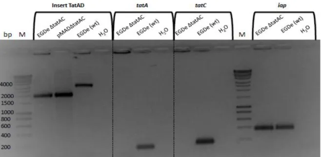
To investigate the importance of the Tat pathway in L. monocytogenes, a deletion mutant for the genes coding for this system was constructed as described before in section 2.3.1. The confirmation of the deletion of the genes was done by polymerase chain reaction (PCR), using internal primers for the genes lmo0361 (tatA) and lmo0362 (tatC) as well as the primers Tat_A and Tat_D, used in the production of the homologous fragment for gene replacement (for primers sequence see Appendix 1). As a PCR control, internal primers for the L. monocytogenes iap gene were used.
The mutant strain showed a ~2000 bp band, the same size as the constructed plasmid (Figure 6), which represents ~1000 bp upstream lmo0362 and ~1000 bp downstream
lmo0361. The wild-type strain EGDe presented a ~3000 bp band. The difference between
the mutant and the wild-type was explained by the deletion of tatA and tatC (911 bp) (Figure 6). The amplification done using the internal primers for the genes lmo0361 (tatC) and
lmo0362 (tatA) resulted in no bands for the mutant and a band of ~200 bp for the wild-type
strain, as expected.
Figure 6 – Polymerase chain reaction results confirming the construction of a tatAC deletion mutant from the parent strain L. monocytogenes EGDe.
Results
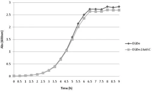
19 In order to investigate if the deletion of tatAC genes was impairing for mutant growth, the mutant strain and the wild-type were grown in BHI at 37 ˚C (Figure 7) and growth rates (µmax)
were calculated. The wild-type showed a µmax = 1.26 h-1 and the mutant showed a µmax = 1.31
h-1, showing that Tat pathway is not essential for L. monocytogenes survival.
Figure 7 – Growth curves of L. monocytogenes EGDe and EGDe ΔtatAC mutant grown in complete medium (BHI) at 37 ˚C.
3.2.
Transcriptional analysis
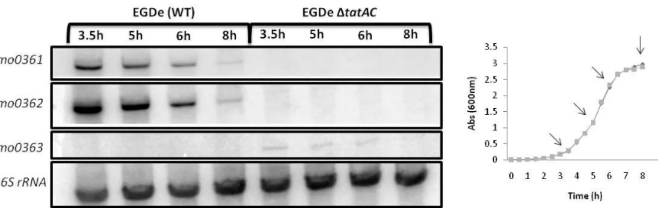
3.2.1. Northern blot
The transcription of the genes lmo0361 (tatC) and lmo0362 (tatA) was analyzed during the exponential and stationary phase. The total RNA was analyzed by Northern blot, by using probes for genes lmo0361, lmo0362, lmo0363 and a control probe for 16S rRNA gene (Appendix 1). The genes lmo0361 and lmo0362 presented the same pattern of expression (Figure 8). The same RNA was observed with both probes indicating that genes lmo0361 and lmo0362 are transcribed in a bicistronic manner. The transcription of tatAC genes was growth-phase dependent, since it seemed to be less expressed in the stationary phase, in comparison to the exponential phase, as shown in Figure 8. As expected, no transcripts were detected in the deletion mutant.
Proteins exported by the Tat pathway have a conserved N-terminus motif which is cleaved when the protein is secreted. The gene lmo0363 upstream the genes coding for the Tat pathway codes for a peptidase and it is located just 96 bp from lmo0362 (see Figure 4 in section 1.3.). To investigate if gene lmo0363 is transcribed together with genes lmo0362 and
Results
20
lmo0361 a Northern blot analysis for this gene was also performed (Figure 8). Transcripts of
the lmo0363 were practically undetectable and showed to be transcribed independently from
lmo0362 and lmo0361.
Figure 8 – Northern blot analysis of lmo0361, lmo0362 and lmo0363 transcripts in wild-type and tatAC mutant strain. RNA was isolated from both strains at different times of growth (3.5, 5, 6 and 8 hours, signed with an arrow in growth curve), corresponding to the exponential and stationary phases.
The locus for lmo0361 and lmo0362 in L. monocytogenes EGDe (serovar 1/2a) is similar to a
locus present in the nonpathogenic L. innocua, but it is absent in the genome of the L. monocytogenes F2365 (serovar 4b) (55).
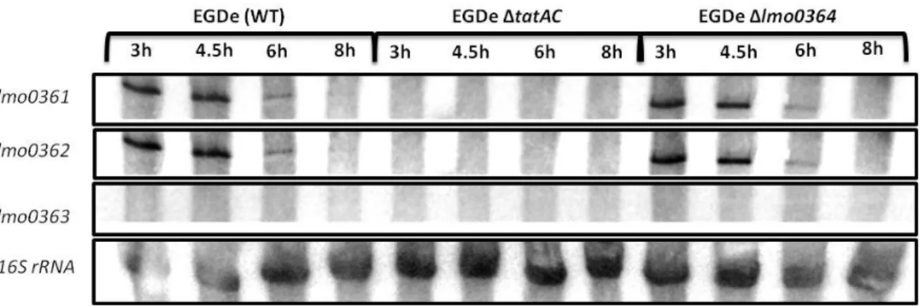
Between the genes coding for the secretion system and the operon containing the gene for the secreted protein, there is a gene coding for a transcription regulator (lmo0364) (see Figure 4 in section 1.3.).
To address the possible involvement of this transcription regulator in transcription regulation of genes tatA and tatC, a new transcription analysis was performed. For that a deletion mutant in this gene was also constructed. In the deletion mutant, the resulting protein lacks in the N-terminal, including the DNA-binding domain, 247 amino acids, out of 313 that constitute Lmo0364.
The results in Figure 9 show that the lmo0364 gene product has no influence in the transcription of tatA and tatC, since no difference between the mutant and the wild-type strain was detected.
Results
21 Figure 9 - Northern blot analysis of lmo0361, lmo0362 and lmo0363 transcripts in wild-type, tatAC and lmo0364 mutant strains. RNA was isolated from both strains at different times of growth (3, 4.5, 6 and 8 hours), corresponding to the exponential and stationary phases.
3.2.2. Promoter activity
The promoter activity of the genes lmo0361, lmo0362, lmo0363, lmo0364 and of the operon
lmo0365-67 was analyzed for the wild-type strain using a β-galactosidase assay.
Interestingly, the promoters showed different levels of activity, with lmo0362 being the most active promoter, followed by the operon lmo0365-67 promoter (Figure 10). Although transcripts of the gene lmo0363 were undetectable in the Northern blot, promoter activity was low but recorded. Even much lower than the activity of the lmo0362 and lmo0365 promoters, the transcription regulator lmo0364 also showed promoter activity (Figure 10). The promoter of lmo0361 gene showed no promoter activity when compared to the empty plasmid demonstrating once more that this gene is not transcribed on its own but in a bicistronic transcript with lmo0362.
Figure 10 – Determination of promoter activity by using lacZ fusion.
Promoter activity was measured for strains containing the empty plasmid and plasmids with the promoter regions of the genes lmo0361,
lmo0362, lmo0363, lmo0364 and
of the operon lmo0365-67,
determined using the β-galactosidase assay method. All samples were collected after five hours of culture growth in BHI, at 37 ˚C. The presented activities are the averages of three independent experiments each conducted in duplicate. Standard deviation bars are shown.
Results
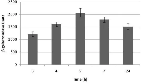
22 The lmo0362 transcript level showed to be growth-phase dependent in the Northern blot analysis, being the gene less expressed during the stationary phase. The promoter activity was investigated at different time points during culture growth (Figure 11). The promoter activity of the lmo0362 gene showed an increased activity during exponential phase, followed by a decrease during the stationary phase.
Figure 11 – Comparison of lmo0362 promoter activity at different time points during growth (3, 4, 5, 7 and 24 hours in BHI, at 37 ˚C).
The presented activities are the averages of three independent experiments each conducted in duplicate. Standard deviation bars are shown.
In order to investigate if the transcription regulator (Lmo0364) was related to the transcription of the operon lmo0365-67, where the last gene codes for the protein hypothetically secreted by the Tat pathway (25), and to confirm the result from the Northern blot for lmo0362, the promoter activity of lmo0362 and lmo0365 (Figure 12 A and B, respectively) was assessed in the wild-type and the deletion mutants ΔtatAC and Δlmo0364.
Results
23 Figure 12 – Determination of promoter activities in the EGDe (wt), ΔtatAC and Δlmo0364 mutant strais. Cells were harvested in the exponential growth phase. (A) Promoter activity measured for the gene lmo0362. (B) Promoter activity measured for the operon lmo0365-67.
The presented activities are the averages of three independent experiments each conducted in duplicate. Standard deviation bars are shown.
No differences were found among the wild-type and the mutant strains both for the activities of lmo0362 and lmo0365-67 promoters, indicating that there is not a direct relationship between the transcription regulator Lmo0364 and the promoter activities of the genes
lmo0362 and lmo0365-67.
The results obtained for the activity of lmo0362 promoter (Figure 12A) also indicate that the presence of this secretion system and the promoter activity of the corresponding genes are not related. The same could be verified for the lmo0365-67 operon. This operon hosts the gene coding for the protein putatively secreted by the Tat pathway. Nevertheless, the promoter activity of the operon was not different in the Tat pathway mutant.
To confirm that the transcriptional regulator was not associated to the transcription of the genes studied and that the results were not disguised by a down regulation of its transcription in the growing conditions used, the promoter activity of the lmo0364 was also assessed. No differences were found among strains (Figure 13) or time points during growth (data not shown). Finally, the influence of Lmo0364 in the promoter activity of lmo0363 gene was also investigated. The obtained results (not shown) also indicated the absence of differences between the wild-type and Δlmo0364 mutant strains.