Universidade de Aveiro 2016
Departamento de Biologia
Raquel do Nascimento
Amaro
Characterization of fungal community associated
with house dust
Caracterização da comunidade fúngica associada ao
pó doméstico
Declaro que este relatório é integralmente da minha autoria, estando devidamente referenciadas as fontes e obras consultadas, bem como identificadas de modo claro as citações dessas obras. Não contém, por isso, qualquer tipo de plágio quer de textos publicados, qualquer que seja o meio dessa publicação, incluindo meios eletrónicos, quer de trabalhos académicos.
Universidade de Aveiro 2016
Departamento de Biologia
Raquel do Nascimento
Amaro
Characterization of fungal community associated
with house dust
Caracterização da comunidade fúngica associada ao
pó doméstico
Dissertação apresentada à Universidade de Aveiro para cumprimento dos requisitos necessários à obtenção do grau de Mestre em Toxicologia e Ecotoxicologia, realizada sob a orientação científica da Doutora Ana Catarina Almeida Sousa, Investigadora em pós-doutoramento do CICECO (Centro de Investigação em Materiais Cerâmicos e Compósitos), no Departamento de Química da Universidade de Aveiro, e co-orientação do Doutor Carlos Miguez Barroso, Professor Auxiliar no Departamento de Biologia da Universidade de Aveiro.
Apoio financeiro da FCT e do FSE no âmbito do III Quadro Comunitário de Apoio, através do projeto PEst-OE/SAU/UI0709/2014.
“Deus Quer, o Homem sonha e a Obra nasce.”
o júri
presidente Prof. Doutora Sónia Alexandra Leite Velho Mendo Barroso
Professora auxiliar com agregação do Departamento de Biologia da Universidade de Aveiro
Doutor João Paulo Fernandes Teixeira
Investigador Auxiliar no Departamento de Saúde Ambiental, Instituto Nacional de Saúde Dr. Ricardo Jorge, Porto
Doutora Ana Catarina de Almeida Sousa
Investigadora em pós-doutoramento do CICECO (Centro de Investigação em Materiais Cerâmicos e Compósitos), no Departamento de Química da Universidade de Aveiro
agradecimentos Este trabalho de investigação não é só meu, mas de um conjunto de pessoas fantásticas, que me apoiaram e proporcionaram o ambiente ideal à realização do mesmo. Desta forma gostaria de agradecer:
À Ana Catarina Sousa, por toda a tua disponibilidade e entusiasmo desde o momento inicial. Por teres sido mais do que uma orientadora: uma amiga! Obrigada por todo o apoio, pelo carinho, pelas noites sem dormir, pela paciência, pela transmissão de conhecimentos, enfim, mil obrigados não chegariam. Não poderia pedir melhor orientadora!
À Sónia Coelho, por teres assumido a responsabilidade de ser o braço direito da Caty aqui em Aveiro. Quem diria que os nossos caminhos se voltariam a cruzar ao final de tantos anos? Obrigada por todo o esforço extra que fizeste para me ajudar e por todo o apoio nas horas de aflição. A tua ajuda foi essencial. Ao Doutor João Paulo Teixeira, por me ter recebido de braços abertos no Departamento de Saúde Ambiental do Instituto Nacional de Saúde Dr. Ricardo Jorge (INSA), no Porto. E à Cristiana Costa Pereira, pela transmissão da tua sapiência no mundo dos fungos, pela tua dedicação e por todo o apoio.
Prof. Doutor Luís Taborda Barata pelo recrutamento e avaliação médica dos voluntários.
Ao projeto 6x60x6 pelas amostras e ao CICS-UBI pelo financiamento. A todo o pessoal do INSA, por me terem acolhido no seio da sua “família”, tornando os meus dias no Porto mais leves. À Filipa Esteves, por seres a minha compincha no laboratório. Por todos os dias/noites de trabalho árduo em conjunto, por todos os sorrisos, por todos os momentos de descontração, por todo o apoio e pela amizade verdadeira, que surgiu de forma tão espontânea. Obrigada por todos os bons momentos que vivi contigo e que venham muitos mais! E à Mai Lan, que desde o primeiro dia te dedicaste a este trabalho a 100%, como se de teu se tratasse. Nunca esquecerei os momentos felizes que passei contigo dentro e fora do laboratório. Desde as canções, ao “God of fungi”, aos sorrisos e todas as brincadeiras. Somos a prova viva que a amizade vai além-fronteiras e consegue quebrar todos os estereótipos. Tão diferentes e tão iguais! Vemo-nos em Paris!
agradecimentos A todos os meus amigos, que ao longo deste longo percurso me apoiaram, se preocuparam e me incentivaram a ir mais além. Obrigada por tudo.
E por fim, o meu maior agradecimento vai para a minha querida mãe. Obrigada por me teres ensinado a lutar pelos meus sonhos com unhas e dentes e por me amparares nas pequenas quedas que dei ao longo deste percurso. Sem ti não teria conseguido, certamente. Não há palavras para descrever a Mãe que és. Espero que um dia consiga ser metade da mulher que és. O meu amor por ti é incondicional.
Obrigada, do fundo do coração, Raquel Amaro
palavras-chave Comunidade fúngica, pó doméstico, asma, qualidade do ar interior.
resumo Com a crescente urbanização, a população mundial passa cada vez mais tempo
no interior de edifícios, onde a exposição a contaminantes pode ser elevada. Este cenário leva a uma degradação da Qualidade do Ar Interior (QAI) que, em casos extremos, pode conduzir ao Síndrome do Edifício Doente (SED). É possível encontrar fungos em todo o tipo de ambientes e a comunidade fúngica encontrada no interior dos edifícios desempenha um papel essencial no estado de saúde dos indivíduos que frequentam esses locais. O pó em específico, atua como um reservatório de todos contaminantes presentes no interior dos edifícios, incluindo fungos, e pode ser utilizado para caracterizar o ambiente interior dos respetivos locais.
A convivência dos indivíduos com os fungos no interior de um edifício nem sempre é benéfica para a saúde. Existe uma forte associação entre os pacientes com doenças respiratórias alérgicas e a sensibilização a fungos, onde os últimos desempenham um papel importante no desenvolvimento, persistência e gravidade das primeiras. Para este tipo de indivíduos imunologicamente sensíveis, a exposição à contaminação fúngica pode desencadear os sintomas respiratórios como a asma.
Deste modo, este trabalho pretendeu numa 1ª fase caracterizar a comunidade fúngica do pó em ambiente doméstico de habitações construídas em diferentes décadas utilizando diferentes técnicas de amostragem de pó e numa 2ª fase caracterizar a comunidade fúngica presente em ambiente doméstico de doentes asmáticos e respetivos controlos por forma a avaliar possíveis associações. Este trabalho foi dividido em dois pontos fulcrais: i) identificar a comunidade fúngica no pó doméstico e a sua abundância ii) associar os géneros fúngicos encontrados com a severidade de asma.
Os géneros fúngicos maioritariamente encontrados foram Aspergillus,
Penicillium, Cladosporium, Alternaria e leveduras. Quanto à associação à
exacerbação da asma, não foi encontrada nenhuma relação. No entanto, dada a natureza preliminar do ponto ii), será necessário um número maior de amostras, por forma a tirar conclusões mais robustas.
keywords Fungal community, house dust, asthma, indoor air quality.
abstract With the increasing urbanization, the world population spends more and more
time indoors, where exposure to contaminants inside buildings can be high. This scenario leads to a degradation of Indoor Air Quality (IAQ), which, in extreme cases, can lead to Sick Building Syndrome (SBS).
Fungi can be found in all types of environments and the fungal community found inside buildings plays an essential role in the health of individuals that use these locations. Dust in particular, acts as a reservoir of all contaminants inside buildings, including fungi and can be used to characterize the indoor environment.
The coexistence of individuals with the fungi in the interior of a building is not always beneficial to health. There are strong associations between the patients with respiratory allergies and sensitization to molds where the latter play an important role in the development, persistence and severity of the former. For this type of immunologically susceptible individuals, exposure to fungal contamination can trigger respiratory symptoms such as asthma.
Thus, this work aims in a first stage to characterize the fungal community in house dust samples from houses built along different decades using different dust sampling procedures and in a second stage to characterize the fungal community in dust from the houses of asthmatic patients and respective controls in order to unravel possible associations. This work was divided into two key points: i) to identify the fungal community in house dust and its abundance ii) to associate the fungal genera found with the severity of asthma.
The most abundant fungal genera found were Aspergillus, Penicillium,
Cladosporium, Alternaria and yeast. As for the association to asthma
exacerbations, no association was found. However, given the preliminary nature of point ii), a larger number of samples will be necessary in order to draw any robust conclusions.
Table of contents
I Table of contents
LIST OF FIGURES ... V LIST OF TABLES ... VII
CHAPTER I: GENERAL INTRODUCTION ... 1
1. Indoor Environment ... 3
1.1. Indoor Air Quality and Sick building Syndrome ... 5
1.2. Fungal community ... 5
1.3. Evaluation of the fungal community in indoor environments ... 9
1.4. Indoor dust ... 9
1.4.1. Characterization of indoor dust ... 10
2. Health impacts of fungal exposure ... 11
2.1. Human Respiratory System ... 11
2.2. Inhalation of organic particulate matter and fungal allergens ... 13
3. Asthma ... 15
4. Aim and organization of the thesis ... 18
5. References ... 19
CHAPTER II: HOUSE DUST FUNGAL COMMUNITIES’ CHARACTERIZATION: A DOUBLE TAKE ON THE SIX BY SIXTY BY SIX PROJECT (6X60X6) ... 27
1. Abstract ... 29
2. Introduction ... 29
3. Materials and Methods... 31
3.1. Sampling ... 31
3.2. Treatment of samples ... 32
3.3. Culture Methods: Fungal Culture and Identification ... 32
4. Results and Discussion ... 33
5. Conclusions and future perspectives ... 38
6. Acknowledgments ... 38
7. References ... 38
CHAPTER III: FUNGAL COMMUNITIES IN HOUSE DUST SAMPLES FROM ASTHMATIC PATIENTS... 41
1. Abstract ... 43
2. Introduction ... 43
3. Materials and methods ... 44
3.1. Sampling collection... 44
3.2. Samples’ treatment... 45
3.3. Fungal characterization ... 45
3.4. Statistical analysis ... 46
Table of contents
III
5. References ... 51
CHAPTER IV: FINAL REMARKS ... 55
1. General Conclusion ... 57
2. Future work ... 58
List of figures
V
LIST OF FIGURES
Figure 1 Representation of a mycelium (A) with septated hyphae, adapted from (Fisher & Cook, 1998); (B) with aseptated hyphae and his apical growth, adapted from Vergara-Fernández et al., (2011) ... 6 Figure 2 Representation of the tree different types of mycelia, adapted from Fisher & Cook (1998) ... 7 Figure 3 Human Respiratory System displayed by upper and lower respiratory tract, retrieved from Tu et al. (2013) ... 12 Figure 4 Pulmonary hematosis on alveoli, retrieved from Netter (2014) ... 13
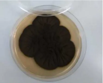
Figure 5 Regional deposition of particles on human respiratory tract, retrieved from Sierra-Vargas & Teran (2012) ... 14 Figure 6 Healthy bronchial tube (A); and Asthmatic Bronchial tube (B), retrieved from American Thoracic Society (2013) ... 16 Figure 7 House dust sampling. (A) Household vacuum cleaner used for active sampling; (B) Petri dishes placed on the top of a shelf; (C) Vacuum cleaner bag retrieved after 60 days of sampling ... 30 Figure 8 Cladosporium sp., 20 days growth on MEA plate ... 32
Figure 9 Most frequent genus detected. A) Alternaria sp.; B) Aspergillus sp.; C) Penicillium sp ... 35 Figure 10 Most common genera found on house dust samples in both group of houses: A) Aspergillus niger; B) Penicillium sp. ; C) Mucor sp.; and D) Alternaria alternata ... 46
List of tables
VII
LIST OF TABLES
Table 1 Main indoor pollutants, related sources and some threats towards human health (International Agency for Research on Cancer, 1987; Kim et al., 2013; Le Cann et al., 2011; Mercier et al., 2011; Tischer & Heinrich, 2013; US EPA, 2016; Viegi et al., 2004; WHO,
2000) ………..4
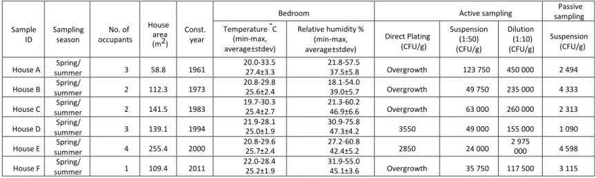
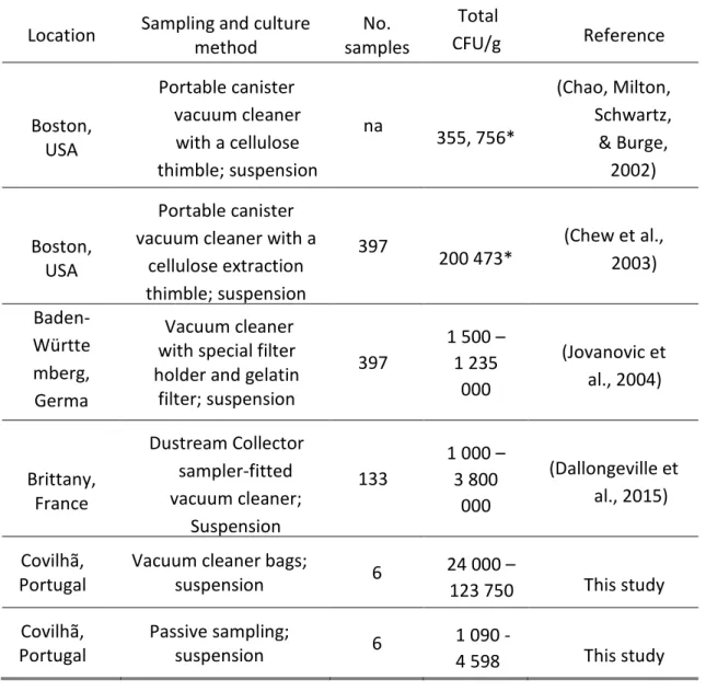
Table 2 Classification of Fungi, adapted from Fisher & Cook, (1998) and Prescott et al., (2005) ... 8 Table 3 Characteristics of the surveyed houses with the indication of: sampling season, number of occupants, area (m2), construction year (Const. year), temperature (ºC) and relative humidity (%) registered in the master bedroom (min-max, average ± stdev) and the number of total Colony Forming Units (CFU) using active and passive sampling methods. For the active sampling method the results are shown for the three different culture techniques used (direct plating, suspension and dilution) ... 33 Table 4 Comparison of the total amount of fungi detected in different surveys worldwide. Total CFU/g: Total number of Colony Forming Units (CFU) per gram of dust. *average values. na: information not available ... 34 Table 5 Identification of fungi found at each house using dust samples from the vacuum cleaner bag (active sampling) and from the deposited dust on petri dishes (passive sampling) ... 36 Table 6 Colony Forming Units of fungi per gram of dust in each sample with the indication of the corresponding dominant fungal genera. ... 45 Table 7 Dust samples analysis: Colony Forming Units per gram of dust (CFU/g) in each dust sample based on direct plating results and respective fungal colony identification. Bold letters denote those species or genera that were more abundant ... 48
Chapter I:
General Introduction
Chapter I
3
1.
Indoor Environment
With modernization of the society, individuals spend more time inside buildings. Currently people spend a large amount of their daily time indoors, from 85 to 90%, wherein 2/3 of this time is spent at home (European Environment Agency, 2013).
Indoor environment is defined by the environment inside homes, workplaces, schools, vehicles and all the other closed environments (Viegi et al., 2004). Its constitution is strongly influenced by the outdoors contaminants (Lee et al., 2000), but usually, indoors have higher concentrations of contaminants than outdoors (Hulin et al., 2012; Sundell, 2004; Viegi et al., 2004). In fact, studies by the United States Environmental Protection Agency (US-EPA) demonstrated that indoor pollutants’ levels could be 2 to 5 times higher than outdoors, therefore indoor contamination now ranks in top five amongst public health risks (US EPA, 2013). Hereupon, a good Indoor Air Quality (IAQ) is an essential determinant to an healthy life (WHO, 2010).
Particulate matter (PM), carbon monoxide, the different ways of exposure to tobacco smoke, pesticides, solvents, Volatile Organic Compounds (VOCs), radon, asbestos, metals, occupation-related contaminants and biological allergens, such mites, allergens and moulds, are some of the indoor contaminants that are possible to address through environmental analysis (WHO, 2008).
Characterization of fungal community associated with house dust
4
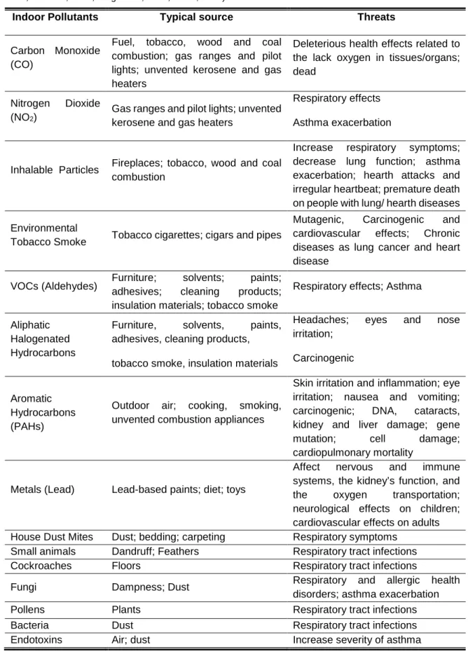
Table 1 Main indoor pollutants, related sources and some threats towards human health (International Agency for Research on Cancer, 1987; Kim et al., 2013; Le Cann et al., 2011; Mercier et al., 2011; Tischer & Heinrich, 2013; US EPA, 2016; Viegi et al., 2004; WHO, 2000).
Indoor Pollutants Typical source Threats
Carbon Monoxide (CO)
Fuel, tobacco, wood and coal combustion; gas ranges and pilot lights; unvented kerosene and gas heaters
Deleterious health effects related to the lack oxygen in tissues/organs; dead
Nitrogen Dioxide
(NO2) Gas ranges and pilot lights; unvented kerosene and gas heaters
Respiratory effects Asthma exacerbation
Inhalable Particles Fireplaces; tobacco, wood and coal combustion
Increase respiratory symptoms; decrease lung function; asthma exacerbation; hearth attacks and irregular heartbeat; premature death on people with lung/ hearth diseases Environmental
Tobacco Smoke Tobacco cigarettes; cigars and pipes
Mutagenic, Carcinogenic and cardiovascular effects; Chronic diseases as lung cancer and heart disease
VOCs (Aldehydes) Furniture; solvents; paints; adhesives; cleaning products; insulation materials; tobacco smoke
Respiratory effects; Asthma
Aliphatic Halogenated Hydrocarbons
Furniture, solvents, paints, adhesives, cleaning products, tobacco smoke, insulation materials
Headaches; eyes and nose irritation;
Carcinogenic
Aromatic Hydrocarbons (PAHs)
Outdoor air; cooking, smoking, unvented combustion appliances
Skin irritation and inflammation; eye irritation; nausea and vomiting; carcinogenic; DNA, cataracts, kidney and liver damage; gene mutation; cell damage; cardiopulmonary mortality
Metals (Lead) Lead-based paints; diet; toys
Affect nervous and immune systems, the kidney’s function, and the oxygen transportation; neurological effects on children; cardiovascular effects on adults House Dust Mites Dust; bedding; carpeting Respiratory symptoms
Small animals Dandruff; Feathers Respiratory tract infections
Cockroaches Floors Respiratory tract infections
Fungi Dampness; Dust Respiratory and allergic health
disorders; asthma exacerbation
Pollens Plants Respiratory tract infections
Bacteria Dust Respiratory tract infections
Chapter I
5 All those contaminants can lead to a poor Indoor Air Quality (IAQ), causing complains about indoor air, which sometimes leads to nonspecific building-related symptoms, commonly called Sick Building Syndrome (SBS).
1.1.
Indoor Air Quality and Sick building Syndrome
Indoor Air Quality is influenced by chemical, physical factors and bioaerosols and an upward of one of those factors can lead to Sick Building Syndrome (Di Giulio et al., 2010). The definition of SBS appeared on early 1970s (Chang et al., 2015) and it includes a range of health symptoms of unclear aetiology associated with the environment of a specific building, among them mucous membrane symptoms (eye, nose, and throat irritation), skin irritation, fatigue and headache (Burge, 2004; Fisk et al., 2009; Tsai et al., 2005). Usually there is a temporal relationship between the individuals’ symptoms and the building, with an improvement in the symptoms after the individuals leave a particular building (Burge, 2004; Crook & Burton., 2010). Traditionally, SBS was an occupational related problem but nowadays this term is commonly applied to domestic dwellings, especially those with water damage problems (Burge, 2004).
There are several parameters evaluated during the monitoring of IAQ: temperature, humidity, formaldehyde, carbon dioxide, particulate matter, volatile organic compounds, odours, bacteria, fungi, amongst others (Chang et al., 2015; WHO, 2010; Wolkoff et al., 2006). The quantity of microorganisms is determinant for air quality. In a normal environment, there are few airborne microorganisms, but an increase in the number of those microorganisms may represent a disease risk factor (Di Giulio et al., 2010). One important group of those microorganisms are fungi. The exposure to airborne fungi can lead to several health effects on humans (Li & Yang, 2004) and there are some studies relating levels of indoor airborne fungi with the prevalence of some SBS symptoms (see eg. Chang et al., 2015; Crook & Burton, 2010).
1.2.
Fungal community
Fungi communities are ubiquitous and easily found in all types of environments. About 100,000 species of fungi have been described including mushrooms, moulds and yeasts (Madigan et al., 2009; Prillinger et al., 2002).
Fungi are non-motile eukaryotic organisms so they have membrane-enclosed nucleus which contains genetic material and several membrane-bound cytoplasmic organelles (Deacon, 2005; Madigan et al., 2009). Therefore, most of them are multicellular. Fungi are classified as heterotrophs – they nourish through different organic means, by absorbing them from substrate. Some are saprophytes – the ones that digest dead organic matter and
Characterization of fungal community associated with house dust
6
wastes – and others are parasites, and obtain nutrients from tissues of other organisms (Black, 2002; Fisher & Cook, 1998).
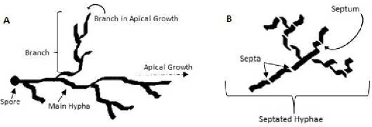
A typical fungus has a body called thallus, consisting of a network termed mycelium formed by a mass of threadlike structures – the hyphae, with an apical growth in branches or by elongation at the tips. These type of organisms are called filamentous fungi (figure 1). But some fungi replaced their typical growth as hyphae for yeasts – usually unicellular cells (Black, 2002; Deacon, 2005; Fisher & Cook, 1998; Madigan et al., 2009).
Figure 1 Representation of a mycelium (A) with septated hyphae, adapted from (Fisher & Cook, 1998); (B) with aseptated hyphae and his apical growth, adapted from Vergara-Fernández et al. (2011).
Hyphae are tubular cell walls that surround the cytoplasmic membrane (Madigan et al., 2009). They can be septated or aseptated (Figures 1a and 1b), when septated, there are cross walls called septa which divide the hyphae into small fragments (Fisher & Cook, 1998).
The septa have pores to help the transition of cytoplasm and nuclei between cells. Some fungi only have one septal pore, with an organelle called Woronin body, this organelle acts like a pore-blocker when a hyphal cell is damaged or old, so the tainted material cannot enter in a healthy cell (Black, 2002).
The mycelium has the function of nourishing the fungus, absorbing small nutrients, molecules through the enzymes released by mycelial cells (Black, 2002). There are three types of mycelia, classified according to where they grow: vegetative, aerial and reproductive mycelium (figure 2). The vegetative ones grow in or on the substrate and their function is to absorb nutrients. Aerial mycelia have aerial hyphae which grow above the surface of the substrate, almost all these constitute the visible colony. Reproductive mycelia develop where the reproductive structures grow (Fisher & Cook, 1998).
Chapter I
7 Figure 2 Representation of the tree different types of mycelia, adapted from Fisher et al. (1998).
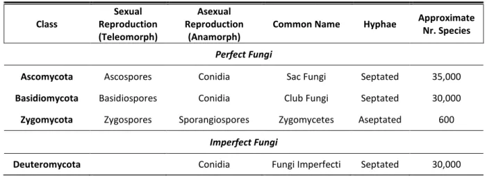
The majority of fungi’s cell walls contain chitin, and only a few fungi have cellulose (Fisher & Cook, 1998). All fungi have lysosomal enzymes to digest damaged cells and also to invade hosts in the case of parasitic fungi. The yeasts, and other types of fungi, may also have plasmids, which have the ability to clone foreign genes into yeasts cells (Black, 2002). Almost all fungi can reproduce both sexually and asexually, however there are a few that only reproduce by asexual way (Black, 2002). These are coined as imperfect fungi, and they only have an anamorph form – just the asexual form of reproduction. Perfect fungi or teleomorphs reproduce by both ways, having more than one sexual form. They can also have at least one anamorph form. If there are more than one anamorph form associated to a teleomorph form, the first one are called synanamorphs and all the forms together are called holomorphs (Fisher & Cook, 1998).
Therefore, the taxonomy of fungi is based on the sexual reproduction and it is possible to separate these organisms into 4 main categories: Zygomycetes, Ascomycetes, Basidiomycetes and Deuteromycetes (table 2) (Fisher & Cook, 1998; Prescott et al., 2005).
Characterization of fungal community associated with house dust
8
Table 2 Classification of Fungi, adapted from Fisher & Cook (1998) and Prescott et al., (2005)
Class Sexual Reproduction (Teleomorph) Asexual Reproduction (Anamorph)
Common Name Hyphae Approximate Nr. Species
Perfect Fungi
Ascomycota Ascospores Conidia Sac Fungi Septated 35,000
Basidiomycota Basidiospores Conidia Club Fungi Septated 30,000
Zygomycota Zygospores Sporangiospores Zygomycetes Aseptated 600 Imperfect Fungi
Deuteromycota Conidia Fungi Imperfecti Septated 30,000
Zygomycetes are multinucleate fungi or called coenocytic. They differ from the others by forming zygospores. These structures, which also produce spores, enclose zygotes which are the result of the fusion of the nuclei from multinucleate cells (Black, 2002; Madigan et al., 2009)
Ascomycetes range from single-celled species, from yeasts, to species which grow as filaments. They produce a sac-like asci cells with diploid nucleus, resulting from the fusion of two haploid cells from different mating during sexual reproduction. These diploid nucleuses go through meiosis and forms haploid ascospores, which are then released into environment. Asci can also be formed in a fruiting body called ascocarp. This type of fungi also reproduces asexually through the production of spores called conidia (Black, 2002; Madigan et al., 2009).
Basidiomycetes are usually the mushrooms, rusts, smuts and toadstools, yet in this group there are also yeasts and plant’s and human’s pathogens. They have a basidium, a single-cell structure in which basidiospores are formed. Basidiospores are sexual spores which germinate forming a septate mycelium. Mycelium cells unite into a dikaryotic form which grows and produces basidium that in turn produces basidiospores (Black, 2002; Madigan et al., 2009).
Deuteromycetes, also called the imperfect fungi, do not have any sexual stage in their life cycle, but due to their vegetative characteristics and their ability to produce asexual spores, most of these fungi are often associated to the Ascomycota group (Black, 2002).
Although fungal communities vary due to several indoor factors, such as occupation or heating ventilation and air-conditioning systems (HAVAC), at a global scale, indoor fungal diversity is also strongly influenced by the outdoor environment (Amend et al., 2010). Thus, the main indoor taxa are species also found in the outside of the dwellings – Cladosporium
Chapter I
9 sp., Aspergillus sp. Alternaria alternata and Penicillium sp. (Augustyniuk-Kram, 2013; Fairs, 2010).
1.3.
Evaluation of the fungal community in indoor environments
There are several methods to collect samples to assess the diversity of the fungal community in indoor environments including: i) air sampling; ii) settled dust sampling and; iii) house dust vacuuming.
Air sampling collection implies a special device – a portable air sampler – to filter the air during a specific time period (Hicks et al., 2005).
Settled dust sampling is considered a passive method consisting in the placement of a sterile empty petri-dish at a specific location during a certain period of time in which dust falls into it. After the sampling period, the petri-dish is sealed and sent to the laboratory to analyse the fungal communities by seeding the dust in appropriate culture media (Adams et al., 2013; Barberán et al., 2015). Other passive methods include the use of Electrostatic Dust Fall Collectors, collectors that consist of electrostatic cloths used as collectors for dust sedimentation (Madsen et al., 2012).
In house dust vacuuming, a vacuum cleaner is used to collect the house dust during a specific time period and a specific area. In some cases the vacuum cleaner may have a special dust collector and filters attached, but most commonly vacuum cleaner used is the one that the house inhabitants’ use to regularly vacuum their houses. (Norbäck et al., 2014; Pitkäranta et al., 2008). After collection, the dust is removed from the vacuum cleaner bag and samples are sieved before being analysed.
1.4.
Indoor dust
Indoor dust works as a long-term reservoir of all kind of indoor contaminants, including fungi, being a useful tool for the characterization of indoor environments (Sousa et al., 2014a). In fact, house dust is a matrix relatively easy to collect. Hence, it’s application on human exposure to environmental contaminants studies has been increasing and recently its usage in large epidemiological studies has been recommended (Rintala et al., 2008; Sousa et al., 2014a; Sousa et al., 2014b; Whitehead et al., 2011) and should also be considered in Indoor Air Quality monitoring (Pan et al., 2000).
Dust sampling is carried out since 1940s (Conant et al., 1936; Rintala et al., 2012) and since then there is no consensus about the best collection methodology. Therefore there is no standard protocol for human exposure validated for the measurement of allergens and other contaminants in house dust (Mansour et al., 2001; Paustenbach et al., 1997). Despite the
Characterization of fungal community associated with house dust
10
knowledge on house dust potentialities, there are only few studies using dust as an indicator of microbial communities.
Exposure to house dust can result in several health effects on humans. In fact, there are several studies that demonstrated an association between certain dust properties and the Sick Building Syndrome symptoms (Gyntelberg et al., 1994; Mendell et al., 2002; Niven et al., 2000; Pan et al., 2000; Skov et al., 1987). Furthermore, there are several reports in the scientific literature relating the human exposure to house dust and the development and/or exacerbation of respiratory symptoms and diseases such as asthma (Calderón et al., 2015; Douwes et al., 2000; Van Dyken et al., 2011).
1.4.1. Characterization of indoor dust
Indoor dust is by definition a complex mixture of particles and biological material, that is found settled in all indoor surfaces, floors and carpets, most of the times brought from outdoors by indoor occupants or carried by indoor aerosols (US EPA, 1997, 2011). In fact, about 45 to 50% of the house dust comes from soil and street dust, 2 to 3% comes from tyre wear, cement and car emissions, 1% is salt and 43% of the house dust is material presumably organic (Fergusson et al., 1986). Recently, indoor sources of dust are also being considered as well as their contribution to the levels of contaminants present in dust. The major indoor sources include microorganisms, furniture, household objects, pollen and other allergens, smoke, the occupants (including pet’s) and their activities (Butte et al., 2002).
House dust works as an adsorbent of organic and inorganic contaminants, so it can be considered one of the most important sources of human exposure to a large number of pollutants. Furthermore, dust can be considered an “archive” of indoor contamination, due to its ability to accumulate all kind of matter, and to its low capacity for degradation of contaminants, which leads to a continuous enrichment of chemicals (Butte et al., 2002; Cizdziel & Hodge, 2000).
There are several dust characteristics that influences the exposure potential to humans: i) the size of the particles; ii) the concentration of the contaminants; iii) the fine particle enrichment; and iv) the bioavailability of the fine particles and the larger particles (Paustenbach et al., 1997). Fine particles adhere more effectively to skin than the biggest ones (Kissel et al., 1996). In fact 50% of house dust particles have less than 50 µm of diameter (Roberts et al., 1991). And the fine particle enrichment has an impact on the amount of contaminants on house dust. The concentration of the contaminants is higher on the smaller size fraction of the particles (Beamer et al., 2012; Paustenbach et al., 1997).
Chapter I
11
2.
Health impacts of fungal exposure
Individuals are continuously exposed to airborne fungi and generally this exposure doesn’t translate into adverse effects in humans. In fact, there are some studies that support the called hygiene hypothesis (Gereda et al., 2000; Slameňová et al., 2003), which holds that a premature exposure to a rich microbial environment can decrease the risk of allergic diseases later in life, due to a stimulation of the immune-system to fight the allergy, so later the individual may have a non-allergic immune response (Strachan, 1989).
However, some fungi are in fact responsible for adverse health effects, especially related with respiratory diseases (Pei-Chih et al., 2000). The most frequent fungi found in indoor environments, especially in house dust, are: Alternaria, Cladosporium, Aspergillus and Penicillium and even yeasts, causing several allergic reactions on humans (Augustyniuk-Kram, 2013). These genera of fungi are consistent with those found outdoors, which suggests that indoors’ fungal communities are influenced by fungal taxa from outdoors (Chew et al., 2003).
2.1.
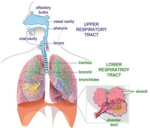
Human Respiratory System
The respiratory system is a complex system responsible for gas exchanges between living beings and the environment (Foster & Costa, 2005; Tu et al., 2013). Oxygen, which is essential for cellular function, enters the organism through nose or mouth during inhalation. Then, the inhaled oxygen passes through pharynx, larynx and reaches trachea that splits into two branches – the bronchi – which penetrate into the lungs. Once in the lungs, each bronchus bifurcates and forms bronchial tubes. These bronchial tubes form a network inside the lungs – the bronchioles – culminating in tinny sacs – the alveoli (see figure 3) (Netter, 2010; Tu et al., 2013).
Characterization of fungal community associated with house dust
12
Figure 3 Human Respiratory System displayed by upper and lower respiratory tract, retrieved from Tu et al. (2013).
Once in the alveoli, the oxygen passes to the bloodstream to reach all the tissues in organism through the blood capillary network aggregated on the alveolar wall. At the same time, carbon dioxide (a waste product derived from cellular function) is excreted into the bloodstream to perform the inverse route and being exhaled from the organism to the environment. This gas exchange on alveoli is called hematosis (see Figure 4) (Tu et al., 2013).
Chapter I
13 Figure 4 Pulmonary hematosis on alveoli, retrieved from Netter (2014).
Due to its function, the respiratory system can be divided into two sections: upper airways and lower airways. The upper airways include all organs that are outside de thorax – mouth, nose, pharynx and larynx -, and the lower ones are the organs located inside the last one – trachea, lungs, bronchi, bronchiole and alveoli (Tu et al., 2013).
Although the main function is gas exchange, this system is still responsible for the filtration, heating and humidification of inhaled air; for the smell, through the passage of air on olfactory bulbs; for the production of the vocal sound on larynx; for homeostasis of the pH of the organism; and for the protection of the organism, against microorganisms and particles (Tu et al., 2013).
2.2.
Inhalation of organic particulate matter and fungal allergens
The surface of the respiratory system is one of the largest interfaces between humans and their environment, being constantly in contact with gaseous contaminants and particulate matter. The inhaled particles usually deposits on respiratory tract, leading to a great number of allergic reactions and cardio respiratory diseases (Foster & Costa, 2005).Characterization of fungal community associated with house dust
14
Particulate matter (PM) availability is an important factor to determine whether it might be dangerous for health or not. The greater the amount of particles in the environment, the greater it is the possibility of particle inhalation and thus, the greater the probability of cardio and respiratory damage, since those are the primary targets of those pollutants (Kodavanti & Watkinson, 2005).
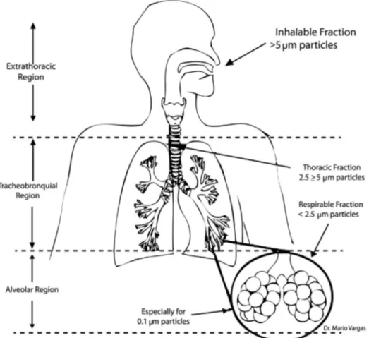
Particle size is a key aspect on particles’ toxicity, as those particles are small enough to be respirable and to penetrate into the lungs are of major concern towards human health. Particles can be divided in three groups, regarding their sizes: coarse particles (from 10.0 to 2.5 µm of diameter); fine particles (from 2.5 to 0.1 µm of diameter); and the ultrafine particles (with <100 nm of diameter). In terms of health impacts, the particles with diameter between 10 µm and 10 nm are the ones of more concern (Heyder, 2004; Kodavanti & Watkinson, 2005; Sierra-Vargas & Teran, 2012).
Figure 5 Regional deposition of particles on human respiratory tract, retrieved from Sierra-Vargas & Teran (2012).
Besides particle size there are other factors which determine the amount of inhaled particles, including their deposition location on respiratory system which is dependent on the particles themselves (chemical and other physical characteristics), and the individual
Chapter I
15 anatomical and physiological characteristics (airways anatomy, inhalation routes and biological factors) (Agnew, 1984).
Once on the respiratory system, there are three types of mechanisms for particle deposition: i) diffusion; ii) impaction; and iii) sedimentation. Diffusion occurs when, after inhalation, the particles target the surface of the airway through Brownian movement - aleatory movement in a fluid. Impaction is related with the inertia, ie, the particles are not able to follow the air flow and reach the airways’ surface. Sedimentation occurs on airways wall (Foster & Costa, 2005). The particles with diameters ranging between 0.5 and 3 µm usually get deposited on the nose by impaction and sedimentation (Heyder et al., 1985; Heyder, 2004). Those that aren’t retained on the nose, pass through larynx, provoking cough and bronchospasms and may reach the tracheobronchial tree (Bennett & Brown, 2005). Once there, the particles can deposit by impaction and by diffusion on bronchi, and/or by sedimentation and diffusion on bronchiole. The ones which diameter ranges between 2 µm and 0.1 µm can reach the alveoli and deposit by sedimentation (Heyder, 2004; Lippmann et al., 1980).
Due to its reduced size, fungal spores can penetrate the airways and cause illicit adverse reactions. The fungal spores size can range between 2 to 500 µm, but the average size range between 2 to 10 µm (Simon-Nobbe et al., 2008) and once on the respiratory tract, the spores germinate and only then release allergens (Burge, 2005). Moreover inhalation of fungal fragments with size in the order of submicrometer can also occur (Green et al., 2006). The presence of allergens on human organism initiates a specific immune response through the linkage between those allergens to antibody IgE (Crameri, 2015). The number of fungal proteins found to be able to bind IgE is around 200 and according to some authors this number is far to be a realistic one (Simon-Nobbe et al., 2008). Most of the fungal allergens are strongly associated to allergic diseases such as asthma (Burge, 2005; Crameri, 2015; Simon-Nobbe et al., 2008).
3.
Asthma
In 2014, and according to the Global Asthma Network (Global Asthma Network, 2014), the number of individuals reported with asthma was over 334 million.
In Portugal, the disease has a great impact on population’s health and the tendency is to increase. In the latest report from the National Observatory of Respiratory Diseases (Observatório Nacional das Doenças Respiratórias, 2016) it was estimated that there are over a million of asthmatic Portuguese with different gravity levels. In the same report, although Portugal was considered the EU country with the lowest rate of hospitalization for asthma, an increase in the number of hospitalizations from 2005 to 2014 was registered
Characterization of fungal community associated with house dust
16
and an increase in the number of deaths with this condition also increased during the same time period. In 2014, the prevalence of the disease varied between regions, with a sharpest increase rate of hospitalizations in the Center, followed by Algarve, North, Lisbon and Vale do Tejo and Alentejo. The rate of hospitalizations by asthma was higher on the group under 18. The group with 40 to 64 years suffered a decrease in the hospitalization rates, as well as the group of 65 to 79. There was a small increase in the hospitalizations for the group above 79, but this could be a consequence of the increase in the average life expectancy (Observatório Nacional das Doenças Respiratórias, 2016).
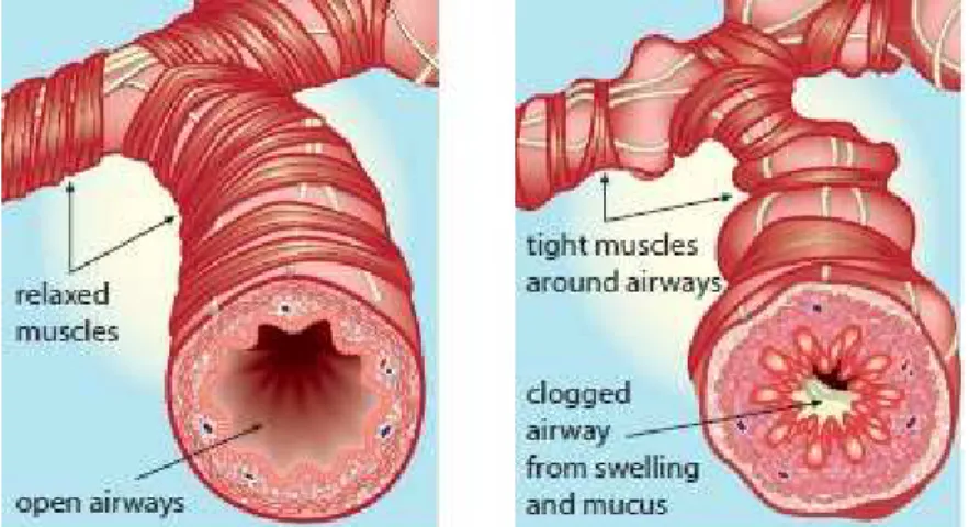
Asthma is an immunologically mediated hypersensitivity disease of the airways (WHO, 2003). It’s a chronic inflammation of the bronchial tubes in the lungs, provoking a reversible obstruction of the flow of air in and out of airways, when exposed to a certain stimuli (see figure 6). The characteristic symptom of asthma is wheezing – a high-pitched whistling sound heard during breathing. However, not always this symptomatology occurs. Other asthma symptoms include breathless, chest tightness and coughing (Global Asthma Network, 2014).
Figure 6 Healthy bronchial tube (A); and Asthmatic Bronchial tube (B), retrieved from American Thoracic Society (2013).
Asthma can affect all ages, but is more common in early childhood (before age 7) and usually the symptoms disappear around the age of 16. The deaths for asthma are uncommon and usually occur in elderly individuals. Even so, it just represents less than 1% of the total deaths worldwide and most of the times can be preventable (Global Asthma Network, 2014).
There are several factors affecting asthma condition, and they can be divided in two major groups: genetic and environmental factors. Individuals with a family history of allergic disease are more prone to develop the same condition (Global Asthma Network, 2014; WHO, 2003) and therefore genetic predisposition is considered a risk factor for asthma.
Chapter I
17 There are strong literature that evidences a small number of genetic variants that influence asthma risk – especially in children.
Concerning the environment, there are according to the Global Asthma Network, several factors to be considered:
• Common cold and exercise: the infection of the upper airways that occurs during a cold can trigger an asthma attack. The same background of infection can occur by effort, during exercise and thus initiate an asthma attack.
• Irritants: exposure to cigarette smoke, different vapours from cooking, heating or from vehicles can lead to an asthma attack. The same can occur by exposure to certain cosmetics and aerosol sprays.
• Second-hand smoke: this air pollutant can induce the appearance of asthma symptoms either in children and adults. It is particularly dangerous during prenatal exposure.
• Pharmaceuticals: Asthma attacks are more common in children who were treated with antibiotics in early stages of life. However this factor is still unclear mostly because wheezing symptoms are usually treated well before the recognition of asthma manifestations with drugs.
• Occupational exposure: bakers, woodworkers, farmers and professionals exposed to laboratory animals and to a certain chemicals are associated with the development of occupational asthma.
• Allergens: allergen exposure causes IgE sensitization, and the continued exposure can result in asthma.
• Animals: there are no consistent evidences that animals are either a risk factor or a protective one.
• Dampness and moulds: according to the Global Asthma Network its contribution is still unclear, because only few individuals are demonstrably allergic to fungal moulds and because dampness can occur in the houses of both allergic and non-allergic forms of asthma patients. Nevertheless, dampness and moulds appears more in asthmatic patients’ homes.
Several studies performed over the last years examined the association between asthma and fungal exposure and provided further evidences on the role of these biological contaminants in the development and progression of this disease.
The first evidences of fungal sensitization of asthmatic patients dates from 1698, when a famous asthmatic physician, John Floyer noticed a deterioration of asthma, when exposed
Characterization of fungal community associated with house dust
18
to damp on houses (Sakula, 1984). Thenceforth, mould sensitivity has been associated all over the years to triggering of asthma attacks, and its consequences.
In a work performed by Zureik et al. (2002), the authors concluded that sensitization to moulds was significantly associated with severity of asthma, for the genera Alternaria and Cladosporium. In a meta-analysis carried out by Mendell et al. (2011), several fungal species were able to induce inflammatory reactions. Sensitivity to fungi of the genus Alternaria and Cladosporium was also associated with persistence and severity of allergic asthma by Knutsen et al. (2012). Markowicz et al. (2014) identified Aspergillus versicolor as a crucial fungus in triggering symptoms of asthma. There was a positive association between the Aspergillus versicolor DNA concentrations and the lack of air during the day in the participants. It was also possible to validate that the concentration of ergosterol (often used as fungal marker) was related to the diagnosis of asthma, proving to be a risk factor for this disease.
The presence of fungi inside the home and the diagnosis of asthma in the house inhabitants was also studied. Blanc et al. (2013) used the Environmental Relative Moldiness Index (ERMI), this index is a quantification method of the exposure to mould inside the buildings in order to assess the effect of fungi in patients with asthma and rhinitis. The authors concluded - that the average value ERMI in homes of adults diagnosed with asthma was significantly higher than the average value of ERMI in households selected at random (control group) in the same geographic area of study in Northern California.
Fungal exposure can require multiple hospital admissions by asthmatic individuals (O’Driscoll et al., 2005). In a study conducted by Black et al. (2000), about fungal allergens sensitization on patients with asthma attacks admitted on Intensive Care Units (ICU), it was concluded that there was an association between life-threatening asthma and sensitization to one or more fungal allergens.
Some fungi, such as Aspergillus, can even act as opportunistic pathogens and attack the respiratory system of patients with previous lung diseases (Ader, 2010; Smarakoon & Soubani, 2008).
In the worst case scenario, fungal allergens derived from environmental moulds can play a role on asthma-related mortality (Targonski et al., 1995).
4.
Aim and organization of the thesis
The aim of this thesis is to characterize the fungal communities in house dust samples from households located in Covilhã municipality. Hence two different studies were performed: the first one aimed to characterize the fungal community in dust samples from houses
Chapter I
19 constructed in different decades (from 1960s to 2010s) from the 6X60X6 project and to compare two dust sampling strategies. The second study aimed to characterize the fungal community in dust samples from the houses of asthmatic patients and controls in order to understand possible associations between the fungal composition and asthma exacerbations.
The present dissertation is organized in 4 different Chapters. Chapter 1 provides a general introduction about fungal communities on house dust and their relationship with asthma exacerbation. Chapter 2 and 3 are structured as research papers and correspond to the 6X60X6 Project and to the asthma study, respectively. In chapter 4, general remarks are presented and the obtained results are discussed and compared with previous works.
5.
References
Adams, R. I., Amend, A. S., Taylor, J. W., & Bruns, T. D. (2013). A Unique Signal Distorts the Perception of Species Richness and Composition in High-Throughput Sequencing Surveys of Microbial Communities: A Case Study of Fungi in Indoor Dust. Microbial Ecology, 66(4), 735–741.
Ader, F. (2010). Invasive pulmonary aspergillosis in patients with chronic obstructive pulmonary disease: an emerging fungal disease. Current infectious disease reports, 12(6), 409–16.
Agnew, J. E. (1984). Physical properties and mechanisms of deposition of aerosols. Em S. W. Clarke & D. Pavia (Eds.), Aerosols and the Lung: Clinical and Experimental Aspects, 49–70.
Amend, A. S., Seifert, K. A., Samson, R., & Bruns, T. D. (2010). Indoor fungal composition is geographically patterned and more diverse in temperate zones than in the tropics. Proceedings of the National Academy of Sciences of the United States of America, 107(31), 13748–53.
American Thoracic Society. (2013). What is asthma? Am J Respir Crit Care Med, 188(1), 7–8.
Augustyniuk-Kram, A. (2013). Spectrum and Concentration of Culturable Fungi in House Dust from Flats in Warsaw, Poland. Aerosol and Air Quality Research, 13, 1438– 1447.
Barberán, A., Dunn, R. R., Reich, B. J., Pacifici, K., Laber, E. B., Menninger, H. L., … Fierer, N. (2015). The ecology of microscopic life in household dust. Proceedings of the Royal Society B: Biological Sciences, 282, 20151139.
Beamer, P. I., Elish, C. A., Roe, D. J., Loh, M. M., & Layton, D. W. (2012). Differences in metal concentration by particle size in house dust and soil. Journal of Environmental Monitoring, 14, 839-844.
Bennett, W. D., & Brown, J. S. (2005). Particulate Dosimetry in the Respiratory Tract Structure of the Respiratory Tract. Em W. M. Foster & D. L. Costa (Eds.), Air pollutants and the Respiratory Tract (pp. 21–73). Taylor & Francis Group.
Characterization of fungal community associated with house dust
20
and Explorations (5th ed., pp. 292–299). Jonh Wiley & Sons JG Black - Inc.
Black, P. N., Udy, A. A., & Brodie, S. M. (2000). Sensitivity to fungal allergens is a risk factor for life-threatening asthma. Allergy, 55, 501–504.
Blanc, P. D., Quinlan, P. J., Katz, P. P., Balmes, J. R., Trupin, L., Cisternas, M. G., … Vesper, S. J. (2013). Higher environmental relative moldiness index values measured in homes of adults with asthma, rhinitis, or both conditions. Environmental research, 122, 98–101.
Burge, H. A. (2005). Biological Airborne Pollutants. Em W. M. Foster & D. L. Costa (Eds.), Air pollutants and the Respiratory Tract (pp. 329–355). Taylor & Francis Group. Burge, P. S. (2004). Sick building syndrome. Occupational and Environmental Medicine,
61(2), 185–190.
Butte, W., & Heinzow, B. (2002). Pollutants in house dust as indicators of indoor contamination. Reviews of Environmental Contamination and Toxicology, 175, 1– 46.
Calderón, M. A., Linneberg, A., Kleine-Tebbe, J., De Blay, F., De Rojas, D. H. F., Virchow, J. C., & Demoly, P. (2015). Respiratory allergy caused by house dust mites: What do we really know? Journal of Allergy and Clinical Immunology, 136(1), 38–48. Chang, C. J., Yang, H. H., Wang, Y. F., & Li, M. S. (2015). Prevalence of sick building
syndrome-related symptoms among hospital workers in confined and open working spaces. Aerosol and Air Quality Research, 15(6), 2378–2384.
Chew, G. L., Rogers, C., Burge, H. a, Muilenberg, M. L., & Gold, D. R. (2003). Dustborne and airborne fungal propagules represent a different spectrum of fungi with differing relations to home characteristics. Allergy, 58, 13–20.
Cizdziel, J. V., & Hodge, V. F. (2000). Attics as archives for house infiltrating pollutants: Trace elements and pesticides in attic dust and soil from southern Nevada and Utah. Microchemical Journal, 64(1), 85–92.
Conant, N. F., Wagner, H. C., & Rackerman, F. M. (1936). Fungi in pillows, mattresses, and furniture. J. Allergy, 7, 234–237.
Crameri, R. (2015). Structural aspects of fungal allergens. Seminars in Immunopathology, 37(2), 117–121.
Crook, B., & Burton, N. C. (2010). Indoor moulds, Sick Building Syndrome and building related illness. Fungal Biology Reviews, 24(3-4), 106–113.
Deacon, J. D. (2005). Introduction: the fungi and fungal activities. Em Fungal Biology (4th ed.). Wiley-Blackwell.
Di Giulio, M., Grande, R., Di Campli, E., Di Bartolomeo, S., & Cellini, L. (2010). Indoor air quality in university environments. Environmental Monitoring and Assessment, 170(1-4), 509–517.
Douwes, J., Zuidhof, A., Doekes, G., van der Zee, S. C., Wouters, I., Boezen, M. H., & Brunekreef, B. (2000). (1->3)-beta-D-glucan and endotoxin in house dust and peak flow variability in children. American journal of respiratory and critical care medicine, 162, 1348–54.
European Environment Agency. (2013). Environment and human health. European Union, Vol. 110 (5), 5-86.
Fairs, A. (2010). Guidelines on Ambient Intramural Airborne Fungal Spores. Investig Allergol Clin Immunol, 20(6), 490–498.
Chapter I
21 composition and sources of house dust and street dust. The Science of the Total Environment, 50, 217–221.
Fisher, F., & Cook, N. B. (1998). The Basics. Em Fundamentals of Diagnostic Mycology (pp. 1–11). Saunders.
Fisk, W. J., Mirer, A. G., & Mendell, M. J. (2009). Quantitative relationship of sick building syndrome symptoms with ventilation rates. Indoor Air, 19(2), 159–165.
Foster, W. M., & Costa, D. L. (2005). Air Pollutants and the Respiratory Tract. Taylor & Francis (pp 1-478).
Gereda, J. E., Leung, D. Y., Thatayatikom, A., Streib, J. E., Price, M. R., Klinnert, M. D., & Liu, A. H. (2000). Relation between house-dust endotoxin exposure, type 1 T-cell development, and allergen sensitisation in infants at high risk of asthma. Lancet, 355(9216), 1680–1683.
Global Asthma Network. (2014). The Global Asthma Report 2014 (Vol. 5).
Green, B. J., Tovey, E. R., Sercombe, J. K., Blachere, F. M., Beezhold, D. H., & Schmechel, D. (2006). Airborne fungal fragments and allergenicity. Medical mycology : official publication of the International Society for Human and Animal Mycology, 44 Suppl 1(September), S245–S255.
Gyntelberg, F., Suadicani, P., Nielsen, J. W., Skov, P., Valbjmn, O., Nielsen, P. A., … Norn, S. (1994). Dust and the Sick Building Syndrome. Indoor Air, 4, 223–238.
Heyder, J. (2004). Deposition of inhaled particles in the human respiratory tract and consequences for regional targeting in respiratory drug delivery. Proc Am Thorac Soc, 1(4), 315–320.
Heyder, J., Gebhart, J., & Scheuch, G. (1985). Interaction of Diffusional and Gravitational Particle Transport in Aerosols. Aerosol Science and Technology, 4(3), 315–326. Hicks, J. B., Lu, E. T., De Guzman, R., & Weingart, M. (2005). Fungal types and
concentrations from settled dust in normal residences. Journal of occupational and environmental hygiene, 2(10), 481–492.
Hulin, M., Simoni, M., Viegi, G., & Annesi-Maesano, I. (2012). Respiratory health and indoor air pollutants based on quantitative exposure assessments. European Respiratory Journal, 40(4), 1033–1045.
International Agency for Research on Cancer. (1987). Overall Evaluations of Carcinogenicity: An Updating of IARC Mongraphs. IARC Monographs on the Evaluation of the Carcinogenic Risks to Humans, 1-42(Supp 7), 1-449.
Kim, K. H., Jahan, S. A., & Kabirl E. (2013). A review on human health perspective of air pollution with respectto allergies and asthma. Environmental International, 59, 41– 52.
Kissel, J. C., Richter, K. Y., & Fenske, R. A. (1996). Field measurement of dermal soil loading attributable to various activities: Implications for exposure assessment. Risk Analysis, 16(1), 115–125.
Knutsen, A. P., Bush, R. K., Demain, J. G., Denning, D. W., Dixit, A., Fairs, A., … Wardlaw, A. J. (2012). Fungi and allergic lower respiratory tract diseases. The Journal of allergy and clinical immunology, 129(2), 280–91; quiz 292–3.
Kodavanti, U. P., & Watkinson, W. P. (2005). Bioavailability of Particle-Associated Air Pollutants and Relationship to Cardiopulmonary Injury. Em M. W. Foster & D. L. Costa (Eds.), Air pollutants and the Respiratory Tract (2.a ed., pp. 75–134). Taylor & Francis Group.
Characterization of fungal community associated with house dust
22
Le Cann, P., Bonvallot, N., Glorennec, P., Deguen, S., Goeury, C., & Le Bot, B. (2011). Indoor environment and children’s health: Recent developments in chemical, biological, physical and social aspects. International Journal of Hygiene and Environmental Health, 215(1), 1–18.
Lee, S., & Chang, M. (2000). Indoor and outdoor air quality investigation at schools in Hong Kong. Chemosphere, 41(1-2), 109–113.
Li, D. W., & Yang, C. S. (2004). Fungal contamination as a major contributor to sick building syndrome. Advances in Applied Microbiology, 55, 31–112.
Lippmann, M., Yeates, D. B., & Albert, R. E. (1980). Deposition and clearance of inhaled particles. British Journal of Industrial Medicine, 37, 337–3662.
Madigan, M. T., Martinko, J. M., Dunlap, P. V., & Clark, D. P. (2009). Methods in Microbial Ecology: Measuring Microbial Activities in Nature. Em Brock Biology of Microorganisms: International Edition (12th ed., pp. 535–543). Pearson International Edition.
Madsen, A. M., Matthiesen, C. B., Frederiksen, M. W., Frederiksen, M., Frankel, M., Spilak, M., … Timm, M. (2012). Sampling, extraction and measurement of bacteria, endotoxin, fungi and inflammatory potential of settling indoor dust. Journal of environmental monitoring : JEM, 14(12), 3230–9.
Mansour, M., Lanphear, B. P., Hornung, R., Khoury, J., Bernstein, D. I., Menrath, W., & Decolongon, J. (2001). A side-by-side comparison of sampling methods for settled, indoor allergens. Environmental research, 87(1), 37–46.
Markowicz, P., Cai, G., Hashim, Z., Ali, F., Zheng, Y., Lai, X., … Hashim, J. H. (2014). Endotoxin , Ergosterol , Fungal DNA and Allergens in Dust from Schools in Johor Bahru , Malaysia- Associations with Asthma and Respiratory Infections in Pupils. PLOS ONE, 9(2), e88303.
Mendell, M. J., Fisk, W. J., Petersen, C. J., Hines, M., Dong, D., Faulkner, J. A., … Boeniger, M. F. (2002). Indoor particles and symptoms among office workers: Result from a double-blind cross-over study. Epidemiology, 13, 296–304.
Mendell, M. J., Mirer, A. G., Cheung, K., Tong, M., & Douwes, J. (2011). Respiratory and allergic health effects of dampness, mold, and dampness-related agents: a review of the epidemiologic evidence. Environmental health perspectives, 119(6), 748–56. Mercier, F., Glorennec, P., Thomas, O., & Bot, B. Le. (2011). Organic Contamination of Settled House Dust , A Review for Exposure Assessment Purposes. Environment: Science and Technology, 45, 6716–6727.
Netter, F. H. (2010). Atlas of Human Anatomy (5th ed.). Saunders.
Netter, F. H. (2014). Atlas of Human Anatomy (6th ed.). Philadelphia: Saunders.
Niven, R. M., Fletcher, A. M., Pickering, C. A, Faragher, E. B., Potter, I. N., Booth, W. B., … Potter, P. D. (2000). Building sickness syndrome in healthy and unhealthy buildings: an epidemiological and environmental assessment with cluster analysis. Occupational and environmental medicine, 57, 627–634.
Norbäck, D., Markowicz, P., Cai, G. H., Hashim, Z., Ali, F., Zheng, Y. W., … Hashim, J. H. (2014). Endotoxin, ergosterol, fungal dna and allergens in dust from schools in johor bahru, malaysia- Associations with asthma and respiratory infections in pupils. PLoS ONE, 9(2), 1-10.
O’Driscoll, B. R., Hopkinson, L. C., & Denning, D. W. (2005). Mold sensitization is common amongst patients with severe asthma requiring multiple hospital admissions. BMC
Chapter I
23 pulmonary medicine, 5(4).
Observatório Nacional das Doenças Respiratórias. (2016). 11o Relatório Prevenir as Doenças Respiratórias Acompanhar e Reabilitar os Doentes.
Pan, Z., Mølhave, L., & Kjaergaard, S. K. (2000). Effects on eyes and nose in humans after experimental exposure to airborne office dust. Indoor Air, 10(4), 237–45.
Paustenbach, D. J., Finley, B. L., & Long, T. F. (1997). The critical role of house dust in understanding the hazardz posed by contaminated soils. International Journal of Toxicology, 16, 339–362.
Pei-Chih, W., Huey-Jen, S., & Chia-Yin, L. (2000). Characteristics of indoor and outdoor airborne fungi at suburban and urban homes in two seasons. Science of the Total Environment, 253(1-3), 111–118.
Pitkäranta, M., Meklin, T., Hyvärinen, A., Paulin, L., Auvinen, P., Nevalainen, A., & Rintala, H. (2008). Analysis of fungal flora in indoor dust by ribosomal DNA sequence analysis, quantitative PCR, and culture. Applied and Environmental Microbiology, 74(1), 233–244.
Prescott, L., Harley, J., & Klein, D. (2005). Microbiology (6th ed.). New York: McGraw- Hill. Prillinger, H., Lopandic, K., Schweigkofler, W., Deak, R., Aarts, H. J. M., Bauer, R., … Maraz, A. (2002). Phylogeny and systematics of the fungi with special reference to the Ascomycota and Basidiomycota. Chemical immunology, 81, 207–295.
Rintala, H., Pitkäranta, M., & Täubel, M. (2012). Microbial communities associated with house dust. Em Advances in Applied Microbiology (1.a ed., Vol. 78, pp. 75–120). Elsevier Inc.
Rintala, H., Pitkäranta, M., Toivola, M., Paulin, L., & Nevalainen, A. (2008). Diversity and seasonal dynamics of bacterial community in indoor environment. BMC Microbiology, 8(56), 1–13.
Roberts, J. W., Budd, W. T., Ruby, M. G., Bond, A. E., Lewis, R. G., Wiener, R. W., & D.E., C. (1991). Development and field testing of a high volume sampler for pesticides and toxics in dust. J Expo Anal Environ Epidemiol, 1(2), 143–55.
Sakula, A. (1984). Sir John Floyer’s A Treatise of the Asthma. Thorax, 39, 248–254. Sierra-Vargas, M. P., & Teran, L. M. (2012). Air pollution: Impact and prevention.
Respirology, 17(7), 1031–1038.
Simon-Nobbe, B., Denk, U., Pöll, V., Rid, R., & Breitenbach, M. (2008). The spectrum of fungal allergy. International archives of allergy and immunology, 145(1), 58–86. Skov, P., ValbjØrn, O., & Disg. (1987). The «sick» building syndrome in the office
environment: The Danish town hall study. Environment International, 13(4-5), 339– 349.
Slameňová, D., Lábaj, J., Križková, L., Kogan, G., Šandula, J., Bresgen, N., & Eckl, P. (2003). Protective effects of fungal (1 → 3)-β-D-glucan derivatives against oxidative DNA lesions in V79 hamster lung cells. Cancer Letters, 198(2), 153–160.
Smarakoon, P., & Soubani, A. (2008). Invasive pulmonary aspergillosis in patients with COPD : a report of five cases and systematic review of the literature. Chronic Respiratory Disease, 5, 19–27.
Sousa, A. C., Takahashi, S., & Tanabe, S. (2014a). Organic Contaminants in House Dust. Current Organic Chemistry, 18(17), 2181.
Sousa, A. C., Almeida, J. R. S. L., Pereira, C. C., Ramiro Pastorinho, M., Pereira, Â. M. C., Nogueira, A. J. a, … Alves, A. (2014b). Characterization of fungal communities in
Characterization of fungal community associated with house dust
24
house dust samples collected from central Portugal-a preliminary survey. Journal of toxicology and environmental health. Part A, 77(14-16), 972–82.
Strachan, D. P. (1989). Hay fever, hygiene, and household size. Br Med J, 299(November), 1259–1260.
Sundell, J. (2004). On the history of indoor air quality and health. Indoor air, 14(Suppl 7), 51–58.
Targonski, P. V., Persky, V. W., & Ramekrishnan, V. (1995). Effect of environmental molds on risk of death from asthma during the pollen season. The Journal of Allergy and Clinical Immunology, 95(5), 955–961.
Tischer, C. G., & Heinrich, J. (2013). Exposure assessment of residential mould, fungi and microbial components in relation to children’s health: achievements and challenges. International journal of hygiene and environmental health, 216(2), 109–14.
Tsai, F. C., & Macher, J. M. (2005). Concentrations of airborne culturable bacteria in 100 large US office buildings from the BASE study. Indoor Air, 15(Suppl 9), 71–81. Tu, J., Inthavong, K., & Ahmadi, G. (2013). Chapter 2: The Human Respiratory System. Em
Computational Fluid and Particle Dynamics in the Human Respiratory System (pp. 19–44).
US EPA. (1997). Exposure Factors Handbook. Exposure, 1(August), 1193.
US EPA. (2011). Exposure Factors Handbook: 2011 Edition. U.S. Environmental Protection Agency, September(Chapter 5), 267–318.
US EPA. (2013). Questions About Your Community: Indoor Air. Retrieved on 30 april 2016 from http://www.epa.gov/region1/communities/indoorair.html
US EPA. (2016). Criteria Air Pollutants. Retrieved on 30 april 2016, from https://www.epa.gov/criteria-air-pollutants
Van Dyken, S. J., Garcia, D., Porter, P., Huang, X., Quinlan, P. J., Blanc, P. D., … Locksley, R. M. (2011). Fungal chitin from asthma-associated home environments induces eosinophilic lung infiltration. Journal of immunology (Baltimore, Md. : 1950), 187(5), 2261–7.
Vergara-Fernández, A., Hernández, S., San Martín-Davison, J., & Revah, S. (2011). Morphological characterization of aerial hyphae and simulation growth of fusarium solani under different carbon source for application in the hydrophobic VOCs biofiltration. Revista mexicana de ingeniería química, 10(2), 225–233.
Viegi, G., Simoni, M., Scognamiglio, a, Baldacci, S., Pistelli, F., Carrozzi, L., & Annesi-Maesano, I. (2004). Indoor air pollution and airway disease. The international journal of tuberculosis and lung disease : the official journal of the International Union against Tuberculosis and Lung Disease, 8(12), 1401–1415.
Whitehead, T., Metayer, C., Buffler, P., & Rappaport, S. M. (2011). Estimating exposures to indoor contaminants using residential dust. Journal of exposure science & environmental epidemiology, 21(6), 549–64.
WHO. (2003). Prevention of Allergy and Allergic Asthma. January, 1-14.
WHO (2008). Indoor Air Pollution - Children’s Health and Environment. WHO Training Package for the Health Sector (Vol. July).
WHO. (2000). Environmental tobacco smoke. Em Air Quality Guidelines (2nd ed., pp. 1– 23). Copenhagen.
WHO. (2010). WHO guidelines for indoor air quality : Selected pollutants. WHO Regional Office for Europe (Vol. 9).