Braz. J. of Develop.,Curitiba, v. 6, n.5, p.27725-27734 may. 2020. ISSN 2525-8761
Bisphosphonate-related osteonecrosis of the jaws, osteomyelitis and
osteoradionecrosis: Comparative immunohistochemical study
Osteonecrose medicamentosa associada ao bisfosfonato, osteomielite e
osteorradionecrose: estudo imunohistoquímico comparativo
DOI:10.34117/bjdv6n5-281
Recebimento dos originais: 25/04/2020 Aceitação para publicação: 15/05/2020
Carlos Cesar Deantoni
Doutor em Cirurgia e Traumatologia Bucomaxilofacial pela Unisagrado. Instituição: Unisagrado.
Endereço: Rua Irmã Arminda 10-50, Jardim Brasil, Bauru, SP. E-mail: ccdeantoni@yahoo.com.br
Luiz Kawai Júnior
Graduado no Curso de Odontologia pela Unisagrado. Instituição: Unisagrado.
Endereço: Rua Irmã Arminda 10-50, Jardim Brasil, Bauru, SP. E-mail: lkawaijr@icloud.com
Beethoven Estevão Costa
Mestrando pelo Hospital de Reabilitação de Anomalias Craniofaciais.
Instituição: Hospital de Reabilitação de Anomalias Craniofaciais (HRAC/Centrinho) Endereço: Rua Silvio Marchione 3-20, Bauru, SP.
E-mail: beethovencosta@usp.br
Carlos Cesar de Antoni Filho
Graduado pela Universidade do Sul de Santa Catarina, Brasil. Instituição: Universidade do Sul de Santa Catarina Endereço: Avenida Pedra Branca 25, Palhoça, SC.
E-mail: carlosdfilho@gmail.com
Marcos Martins Curi
Doutor em Oncologia pelo Hospital AC Camargo. Instituição: Hospital Santa Catarina. Endereço: Av Paulista 218, São Paulo, SP.
E-mail: mmcuri@terra.com.br
Laurindo Moacir Sassi
Doutor pelo Programa de Pós-graduação em Cirurgia de Cabeça e Pesçoço do Depto de Otorrinolaringoloiga da Universidade Federal de São Paulo/Escola Paulista de Medicina.
Instituição: Hospital Erasto Gaertner, Curitiba, Paraná, Brasil.
Endereço: R. Dr. Ovande do Amaral, 201 - Jardim das Américas, Curitiba – PR. E-mail: sassilm@onda.com.br
Braz. J. of Develop.,Curitiba, v. 6, n.5, p.27725-27734 may. 2020. ISSN 2525-8761
Joel Ferreira Santiago Júnior
Doutor em Reabilitação Oral pela Faculdade de Odontologia de Bauru, USP. Instituição: Unisagrado.
Endereço: Rua Irmã Arminda 10-50, Jardim Brasil, Bauru, SP. E-mail: jf.santiagojunior@gmail.com
Camila Lopes Cardoso
Doutora em Estomatologia pela Faculdade de Odontologia de Bauru, USP. Instituição: Unisagrado.
Endereço: Rua Irmã Arminda 10-50, Jardim Brasil, Bauru, SP. E-mail: cardoso_lopes@yahoo.com.br
RESUMO
A osteonecrose medicamentosa associada ao uso de bisfosfonatos (OMMBF), osteomielite (OM) e osteorradionecrose (ORN) apresentam muita similaridade nos aspectos clínicos e radiográficos. No intuito de investigar os aspectos microscópicos de cada doença, e diante de poucos estudos comparativos, em especial análises imunohistoquimica, o objetivo deste trabalho foi comparar a imunoexpressão de marcadores relacionados ao ritmo da remodelação óssea e a vascularização entre as três doenças. Foram selecionados prontuários de pacientes que realizaram biópsia prévia com o diagnóstico das três doenças: OM, OMMBF e ORN. As lâminas de cada espécime foram submetidas às imunomarcações utilizando os anticorpos OPG, RANKL, VEGF e CD31. A análise semiquantitativa cega foi realizada por um examinador, sendo conferida por um supervisor. A amostra obtida foi de 34 espécimes oriundos de 34 pacientes, sendo 16 pacientes com OMMBF, 6 com OM crônica e 12 com ORN. Considerando o valor de p<0,05, não foi identificada diferença significante na expressão dos marcadores entre as doenças. Através deste estudo, pode ser afirmado que considerando os marcadores relacionados ao ritmo de remodelação óssea e a vascularização, as doenças analisadas não se diferenciaram.
Palavras-Chave: Osteonecrose medicamentosa dos maxilares. Bisfosfonatos. Osteomielite.
Osteorradionecrose. VEGF. CD31. OPG. RANKL.
ABSTRACT
Bisphosphonate-related osteonecrosis of the jaws (BRONJ), osteomyelitis (OM) and osteoradionecrosis (ORN) present a great deal of similarity in clinical and radiographic aspects. In order to investigate the microscopic aspects of each disease, and with few comparative studies, especially immunohistochemical analyzes, the objective of this study was to compare the immunoexpression of markers related to the rhythm of bone remodeling and vascularization among the three diseases. Patients were selected from patients who underwent previous biopsy with the diagnosis of the three diseases: OM, BRONJ and ORN. The slides of each specimen were subjected to immunoblots using the OPG, RANKL, VEGF and CD31 antibodies. Blind semiquantitative analysis was performed by an examiner, and was checked by a supervisor. The sample obtained was 34 specimens from 34 patients, 16 patients with BRONJ, 6 with chronic OM and 12 with ORN. Considering the value of p <0.05, no significant difference in the expression of the markers between the diseases was identified. Through this study, it can be affirmed that considering the markers related to the rhythm of bone remodeling and vascularization, the analyzed diseases were not differentiated.
Braz. J. of Develop.,Curitiba, v. 6, n.5, p.27725-27734 may. 2020. ISSN 2525-8761
Keywords: Medication-related osteonecrosis of the jaws. Bisphosphonates. Osteomyelitis.
Osteoradionecrosis. VEGF. CD31. OPG. RANKL.
1 INTRODUCTION
Among the bone pathologies of the maxillaries, Bisphosphonate-related osteonecrosis of the jaws (BRONJ) has been the subject of scientific discussion over the last decade, its etiopathogenesis being considered a broad line of current research. Considering that its clinical and radiographic aspects are similar to those of Osteomyelitis (OM) and Osteoradionecrosis (ORN), very often undistinguishable from one another, some studies have raised hypotheses of variations in the microscopic findings, which could unearth a characteristic peculiar to each disease, thereby contributing to its etiopathogenesis.1,2
On analyzing the three diseases (BRONJ, ORN, OM), the common clinical aspect is one of an exposed bone necrosis in the oral environment, most commonly in the mandible, which may be related to phlogistic signs and symptoms such as erythema, suppuration, pain or edema, which, depending on the case, will be either an acute or chronic process. Moreover, they may generate complications and sequelae such as paresthesia and fractures of the mandible. Common imaging aspects reveal areas of diffuse osteolysis and, potentially, well-defined bone sequestration, which relate to sclerotic portions of avascular bone surrounded by a fibrous tissue, which appears radiolucent under radiography. One peculiar radiographic aspect of BRONJ is a more generalized change in bone sclerosis and thickening of the lamina dura involving a number of teeth.3-5
However, when comparing the history of the three diseases, OM is commonly an inflammatory bone condition which is almost always infected, the consequence of an acute dentoalveolar abscess not properly treated. It exhibits more or less severe signs and symptoms depending on whether it is an acute or chronic condition.6 With BRONJ, the patient reports,
in his/her previous medical history, having used bisphosphonates, administered either orally or intravenously.7 As for ORN, the patient must present a history of prior radiotherapy in the region of the head and neck. Accordingly, the anamnesis is fundamental to the diagnostic process. So fundamental that the anatomopathological examination is often just supplementary to the process of diagnosis and is not so critical.
With the aim of investigating the three diseases (OM, BRONJ and ORN) microscopically, several authors have found both similarities and differences between them.1 Subsequently, a very similar study was conducted, inspired by the authors’ methodology, in order to provide answers to two questions: if there would be a microscopic difference between
Braz. J. of Develop.,Curitiba, v. 6, n.5, p.27725-27734 may. 2020. ISSN 2525-8761 the three diseases (OM, BRONJ and ORN) and if a pathologist would be capable of diagnosing them without previous knowledge of the diseases. The authors concluded that the three diseases are very similar and not capable of being distinguished without clinical information being made available.8
The proposal for the present study, namely to compare the process of bone metabolism and vascularization between OM, BRONJ and ORN, was justified by the paucity of immunohistochemical analyses comparing the three aforementioned diseases.
2 CASE STUDY AND METHODS
An immunohistochemical evaluation (IHC) was performed on slides obtained from specimens previously subjected to a biopsy of the maxillary bones affected by chronic osteomyelitis (OM), bisphosphonate-related osteonecrosis (BRONJ) and osteoradionecrosis (ORN). Sample selection was performed using case records for the period from 2000 to 2017, obtained from three separate institutions: Stomatology Services at the Oncology Center at the Santa Catarina Hospital, in the city of São Paulo; the database of the anatomopathological services of the Unisagrado in Bauru; and the Erasto Gärtner Hospital in Curitiba, in the federal state of Paraná.
The IHC analysis of the specimens was used to evaluate vascularization between the groups. To this end, the following primary antibodies were used: VEGF: Rabbit Anti-Human VEGF (A-20): 152, Santa Cruz Biotechnology®, CD31: Pecam-1 Antibody (M-20): sc-1506, Santa Cruz Biotechnology®, OPG: Antibody OPG (P-17): sc-21038, Santa Cruz Biotechnology®, RANKL: Santa Cruz Biotechnology®, (Receptor Activator for Nuclear Factor κ B Ligand). The IHC protocol was the one established in the USC laboratory based on the recommendations of the Vector Laboratories ImmPRESS Universal Reagent Kit (Peroxidase).
For the immunohistochemical reactions, silanized slides were prepared with specimens 3µm thick. The samples were deparaffinized in xylene and rehydrated in a sequence of treatments with ethanol and distilled water. After the initial treatment of the sections in 15-minute xylene baths, they were dipped in absolute alcohol, alcohol 95% and alcohol 70% and left under running water for 5 minutes; antigen recovery was performed using a citrate buffer (pH 6.0) for 40 minutes, employing the steamer method. Next, the endogenous peroxidase blocking took place, carried out employing 4 hydrogen peroxide 3% baths lasting 5 minutes each, then rinsed in running water. Following this procedure, the slides were pipetted with
Braz. J. of Develop.,Curitiba, v. 6, n.5, p.27725-27734 may. 2020. ISSN 2525-8761 2.5% Normal Horse Serum for 20 minutes, followed by incubation of the primary antibodies for one hour (using PBS with 1% BSA in accordance with the prescribed heading). In the next stage, they were washed in PBS for 5 minutes with incubation of the Immpress Reagent for 30 minutes and, once again, they were double-washed in the PBS for 5 minutes. Lastly, they were processed using the DAKO chromogen DAB kit, pipetted on to the sample and left for several seconds until turning a brownish color. The samples were immersed twice in distilled water, for 2 minutes each. Counterstaining was then performed using Harris hematoxylin for 10 to 15 seconds, washed in running water, stored in distilled water and hydrated with alcohol 95% and 100% for 2 minutes each, in two xylene baths, 15 minutes each, and positioning of the cover slide.
The immunolabeling of the antibodies evaluated was determined through a semi-quantitative analysis based on the work of the authors Luvizuto et al. (2010)9, with scores ranging from “-” to “+++”, where “-” signifies an absence of marking; “+”: slight; “++”: moderate; “+++”: severe. The study was conducted in blind mode, i.e. without the evaluator knowing what was being evaluated. The blind, semi-quantitative analysis was conducted by an examiner and checked by a supervisor.
The scores resulting from the semi-quantitative analysis were input to a table in Excel (Microsoft Excel, Redmond, WA, USA). Statistical analysis was carried out using the Kruskal-Wallis test (nonparametric data, ordinal qualitative variable) to evaluate the statistically significant difference in the expression of all the markers between the three groups, with a level of significance of 5%, using the statistical software application PAST, version 3.11 (HAMMER; HARPER; RYAN, 2001).
3 RESULTS
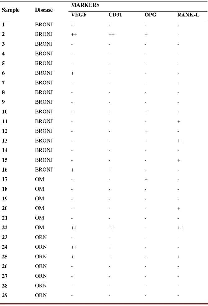
Considering the inclusion criteria for the present study, the sample obtained was 34 specimens from 34 patients, of which 16 had BRONJ, 6 chronic OM and 12 ORN. The results of the semi-quantitative analysis of each sample can be found below (Table 1). In addition,
Table 2 illustrates the percentage immunolabeling of each marker for each disease. Given a
p-value of p<0.005, no significant difference was identified with the expression of the markers between the diseases (Table 1 and 2).
Braz. J. of Develop.,Curitiba, v. 6, n.5, p.27725-27734 may. 2020. ISSN 2525-8761
4 DISCUSSION
The present study observes a line of research into osteonecrosis and, more specifically, follows on from the work of De Antoni et al. (2018)8, which evaluated under the microscope the same samples as this study, in specimens stained using the hematoxylin and eosin technique. The author investigated the three diseases (OM, BRONJ, ORN) taking into account the following variables: bone tissue, inflammatory infiltrate, blood vessels and the presence of microorganisms. In addition, the examiners issued a hypothetical diagnosis for each specimen. Considering the structures evaluated, there was total agreement between examiners (Kappa=1). The only areas to reflect a significant statistical difference were the presence of empty channels, which was less frequent in the case of ORN (p=0.042) and the presence of neutrophils, which was far lower in the BRONJ group (p≤0.001). There was no indication of a concurring diagnosis for the disease among the examiners (Kappa=0.23). From the analyses in this study, it was possible to conclude that the diseases being compared were microscopically very similar, mainly with regard to the presence of bone necrosis, inflammation and microorganisms. Moreover, it was not possible to obtain a diagnosis of the evaluated diseases using microscopic analysis alone.
The motivation for the present study was the intention to accrue more detail in respect of the similarities between the abovementioned diseases, as well as the fact that no comparative study was found that uses vascularization antibodies and bone metabolism. When comparing the samples, there was no significant difference in the expression of markers, i.e. there was a good deal of similarity between the tissues evaluated, corroborating other studies that compared the three diseases.1,8
The antibodies used to evaluate vascularization were for the proteins VEGF and CD31, as these plays an important role in angiogenesis. Their use in the present study aimed to reveal if there would be different numbers of vessels and endothelial cells, respectively, with each disease. Only a few samples revealed positive scores for the markers and, when marking was observed, there was coincidence between both the markers in the same sample. The absence of marking was very important in the three diseases, a characteristic fact that goes hand in hand with necrotic bone tissue.
Reduced vascularization in bone tissue and soft tissue is to be expected with BRONJ. In 2012, MARX1 found empty channels and a complete absence of blood vessels in 100% of BRONJ samples. The osteomyelitis samples showed more blood vessels than the BRONJ samples, though there was no statistically significant difference. By contrast, in the present
Braz. J. of Develop.,Curitiba, v. 6, n.5, p.27725-27734 may. 2020. ISSN 2525-8761 study, just one OM sample exhibited moderate marking in areas of soft tissue, while three BRONJ samples also demonstrated this, two of which have slight marking and the other, moderate. Only one sample revealed moderate expression, however, it should be emphasized that the number of OM samples was almost two-thirds lower than for BRONJ.
When the signalers OPG and RANK-L were evaluated, the absence of expression was striking in the majority of the sample evaluated. Moreover, it did not reveal any significant differences between the three diseases. No studies could be found that compared the expression of these signalers across the three diseases.
It is important to remember that the specimens obtained in this study come from different institutions and, consequently, the patients’ profiles as well as the dental surgeon carrying out the biopsy are not standardized. Necrotic bone is a standard characteristic constant to the diseases investigated. Nevertheless, depending on factors such as the length of time the bone necrosis remains exposed, whether or not they are infected, and if antibiotics were used, among other factors such as surgical manipulation, they may produce more or less clear microscopic change. Irrespective of the cognizance of these factors in the evaluated sample, the presence of nonvital bone and signs of a chronic condition were common to all the samples in this study.
As for the methodology employed, semi-quantitative analysis was selected, mainly as a result of changes in size among the samples, making qualitative analysis more difficult, as the size varied significantly. In addition, some samples exhibited portions of soft tissue on the lamina.
Although this study only comprises a few samples, the limitations of which have been discussed above, the results do corroborate other analyses. In other words, the three diseases, besides presenting clinical and radiographic signs and symptoms, also bear microscopic similarity, making the history of the present disease fundamental to the diagnostic process.
From this study, it can be said that, considering the markers related to the rate of bone metabolism and vascularization, the analyzed diseases exhibited no differences between one another.
Braz. J. of Develop.,Curitiba, v. 6, n.5, p.27725-27734 may. 2020. ISSN 2525-8761
REFERENCES
1. Marx RE, Tursun R. Suppurative osteomyelitis, bisphosphonate induced osteonecrosis, osteoradionecrosis: a blinded histopathologic comparison and its implications for the mechanism of each disease. Int J Oral Maxillofac Surg. 2012;41(3):283-289.
2. Hansen T, Kunkel M, Weber A, James Kirkpatrick C. Osteonecrosis of the jaws in patients treated with bisphosphonates – histomorphologic analysis in comparison with infected osteoradionecrosis. J Oral Pathol Med. 2006;35(3):155-160.
3. Treister N, Sheehy N, Bae EH, Friedland B, Lerman M, Woo S. Dental panoramic radiographic evaluation in bisphosphonate- associated osteonecrosis of the jaws. Oral Dis. 2009;15(1):88-92.
4. Rocha GC, Jaguar GC, Moreira CR, Neves EG, Fonseca FP, Pedreira EN. Radiographic evaluation of maxillofacial region in oncology patients treated with bisphosphonates. Oral Surg Oral Med Oral Pathol Oral Radiol. 2012;114(5):S19-S25.
5. Cardoso CL, Barros CA, Curra C, Fernandes LM, Franzolin SO, Júnior JS, et al. Radiographic Findings in Patients with Medication-Related Osteonecrosis of the Jaw. Int J Dent. 2017:1-6.
6. Lew, DP, Waldvogel FA. Osteomyelitis. Lancet. 2004;364:369-379.
7. Ruggiero SL, Dodson TB, Fantasia J, Goodday R, Aghaloo T, Mehrotra B, et al. F. American Association of Oral and Maxillofacial Surgeons. American Association of Oral and Maxillofacial Surgeons position paper on medication-related osteonecrosis of the jaw--2014 update. J Oral Maxillofac Surg. 2014;72(10):1938-56.
8. De Antoni CC, Matsumoto MA, Silva AAD, Curi MM, Santiago Júnior JF, Sassi LM, et al. Medication-related osteonecrosis of the jaw, osteoradionecrosis, and osteomyelitis: A comparative histopathological study. Braz Oral Res. 2018;32:1-7.
9. Luvizuto ER, Dias SM, Queiroz TP, Okamoto T, Garcia IR Jr, Okamoto R, et al. Osteocalcin immunolabeling during the alveolar healing process in ovariectomized rats treated with estrogen or raloxifene. Bone. 2010;46(4):1021-29.
Braz. J. of Develop.,Curitiba, v. 6, n.5, p.27725-27734 may. 2020. ISSN 2525-8761
Table 1 – Relationship of specimens evaluated for each disease and the score arrived at in the semi-quantitative analysis. Immunolabeling absent (-); slight (+); moderate (++) and severe (+++)
Sample Disease MARKERS
VEGF CD31 OPG RANK-L
1 BRONJ - - - - 2 BRONJ ++ ++ + - 3 BRONJ - - - - 4 BRONJ - - - - 5 BRONJ - - - - 6 BRONJ + + - - 7 BRONJ - - - - 8 BRONJ - - - - 9 BRONJ - - - - 10 BRONJ - - + - 11 BRONJ - - - + 12 BRONJ - - + - 13 BRONJ - - - ++ 14 BRONJ - - - - 15 BRONJ - - - + 16 BRONJ + + - - 17 OM - - + - 18 OM - - - - 19 OM - - - - 20 OM - - - + 21 OM - - - - 22 OM ++ ++ - ++ 23 ORN - - - - 24 ORN ++ + - - 25 ORN + + + + 26 ORN - - - - 27 ORN - - - - 28 ORN - - - - 29 ORN - - - -
Braz. J. of Develop.,Curitiba, v. 6, n.5, p.27725-27734 may. 2020. ISSN 2525-8761 30 ORN - - - - 31 ORN - - - 32 ORN - - - - 33 ORN - - - - 34 ORN - - - - Value of p* 0,99 0,98 0,74 0,39
Table 2 – Percentage of results found in the semi-quantitative analysis. Immunolabeling absent (-), slight (+) and moderate (++)
Disease Markers
VEGF CD31 OPG RANK-L
BRONJ 81,25% (-) 12,5% (+) 6,25% (++) 81,25% (-) 12,5% (+) 6,25% (++) 81,25% (-) 18,75% (+) 81,25% (-) 12,5% (+) 6,25% (++) OM 83,3% (-) 16,6% (++) 83,3% (-) 16,6% (++) 83,33% (-) 16,66% (++) 66,67% (-) 16,66% (+) 16,66% (++) ORN 83,33% (-) 8,33% (+) 8,33% (+) 83,33%(-) 16,66% (+) 91,67% (-) 8,33% (+) 91,67% (-) 8,33% (+) p* Value 0,96 0,97 1 0,73 * Teste Kruskal-Wallis.