Carlos Cesar DE ANTONI(a) Mariza Akemi MATSUMOTO(b) Andréia Aparecida da SILVA(a) Marcos Martins CURI(c)
Joel Ferreira SANTIAGO JÚNIOR(a) Laurindo Moacir SASSI(d)
Camila Lopes CARDOSO(a)
(a) Universidade do Sagrado Coração, Department of Oral Surgery, Bauru, SP, Brazil.
(b) Universidade Estadual Paulista, Department of Histology, Araçatuba, SP, Brazil.
(c) Hospital Santa Catarina, Department of Stomatology, São Paulo, SP, Brazil.
(d) Hospital Erasto Gaertner, Department of Oral Surgery, Curitiba, PR, Brazil.
Medication-related osteonecrosis
of the jaw, osteoradionecrosis,
and osteomyelitis: A comparative
histopathological study
Abstract: It is largely known that some oral diseases can be diagnosed based upon their clinical manifestation combined with the patient’s medical history and generally not depending on histopathological examination. This is the case of some bone diseases such as osteoradionecrosis of the jaw (ORNJ), osteomyelitis of the jaw (OMJ), and medication-related osteonecrosis of the jaw (MRONJ). The present study aimed to analyze the histopathological features of these
specific bone diseases in order to evaluate similarities and differences.
Forty-four bone specimens resected from each bone disease (22 cases of ORNJ, 6 cases of OMJ, and 16 cases of MRONJ) were analyzed by two experienced oral pathologists without prior knowledge of the diagnosis,
considering bone tissue condition, inflammation, vascularization, and
the presence of microorganisms. In addition, the examiners formulated a diagnostic hypothesis for each specimen. Many histopathological similarities were found among the diseases, especially considering
the presence of necrotic bone, inflammation, and microorganisms. Statistically significant differences were detected in empty bone
lacunae, which was decreased in ORN (p = 0.042), and considering
neutrophil count, which was low in the MRONJ group (p ≤ 0.001). The Kappa coefficient was calculated and agreement was detected based
on the histopathological parameters, but not for diagnostic suggestion (p=0.23). In conclusion, histopathological aspects of ORNJ, OMJ,
and MRONJ do not permit a conclusive diagnosis, emphasizing the
necessity of a detailed clinical report.
Keywords: Osteonecrosis; Osteomyelitis; Osteoradionecrosis.
Introduction
Histopathological analysis may be critical for confirming the diagnosis of many craniomaxillofacial diseases. However, ideally, the combination of clinical findings and complementary exams should be followed, since specific conditions such as those affecting bone tissue may present quite similar histological features. Among them, particular attention is given to
osteomyelitis (OMJ), osteoradionecrosis (ORNJ), and medication-related osteonecrosis of the jaw (MRONJ).1,2,3 However, it is important to emphasize
Declaration of Interests: The authors certify that they have no commercial or associative interest that represents a conflict of interest in connection with the manuscript.
Corresponding Author: Camila Lopes Cardoso
E-mail: [email protected]
https://doi.org/10.1590/1807-3107bor-2018.vol32.0023
Submitted: September 19, 2017
that these are quite distinct clinical pathologies with
different etiologies.2
Osteomyelitis (OMJ) is a bone pathology which generally occurs due to complications arising
from an acute dentoalveolar abscess and is caused by the microbial infection with eventual tissue
destruction and necrosis, extending beyond the
initial area of involvement.4 Regarding bone necrosis,
osteoradionecrosis of the jaw (ORNJ) is a common and serious complication following head and neck radiation therapy,5 and medication-related osteonecrosis (MRONJ) is considered the most common complication associated with bisphosphonate therapy.6
Bone necrosis is a common characteristic of
these bone diseases, which also have clinical signs
and symptoms.2 Despite histopathological and clinical similarities, the above-mentioned pathologies
also exhibit very similar image characteristics.7,8
Authors have presented a detailed image description
comparing BRONJ and ORNJ from dental panoramic
radiographs and computed tomography and have
pointed out a number of similarities between the two pathologies, such as osteolysis, osteosclerosis,
sequestration, and dissemination of soft tissue inflammation, emphasizing only two main differences
between them, namely predominant osteolysis in ORNJ and osteosclerosis in MRONJ cases.7
There are few pathological studies comparing OMJ, MRONJ, and ORNJ in the literature.1,2,3,9 Therefore, the present study aimed to compare the histopathological features of these bone diseases in order to detect
some common or prevalent aspects.
Methodology
A retrospective histopathological evaluation was
performed on specimens selected from patients’
records from 2000 to 2017, previously subjected to
biopsy of the maxillary bones and diagnosed as OMJ, ORNJ, and MRONJ associated with bisphosphonate.
The final diagnosis of each disease was recorded
in the patients’ medical records and it was based on clinical, radiographic, and microscopic aspects, combined with the medical history. This study
was approved by the institutional review board
(process no. 1.249.820). The patients’ records were
reviewed and the following clinical criteria were
used for the diagnosis of OMJ: presence of bone
infection, suppuration and radiographic evidence
of osteolysis, absence of head and neck radiation therapy, and no use of bisphosphonates.2,4 The diagnostic criteria for MRONJ were the presence of exposed necrotic bone in the jaws for at least 8 weeks associated with bisphosphonate use and absence of head and neck radiation therapy.2,6 The diagnostic criteria for ORNJ were the presence of exposed necrotic bone in the jaws for at least
8 weeks in a patient who received at least 50 Gy
of head and neck radiation therapy and absence of bisphosphonate therapy.5
The specimens were obtained from existing
paraffin-embedded blocks of each bone disease from
three different institutions. Microscope slides were cut in 6-μm sections and stained with hematoxylin and eosin (HE), and analyzed by two experienced oral pathologists, who were blinded to the diagnosis. The following parameters were considered for the
histopathological analysis: overall bone tissue status (vitality, necrosis, reactivity), bone cells (osteoblasts, osteoclasts), inflammation, vascularization (blood vessels, thrombosis), and presence of biofilm. The
detailed criteria are displayed in Table 1, where the examiners marked the absence or presence of each item, and at the end, each pathologist made a
presumptive diagnosis, limited to the three diseases. After the evaluation, the diagnoses were compared
with the original ones.
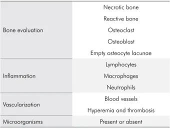
Table 1. Microscopic evaluation criteria.
Bone evaluation
Necrotic bone
Reactive bone
Osteoclast
Osteoblast
Empty osteocyte lacunae
Inflammation
Lymphocytes
Macrophages
Neutrophils
Vascularization Blood vessels Hyperemia and thrombosis
The data were fed into an Excel spreadsheet
(Microsoft Office Excel, Redmond, WA, USA), analyzed
by SigmaPlot software (SigmaPlot, San Jose, CA,
USA) version 12.3, and their normal distribution and equal variance were checked by the Shapiro-Wilk test, followed by the Kruskal-Wallis test (one-way
Figure 1. a, b) MRONJ – extensive biofilms on the surfaces of non-viable bone tissue; c) OMJ – Non-viable bone tissue presenting basophilic reversal lines, d) biofilm with leukocyte infiltration; and, e, f) ORNJ – Avascular and acellular bone tissue showing areas with osteoclastic activity (HE; bar = 100 µm).
A B
C D
analysis of variance on ranks (variable: Diagnosis)) and Dunn’s post-hoc test, in order to verify the differences between groups at a 5% significance level. The agreement between the different factors evaluated
by examiners (1 and 2) was interpreted by a Kappa
interrater coefficient. The actual diagnoses made
by examiners were compared by a Kappa statistic, and the results were shown according to the actual
clinical condition or final diagnosis.
Results
Forty-four bone specimens resected from each bone disease comprising 22 cases of ORNJ, 6 cases of OMJ, and 16 cases of MRONJ were included. Thickness
and percentage of bone tissue and connective tissue varied considerably, as the specimens were obtained
from incisional biopsies. Minimum thickness was
5 mm. All specimens had the ideal thickness required
for diagnosis.
The demographic data are displayed in Table 2 The concordance between the different factors
evaluated by examiners (1 and 2) was interpreted by a Kappa interrater coefficient, which was equal
to 1, that is, there was no interrater difference
in the evaluation of the presence or absence of each variable. Most of the analyzed criteria were
detected in MRONJ, ORNJ, and OMJ, as shown
in Table 1, resulting in no statistically significant differences in their prevalence rates. The percentages
of all evaluated structures are described in Table
3, including general aspects of bone tissue, such as
necrotic (p = 0.211) and reactive bone (p = 0.723),
along with osteoclastic (p = 0.146) and osteoblastic
cells (p = 0.432). In addition, the rate inflammation
(p = 0.672), especially chronic, was the same for all the three diseases, considering lymphocytes (p = 0.054) and macrophages (p = 0.490). Also, the
presence of blood vessels (p = 0.870) and hyperemia
and thrombosis (p = 0.472) was similar. Another
common finding was the presence of biofilm-forming
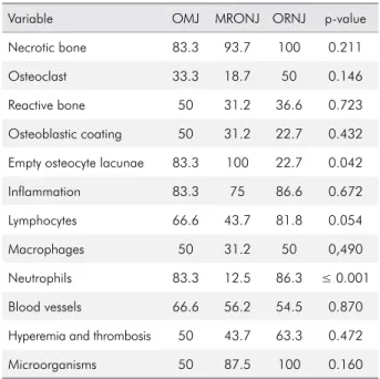
microorganisms (p = 0.160) (Table 3).
Significant differences were detected in empty osteocyte lacunae, which were less frequent in ORNJ
(p = 0.042) than in MRONJ and OMJ, and neutrophils,
which were less frequent in MRONJ (p < 0.05) than
in ORNJ and OMJ (Table 3).
There was no consistent histopathological diagnosis between examiners (Kappa = 0.23). The
tentative diagnosis made by each examiner is
described in Table 4.
Discussion
This study aimed to evaluate the histopathological
aspects of OMJ, ORNJ, and MRONJ considering close
Table 2. Table showing incidence rates by gender, age, and lesion sites.
Bone pathologies Gender Age (years) Site
6 OMJ samples
5 Males -
-1 Females 53 to 56
(mean: 54.3) 6 Mandible
22 MRONJ samples
4 Males -
-18 Females 43 to 89
(mean: 58.5) 19 Mandible
- - 3 Maxilla
16 ORN samples
15 Males -
-1 Females 42 to 75
(mean: 58.1) 16 Mandible
Table 3. Table showing the percentage of all variables compared among the diseases.
Variable OMJ MRONJ ORNJ p-value
Necrotic bone 83.3 93.7 100 0.211
Osteoclast 33.3 18.7 50 0.146
Reactive bone 50 31.2 36.6 0.723
Osteoblastic coating 50 31.2 22.7 0.432
Empty osteocyte lacunae 83.3 100 22.7 0.042
Inflammation 83.3 75 86.6 0.672
Lymphocytes 66.6 43.7 81.8 0.054
Macrophages 50 31.2 50 0,490
Neutrophils 83.3 12.5 86.3 ≤ 0.001
Blood vessels 66.6 56.2 54.5 0.870
Hyperemia and thrombosis 50 43.7 63.3 0.472
similarities among them and, consequently, to make
clinicians aware of the importance of understanding the etiopathogenesis and clinical manifestations of
each disease. Few studies with the same objectives
were found in a recent review of the literature;1,3,10,11
but more information was needed in order to clarify histological differences between the diseases.
It is important to emphasize that the pathologies chosen in the present study had already been diagnosed
based upon the patients’ clinical information given by the clinicians. Since the information was retrieved from the files of an oral pathology service, the number of
cases was not identical, but all cases were maintained
in the study, thus avoiding the significant reduction
of sample size to a minimum. Another important aspect refers to OMJ. It is true that MRONJ and ORNJ
can easily evolve into osteomyelitis once secondary
infection occurs,12 which can hinder the final diagnosis as well as the comprehension of the etiological process
of each disease. However, according to the patients’
records, only those cases originating from chronic or
acute dentoalveolar abscesses were chosen, with no
association with radiation therapy or bisphosphonates,
or with any other antiresorptive drug.
The analysis of the histological criteria was based on their presence or not, and no morphometric
method was used to quantify cells or structures, as
this is not a routine practice for the analyzed diseases.
Nevertheless, most of the analyzed criteria did not reveal significant differences, but it was clear that some of them were more prevalent in one or other
disease. These aspects will also be discussed in order to elucidate some subtleties of each disease. Also, regarding the methodology, the morphometric analysis
was not performed mainly due to the large variability
in sample size, since the study was performed with biopsy specimens from different patients performed by different surgeons.
The presence of necrotic bone was fairly common among the clinical diseases. Marx and Tursun2 identified necrotic bone in 100 of all OMJ, ORNJ, and
MRONJ specimens, considering a set of characteristics such as the presence of empty osteocyte lacunae,
absence of osteoblastic rimming, and empty Haversian
and Volkmann canals. Although these characteristics
were individually analyzed in our study, they were
Table 4. Tentative diagnosis of each examiner per sample, indicating lack of consistency between them (Kappa=0.23).
Examiner 1 Examiner 2 Final Diagnosis
OM OMMBF ORN
OM OM ORN
OMMBF OM ORN
OM OM ORN
OMMBF OMMBF ORN
OMMBF OM ORN
OMMBF ORN ORN
OMMBF ORN ORN
OM OMMBF ORN
OMMBF OMMBF ORN
OMMBF OMMBF ORN
OM OM ORN
OM ORN ORN
OM OM ORN
OM OM ORN
OM OM ORN
ORN ORN ORN
OM OM ORN
OM OM ORN
OM ORN ORN
OM OMMBF ORN
OM ORN ORN
OM OM OM
OM ORN OM
ORN ORN OM
OMMBF ORN OM
OMMBF OMMBF OM
OM ORN OM
OMMBF ORN OMMBF
OM OMMBF OMMBF
ORN OMMBF OMMBF
OM OMMBF OMMBF
OM ORN OMMBF
OM OM OMMBF
OM OMMBF OMMBF
OM ORN OMMBF
OMMBF OMMBF OMMBF
OM OM OMMBF
ORN ORN OMMBF
OM ORN OMMBF
OM OM OMMBF
OMMBF OMMBF OMMBF
OM OMMBF OMMBF
also taken as characteristics that compounded the general picture of bone necrosis.
Although there was no significant difference in
the presence of osteoclasts among the three clinical diseases, it was clear that they were lower in MRONJ specimens than in the other diseases, possibly due to
the mechanism of action of the drug involved in the
osteonecrotic process. Interestingly, in the study of Marx and Tursun,2 osteoclasts were seen only in OMJ specimens. The same occurred in reactive bone and osteoblastic coating. Despite the lack of significant
difference among the diseases, OMJ specimens showed larger amounts of both criteria.
When inflammation was assessed, more than 75 of the samples revealed presence of some kind of leukocytes, and neutrophils were more frequent in OMJ (83.3) and ORNJ specimens (86.3). Significant lack
of leukocytes was detected in MRONJ, in agreement with Marx and Tursun.2
Compromised vascularization was expected to be seen, especially in ORNJ and MRONJ, given the etiological factors of both diseases. When the diseases were compared, no significant differences were detected.
Hyperemia and thrombosis predominated in ORNJ (63.3), followed by OMJ (50) and MRONJ (43.7). There
was absence of blood vessels in MRONJ samples.10
Microorganisms were found in many samples,
revealing no significant differences among the diseases. In OMJ, microorganisms were observed
in 50 of the samples, compared to 87.5 and 100 in
ORNJ and MRONJ, respectively. Microorganisms
of the genus Actinomyces have been found in these
diseases by several authors,6,13,14,15,16 including by Marx
and Tursun2 The microorganisms were detected on trabecular bone surfaces in 76 of MRONJ and in 58 of ORNJ cases. In OMJ samples, these microorganisms were found in medullary spaces.2 On the other hand, in our study, OMJ samples showed a lower amount of microorganisms, compared to the other samples. It is important to emphasize that only 6 OMJ samples
were evaluated in the present study.
Another aim of this study was to test oral pathologists’ capacity to establish a histopathological diagnosis of the proposed diseases in the absence of clinical information. Pathologists who work with diagnoses and know the three entities know that
anamnesis is crucial for the diagnosis, given that clinical and radiographic features are very similar. With regard to the first research question, the blinded evaluation by two oral pathologists confirmed the hypothesis that it is not possible to establish a final
diagnosis only by way of microscopic analysis. The
diagnoses of the diseases mentioned above tended to be quite similar microscopically, as unanimously
endorsed by experts.
Conclusions
The histopathological aspects of ORNJ, OMJ,
and MRONJ do not permit a conclusive diagnosis,
emphasizing the necessity of a detailed clinical report and of clinicians’ expertise.
1. Guimarães EP, Pedreira FR, Jham BC, Carli ML, Pereira AA, Hanemann JA. Clinical management of suppurative osteomyelitis, bisphosphonate-related osteonecrosis, and osteoradionecrosis: report of three cases and review of the literature. Case Rep Dent. 2013;2013:402096. https://doi.org/10.1155/2013/402096
2. Marx RE, Tursun R. Suppurative osteomyelitis, bisphosphonate induced osteonecrosis, osteoradionecrosis: a blinded histopathologic comparison and its implications for the mechanism of each disease. Int J Oral Maxillofac Surg. 2012 Mar;41(3):283–9. https://doi.org/10.1016/j.ijom.2011.12.016
3. Mitsimponas KT, Moebius P, Amann K, Stockmann P, Schlegel KA, Neukam FW et al. Osteo-radio-necrosis (ORN) and bisphosphonate-related osteonecrosis of the jaws (BRONJ): the histopathological differences under the clinical similarities. Int J Clin Exp Pathol. 2014 Jan;7(2):496–508.
4. Lew DP, Waldvogel FA. Osteomyelitis. Lancet 2004;364(9431):369-79.
https://doi.org/10.1016/S0140-6736(04)16727-5 5. Marx RE. Osteoradionecrosis: a new concept of its
pathophysiology. J Oral Maxillofac Surg. 1983 May;41(5):283– 8. https://doi.org/10.1016/0278-2391(83)90294-X
6. Ruggiero SL, Dodson TB, Fantasia J, Goodday R, Aghaloo T, Mehrotra B et al. American Association of Oral and Maxillofacial Surgeons position paper on medication-related osteonecrosis of the jaw: 2014 update. J Oral Maxillofac Surg. 2014 Oct;72(10):1938–56. https://doi.org/10.1016/j.joms.2014.04.031
7. Obinata K, Shirai S, Ito H, Nakamura M, Carrozzo M, Macleod I et al. Image findings of bisphosphonate related osteonecrosis of jaws comparing with osteoradionecrosis. Dentomaxillofac Radiol. 2017 Jul;46(5):20160281. https://doi.org/10.1259/dmfr.20160281
8. Cardoso CL, Barros CA, Curra C, Fernandes LM, Franzolin SO, Ferreira Júnior JS et al. Radiographic findings in patients with medication-related osteonecrosis of the jaw. Int J Dent. 2017;2017:3190301. https://doi.org/10.1155/2017/3190301 9. Curi MM, Cardoso CL, Lima HG, Kowalski LP, Martins
MD. Histopathologic and histomorphometric analysis of irradiation injury in bone and the surrounding soft tissues of the jaws. J Oral Maxillofac Surg. 2016 Jan;74(1):190–9. https://doi.org/10.1016/j.joms.2015.07.009
10. Hansen T, Kunkel M, Weber A, James Kirkpatrick C. Osteonecrosis of the jaws in patients treated with bisphosphonates - histomorphologic analysis in comparison with infected osteoradionecrosis. J Oral Pathol Med. 2006 Mar;35(3):155–60. https://doi.org/10.1111/j.1600-0714.2006.00391.x
11. Carmagnola D, Canciani E, Sozzi D, Biglioli F, Moneghini L, Dellavia C. Histological findings on jaw osteonecrosis associated with bisphosphonates (BONJ) or with radiotherapy (ORN) in humans. Acta Odontol Scand. 2013 Nov;71(6):1410–7. https://doi.org/10.3109/00016357.2013.765592
12. Tardast A, Sjöman R, Løes S, Abtahi J. Bisphosphonate associated osteomyelitis of the jaw in patients with bony exposure: prevention, a new way of thinking. J Appl Oral Sci. 2015 May-Jun;23(3):310–4. https://doi.org/10.1590/1678-775720140506 PMID:26221926 13. Aftimos V, Zeinoun T, Bou Tayeh R, Aftimos G. Bisphosphonate
related osteonecrosis of the jaw: a study of 18 cases associated with fungal infection. Int J Dent. 2014;2014:869067.
https://doi.org/10.1155/2014/869067 PMID:24696686 14. Boff RC, Salum FG, Figueiredo MA, Cherubini K. Important aspects
regarding the role of microorganisms in bisphosphonate-related osteonecrosis of the jaws. Arch Oral Biol. 2014 Aug;59(8):790–9. https://doi.org/10.1016/j.archoralbio.2014.05.002
15. Curi MM, Dib LL, Kowalski LP, Landman G, Mangini C. Opportunistic actinomycosis in osteoradionecrosis of the jaws in patients affected by head and neck cancer: incidence and clinical significance. Oral Oncol. 2000 May;36(3):294–9. https://doi.org/10.1016/S1368-8375(99)00080-9 16. De Ceulaer J, Tacconelli E, Vandecasteele SJ. Actinomyces