Rómulo Sacramento Sobral
Universidade do Minho
Escola de Ciências
Characterisation of the molecular
interaction between MYB-like transcription
factors in the establishment of floral
dorsoventral asymmetry in
Antirrhinum majus
in ho | 20 10 R óm ul o Sa cr am en to S ob ra l C h a ra ct e ri sa ti o n o f th e m o le cu la r in te ra ct io n b e tw e e n M Y B -li ke t ra n sc ri p ti o n fa ct o rs in t h e e st a b lis h m e n t o f fl o ra l d o rs o ve n tr a l a sy m m e tr y in A n ti rr h in u m m a ju sRómulo Sacramento Sobral
Universidade do Minho
Escola de Ciências
Characterisation of the molecular
interaction between MYB-like transcription
factors in the establishment of floral
dorsoventral asymmetry in
Antirrhinum majus
Trabalho efectuado sob a orientação da
Professora Doutora Maria Manuela Ribeiro Costa
Mestrado de Genética Molecular
Dissertação de Mestrado
Acknowledgements
I would like to thank to Prof. Manuela for all her teachings and for being an excellent and supportive supervisor.
To all the people in the lab, I would like to thank for creating an enjoyable environment that allowed me to take pleasure in my work time. In particular, I would like to thank Sara, Juliana, Herlânder and João for all the technical support they gave me since I started my work.
To my family and friends, thank you all for the patience, encouragement and friendship, for being present in the best and worst moments.
Finally to Sara, without her I definitively wouldn’t be writing these words. Thank you for all your support, dedication, for the excellent example, and especially for your love…
Abstract
Characterisation of the molecular interaction between MYB-like transcription factors in the establishment of floral dorsoventral asymmetry in Antirrhinum majus.
Transcription factor interactions are the cornerstone of combinatorial control, which is a crucial aspect of the gene regulatory system that underlies many developmental processes. In Antirrhinum majus, floral dorsoventral asymmetry is controlled by the combined activity of four transcription factors: CYCLOYDEA (CYC),
DICHOTOMA (DICH), RADIALIS (RAD) and DIVARICATA (DIV). CYC, DICH and RAD are expressed dorsally in the floral primordium and promote dorsal petal identity. DIV is expressed in the entire floral primordium, even though it only has a phenotypic
effect in more ventral regions. Genetic and molecular studies have revealed that RAD is a direct target of CYC and that that RAD antagonises the activity of DIV. Previous yeast two-hybrid screens led to the identification of two new MYB-like proteins (RAD-INTERACTING PROTEIN 1 and 2, RIP1 and RIP2) that interact with both RAD and DIV. Therefore, the RIP proteins might also be involved in the antagonism that RAD has on DIV activity, essential for the establishment of flower asymmetry in Antirrhinum. The aim of this thesis was to characterise the molecular antagonism between RAD and DIV, either through direct interaction or indirectly by competition for the RIP proteins.
The results from a DNA-binding assay revealed that RAD does not target directly DIV or the DNA targets of DIV, suggesting that the RIP proteins might be bridging the molecular antagonism between RAD and DIV. A sub-cellular localization assay revealed that the RIP proteins co-localised with RAD and DIV in the nucleus, which means that these proteins are able to interact in the same sub-cellular compartment. A quantitative yeast-two hybrid result indicated that RAD interacts more strongly with the RIP proteins than DIV. This result suggest that the RIP proteins might be co-factors essential for DIV function that are sequestered in the dorsal domain by RAD, restricting DIV activity to the ventral petal. The role of the RIP proteins in the molecular antagonism between RAD and DIV will be further addressed using protein-protein interaction assays in vitro and in planta.
Resumo
Caracterização da interacção molecular entre factores de transcrição do tipo MYB-like no desenvolvimento da assimetria dorsoventral da flor de Antirrhinum majus.
A interacção entre factores de transcrição é a base do sistema de regulação genética e em maior instância do desenvolvimento. Em Antirrhinum majus a assimetria dorsoventral da flor é controlada pela acção combinada de quatro factores de transcrição: CYCLOYDEA (CYC), DICHOTOMA (DICH), RADIALIS (RAD) e DIVARICATA (DIV). CYC, DICH e RAD são expressos dorsalmente no primordio floral promovendo a
identidade dorsal das pétalas dorsais. DIV é expresso em todo o meristema floral, no entanto, os seus efeitos fenotípicos são evidentes apenas nas regiões mais ventrais. Estudos genéticos e moleculares revelaram que RAD é responsável pelo antagonismo da actividade de DIV. Recentemente num yeast two-hybrid foram identificados dois novos factores de transcrição do tipo MYB (RAD-INTERACTING PROTEIN 1 e 2, RIP1 e RIP2) que interagem com RAD e DIV. É possível que as proteínas RIP também estejam envolvidas no antagonismo entre RAD e DIV.
O principal objectivo desta tese é a caracterização do antagonismo molecular entre as proteínas RAD e DIV, quer por interacção directa entre RAD e DIV ou por competição pelas proteínas RIP.
O resultado de um ensaio de ligação ao DNA revelaram que RAD não interage directamente com DIV, nem com os alvos de DNA de DIV, sugerindo que as RIP poderão ter um papel na mediação do antagonismo molecular entre RAD e DIV. Um ensaio de sub-localização celular demonstrou que as proteínas RIP co-localizam com RAD e DIV no núcleo, o que indica que as proteínas tem a possibilidade de interagir no mesmo compartimento subcelular. Um ensaio yeast-two hybrid quantitativo revelou que RAD tem maior afinidade para as proteínas RIP que DIV. Os resultado sugerem a hipótese que as proteínas RIP poderão ser cofactores essenciais para a função de DIV e que na presença de RAD, as RIP são sequestradas, restringindo a actividade de DIV ao domínio ventral onde RAD não está presente. O papel das proteínas RIP no antagonismo
Table of Contents
Acknowledgements iii
Abstract v
Resumo vi
Table of Contents vii
Abbreviations and Acronyms x
1. Introduction 1
1.1 Genetic Regulatory Networks 1
1.2 Genetic Regulatory Networks in the Evolution and Diversity of Plants 2
1.3 Flower Symmetry 4
1.3.1 A. majus as model species for flower symmetry studies 7
1.3.1.1 CYCLOYDEA and DICHOTOMA 8
1.3.1.2 RADIALIS 10
1.3.1.3 DIVARICATA 11
1.3.2 MYB family of transcription factors 13
1.3.2.1 RAD and DIV as MYB-like transcription factors 15
1.3.3 A model for Genetic Regulation of Flower Asymmetry 17
1.3.4 Genetic basis for flower asymmetry among other taxa 20
1.4 Aims of this study 23
2. Material and Methods 25
2.1 Biological material 25 2.1.1 Plant material 25 2.1.1.1 Arabidopsis thaliana 25 2.1.1.2 Nicotiana benthamiana 25 2.1.2 Bacterial material 26 2.1.2.1 Escherichia coli 26 2.1.2.2 Agrobacterium tumefaciens 27 2.1.3 Saccharomyces cerevisiae 27 2.2 DNA Methods 28 2.2.1 Bacterial Transformation 28 2.2.1.1 E. coli 28 2.2.1.2 A. tumefaciens 28
2.2.2 Isolation of plasmid DNA 29
2.2.2.1 P1 P2 P3 method 29
2.2.2.2 Small-scale (mini) isolation of purified plasmid DNA 29
2.2.2.3 Estimation of DNA concentration 29
2.2.3 Agarose Gel Electrophoresis 30
2.2.4 Polymerase chain reaction (PCR) methods for DNA fragment amplification 30 2.2.4.1 Amplification of DNA fragments from plasmid templates 31 2.2.4.2 Amplification of DNA fragments from plasmid templates using Pfu DNA
Polymerase 31
2.2.4.3 Amplification of DNA fragments for Gateway® system based cloning 31
2.2.5 DNA purification 33
2.2.5.1 Wizard SV Gel and PCR Clean-Up System 33
2.2.5.2 Phenol/Chloroform DNA extraction method 33
2.2.6 Restriction digestion with endonucleases 33
2.2.6.1 Plasmid DNA 33
2.2.6.2 PCR product 34
2.2.7 Ligation reaction 34
2.3 Cloning procedures 36
2.3.1 Cloning into the pRSET vector 36
2.3.1.1 RIP1 and RIP2 amplification and digestion 36
2.3.1.2 pRSET digestion 37
2.3.1.3 Ligation and transformation 37
2.3.2 Cloning into pMDC43 and pMDC32 37
2.3.2.1 BP reaction 38
2.3.2.2 LR reaction 38
2.3.3 Cloning into pH7WGF2 39
2.3.3.1 BP reaction 39
2.3.3.2 LR reaction 39
2.3.4 Cloning into pDH51-GW-YFPn and pDH51-GW-YFPc 40
2.3.4.1 BP reaction 40
2.3.4.2 LR reaction 40
2.4 Yeast Methods 41
2.4.1 Saccharomyces cerevisae transformation 41
2.4.2 3-amino-1, 2, 4-triazole assay 41
2.4.3 Colony-lift filter assay 42
2.4.4 Liquid Culture assay using o-nitrophenyl-β-D-galactopyranoside (ONPG) as
substrate 42
2.5 Protein Methods 43
2.5.1 Heterologous pilot expression in E. coli 43
2.5.2 Protein electrophoresis 44
2.5.3 Protein gel staining 45
2.5.4 Protein purification 45
2.6 Electrophoretic Mobile Shift Assay (EMSA) 46
2.6.1 Probe preparation and labelling 46
2.6.2 EMSA reaction and electrophoretic separation 46
2.6.3 Electroblotting 47
2.6.4 Detection 48
2.7 Generation of Arabidopsis transgenic lines 48
2.7.1 A. tumefaciens transformation 48
2.7.2 Plant transformation 48
2.7.3 Selection of primary transformants (T1 generation) 49
2.8 N. benthamiana transient transformation 49
3.2.1 Optimization of the expression and purification of RAD recombinant protein 55 3.2.2 Optimization of the expression and purification of DIV recombinant protein 59
3.3 Expression of RIP1 and RIP2 recombinant protein in E. coli 63
3.3.1 Cloning of the RIP1 and RIP2 cDNA into the pRSET plasmid 63 3.3.2 Optimisation of the expression of the RIP1 and RIP2 recombinant proteins 66
3.4 DNA-binding affinity assay 71
3.5 Yeast two-hybrid based assays 75
3.5.1 3-amino-1,2,4 triazole assay 77
3.5.2 RIP1 and RIP2 interact with RAD and DIV in a yeast two-hybrid assay 79 3.5.3 Determination of the interaction strength by the β-galactosidase liquid assay
81
3.6 Sub-cellular localisation of RAD, DIV and RIP proteins 84
3.6.1 Plasmids construction 84
3.6.2 Transient expression in N. benthamiana 89
3.6.3 Co-localization assay 93
3.7 Testing the interaction between RAD-RIP and DIV-RIP proteins in planta by
Bimolecular Fluorescence Complementation 96
3.7.1 Plasmids construction 96
3.8 Ectopic expression of the RIP proteins in Arabidopsis 103
4. Discussion 105
4.1 RAD does not bind directly to DIV or indirectly to DIV DNA targets. 106
4.2 RAD and DIV interact strongly with the RIP proteins 108
4.3 RAD and DIV co-localise with the RIP proteins in the nucleus 112
4.4 Testing the RAD, DIV and RIPs interactions in planta 114
4.5 The RIPs biological function 116
4.6 Concluding Remarks 116
5. Bibliography 118
Abbreviations and Acronyms
AD BD bHLH BiFC Bp BRET CaMV cDNA Col CYC dH20 DICH DIG DIV dNTP DTT DUF EDTA EMSA EYFP FRET g GFP His HIS3 IPTG iRNA kDa LACZ LB Leu M mJ MM mJ MM mRNA MS NLS OD ONPG ORF PAGE PCR PEG RAD RIP1 RIP2 RNA rpm SAP SD SDS ssDNA TAE TBE TE TEN tRNA UV W X-GAL YFP YPDA 3-AT 6xHisactivation domain of GAL4 binding domain of GAL4 basic helix loop helix
bimolecular fluorescence complementation base pairs
bioluminescence resonance transfer energy cauliflower mosaic virus
complementary DNA
A. thaliana ecotype Columbia CYCLOIDEA deionised water DICHOTOMA digoxigenin DIVARICATA any deoxyribonucleotide dithiothreitol
domain of unknown function ethylenediaminetetraacetic acid electrophoretic mobile shift assay
ENHANCED YELLOW FLUORESCENT PROTEIN
fluorescence resonance energy transfer relative centrifugal force
GREEN FLUORESCENT PROTEIN
histidine HISTIDINE3 Isopropyl β-D-1-thiogalactopyranoside interference RNA kilodalton β-GALACTOSIDASE Luria-Bertani medium leucine molar millijoule molecular marker millijoule molecular marker messenger RNA Murashige and Skoog nuclear localization signal optical density
ortho-Nitrophenyl-β-galactoside
open reading frame
Polyacrylamide gel electrophoresis polymerase chain reaction
Polyethylene glycol
RADIALIS
RAD INTERACTING PROTEIN1 RAD INTERACTING PROTEIN2
ribonucleic acid revolutions per minute
shrimp alkaline phosphatase enzyme synthetic minimal medium
sodium dodecyl sulphate single stranded DNA tris-acetate-EDTA buffer tris-borate-EDTA buffer tris-EDTA buffer tris-EDTA-NaCl buffer transfer RNA ultraviolet triptofan bromo-chloro-indolyl-galactopyranoside
YELLOW FLUORESCENT PROTEIN
yeast peptone dextrose adenine 3-Amino-1,2,4-triazole polyhistidine tag
1. Introduction
Transcriptional regulation lies at the basis of most biological processes in living organisms. It can be defined as the processing of extracellular and/or intracellular signals that lead to changes in the transcription rate of target genes, and is often characterised by a complex inter-play of protein-protein interactions, sub-cellular translocations and post-translational modifications (Carrol et al., 2001). The study of the evolution of transcription regulation is a common theme for those who are interested in understanding the principles and driving forces whereby new protein-coding genes, new protein structures and new biological functions emerge as a consequence of changes in the genetic material during the course of evolution (Patthy, 2008)
1.1 Genetic Regulatory Networks
The organ or body morphology and functionality is the outcome of complex interactions within genetic regulatory networks. It is upon these networks that evolutionary forces act and whose modifications give rise to the vast diversity in morphological form among species (Carroll et al. 2001). One particular component of these genetic regulatory networks, which contribute with the most global effects on development, is a family of proteins called transcription factors (Gellon and McGinnis, 1998).
Transcription factors are an important class of proteins involved in regulating the production of mRNA transcripts from genes by binding to DNA cis-acting elements. Transcription factors can be a part of the basal transcription machinery or regulatory in nature, whereby they control specific groups of genes in particular cell types, time periods or environmental conditions (Latchman, 1997). Transcription factors are modular in structure and contain distinctive domains: the DNA-binding domain, which attaches to specific sequences of DNA (cis-elements) adjacent to regulated genes; and the trans-activation domain that contains binding sites for other proteins such as transcription co-regulators (Mitchel and Tjian, 1989).
Transcription factors use a variety of mechanisms to regulate gene expression: stabilise or block the binding of RNA polymerase to DNA; catalyse the acetylation or
deacetylation of histones; or recruit co-activators or co-repressors proteins to the transcription factor complex (Mitchel and Tjian, 1989).
There is ample evidence for the role of transcription factors in developmental modifications during evolution. In the Animal Kingdom, comparative analyses of Hox (group of homeodomain transcription factors) gene expression in arthropods, annelids and vertebrates have revealed a consistent correlation between major differences in axis morphology and differences in the spatial and temporal regulation of Hox genes (Holland, 1999). As an example, Hox genes are expressed at different relative positions
along the rostrocaudal axis in the mouse, chick and python (Belting et al. 1998; Cohn and Tickle, 1999). Belting et al. (1998) provide direct evidence that change in cis-regulatory elements has led to the diversification in Hoxc8 expression that account for part of the morphologic differences between chicks and mice. In the Plant Kingdom a clear example is the teosinte branched 1 locus (TB1) and its role in the domestication of maize. In maize, tb1 mutants resemble the wild ancestor teosinte with a highly branched architecture that ends in male tassels rather than female ears. Strong selection on the upstream regulatory regions and the resulting altered expression pattern resulted in this change in architecture (Wang et al., 1999; Rosin and Kramer, 2009).
1.2 Genetic Regulatory Networks in the Evolution and
Diversity of Plants
An important goal for research is to elucidate how complex gene regulatory networks evolve and how their evolution results in phenotypic change and speciation (Carrol et al., 2005). Two trends seem to be important for plant evolution and diversity: gene duplication and convergent co-option of pre-existent genetic modules.
Duplication of individual genes, chromosomal segments or entire genomes is considered a major source of morphological novelties (Ohno, 1970). Ohno (1970) evolutionary theory suggests three possible outcomes for the duplicated gene as described in figure 1. Copy can be silenced by degenerative mutations (non-funcionalization); copy acquires a new function (functional divergence) and is positively
compromised in a way that the overall capacity is similar to the function of the ancestral gene (Hughes, 1994; He and Zhang, 2005).
Figure 1- Possible outcomes of duplicated genes. Copy can be silenced by degenerative mutations
(non-funcionalization); copy acquires new function (functional divergence) and is positively selected by natural selection with the other copy maintaining original function (neo-funcionalization); or both copies suffer degenerative mutations becoming partially compromised in a way that the overall capacity is similar to the function of the ancestral gene (Hughes, 1994; He and Zhang, 2005).
Lineage-specific gene duplications that led to the expansion of transcription-factor families are widely believed to have an important role in plant diversification and complexity (Levine and Tjian, 2003). Interestingly, there is evidence that the expansion of transcription-factor families is greater in plants than in animals (Shiu et al., 2005). Transcription factors typically contain multiple functional domains, which mediate binding to DNA, interactions with other proteins and the sub-cellular localization of the transcription factor (Latchman, 1997). A transcription factor with a new binding specificity can be created by duplication of an existing transcription factor followed by mutations, often, although not always, in the DNA-binding domain. Transcription factors can also evolve by the acquisition or loss of one of its other functional domains. For example, the loss of a transcriptional activation domain could turn an activator into a repressor, whereas the acquisition of a new protein–protein interaction domain, that facilitates heterodimerisation with a novel binding partner, could significantly alter the targets of the transcription factor (Chen and Rajewsky, 2007).
The other repeated tendency over the course of evolution, which can account for the diversification of several plant traits, is the evolution of convergent morphological features, even at recent time scales through co-option of the same regulatory networks (Rosin and Kramer, 2009). Several cases have now been characterized that show numerous parallel recruitments of homologous genetic pathways to control convergent features. One such case is the independent recruitment of the specific interaction between KNOTTED-1- like homeobox (KNOX) transcription factor and ASYMMETRIC
LEAVES1/ROUGH SHEATH2/PHANTASTICA (ARP) genes in the independent
evolution of leaves in lycophytes and in seed plants (Harrison et al., 2005).
Plant diversification appears to be closely correlated with modifications in the genetic regulatory networks. There are several described genetic regulatory networks whose function was associated with the control of specific plant features, and among those, is the regulatory network that controls flower symmetry.
1.3 Flower Symmetry
Flower symmetry is an architectural trait that accounts for innumerable variations in floral shape (figure 2). Flower symmetry has been chosen as a model for genetic regulatory studies because it is a multiform and integrative morphological trait, genetically determined and set up at various stages during flower development (Endress, 2001). Moreover, mutants for floral symmetry are available in nature and can be obtained in the lab, enabling extensive molecular genetics analysis (Jabbour et al., 2009). But more importantly, is the fact that flower symmetry evolution encompassed not only gene duplications, but also co-option of regulatory networks to a new function.
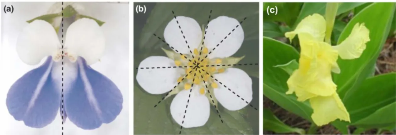
It is possible to distinguish three types of floral symmetry: actinomorphy (or radial symmetry) where the flower can be divided into symmetrical halves by more than one longitudinal plane passing through the axis; zygomorphy (or bilateral symmetry), when a flower is divided by only a single plane into two mirror-image halves; and asymmetry where no symmetry planes are observed (Figure 3) (Endress, 2001).
Figure 3 - Types of floral symmetry: (a) Zygomorphic flower (one symmetry plane); (b) Actinomorphic
flower (more than one symmetry planes); and (c) Asymmetric flower (no symmetry planes). Modified from Kalisz et al. (2006).
Flower symmetry is generally associated with the perianth whorl but by definition should extend to all floral whorls, however perfect whorl and organ symmetry (actinomorphy, sensu strictu) is not very common (Rudall and Bateman, 2004). Floral zygomorphy (flowers with visible differentiation between dorsal and ventral parts) could be the result of a differential organ elaboration by suppression/reduction of the developing flower organs (Endress, 1999). Floral zygomorphy could also the result of colour alterations or organ orientation but according to Rudall and Bateman (2004) these late secondary changes are not phylogenetically relevant. Fossil and phylogenetic data allowed Endress (1999) to infer that in Angiosperms, actinomorphic flowers predate zygomorphic flowers. The first clearly bisexual flower appears in the fossil record in the beginning of the Cretaceous period (113-90 million years ago.). This flower was completely actinomorphic, with identical organs in each floral whorl (Crane et al. 1995). The first distinctive zygomorphic flower is known from the Upper Cretaceous period (Turonian age), 30-40 millions of years after the above mentioned actinomorphic floral fossil (Crepet, 1996). The fact that the basal angiosperms and monocots are almost predominantly actinomorphic, emphasise even more the idea that zygomorphy is a
derived condition (Endress, 1999). Coen and Nugent (1994) suggested a multiple and independent origin of zygomorphy from an actinomorphic ancestor, admitting also the possibility of an independent number or reversions to the ancestral form.
The subclass Asteridae with approximately 65000 species is a particularly good taxonomic group to study the genetic mechanisms underlying floral symmetry transition with about half of the species having actinomorphic corollas and the other half having zygomorphic ones (Ree and Donoghue, 1999) (figure 4).
Figure 4 - Angiosperm simplified phylogenetic tree: a) Eudicots phylogeny; b) Lamiales phylogeny; and c) Solanales phylogeny. Bold branches represent zygomorphic taxa. Adapted from Reeves and Olmstead
(2003).
According to Donoghue et al., 1998, species deep inside the Asteridae subclass from Rubiaceae, Apocynaceae, Apiales and Ericales, appear to have maintained the actinomorphic ancestral condition, however, phylogenetic data also indicate that some actinomorphic species are derived from zygomorphic ancestors (Endress, 2001), whereas species with zygomorphic flowers (Plantago, Veronica, Antirrhinum) appear to have descended from an actinomorphic ancestral. Floral zygomorphy was probably a preponderant factor in Angiosperms prolific diversification in part because many numerous and complex species (Asteraceae, Fabaceae and Orchidaceae) are predominantly zygomorphic (Cronk, 2001). Sargent (2004) suggested that the high
zygomorphic background because a zygomorphic flower attracts the pollinator in a particular and precise orientation (Neal, 1998). Moreover, bees, by innate or acquire behaviour, have preference for a monosymmetric pattern (West, 1998). There are, however, several other zygomorphic associated modifications that also enhance pollination efficiency: ventral petals usually exhibit appealing visual features being ideal landing platforms for pollinators; aborted or significantly reduced stamens facilitate pollinator access to nectar and pollen with the other stamens and carpels repositioned to maximize contact with pollinator. Accordingly, it appears that plant and animal co-evolved leading to the development of highly efficient plant/animal groups that eventually originated reproductive isolation (formation of sexual barriers) and initiated the extensive speciation that characterizes Angiosperm radiation (Neal, 1998; Endress, 2001).
The question remaining is to identify the genetic mechanisms that sustain the parallel independent evolutionary shifts between actinomorphy and zygomorphy. At the genetic level the multiple origin of zygomorphy could be explained by non-homologous gene recruitment to a similar function or by repetitive modification of a pre-existent genetic pathway through parallel evolution of cis-regulatory elements (Preston and Hileman, 2009).
1.3.1
A. majus as model species for flower symmetry studies
Extensive comprehension of the molecular mechanisms underlying floral zygomorphy evolution has been successfully unfolded using Antirrhinum majus (Lamiales, Veronicaceae, tribe Antirrhinae) as a model species. In A. majus, floral zygomorphy is visible in the corolla and in the stamens (Endress, 2001). The corolla has five petals fused in a tube-like shape where it is possible to differentiate two dorsal petals, two lateral petals and one ventral according to their position, size, shape and epidermal cellular characteristics. The two dorsal petals have bigger lobes than the two lateral petals and ventral petal. The androecium is formed by five stamens (four fertile stamens and a fifth stamen in the dorsal portion that does not develop properly) (Endress, 1994) (figure 5).
Figure 5 - A. majus floral structure. (a) A. majus flower: five petals exhibit clear shape and size
differences along the symmetry axis (indicated by dashed line). The upper lip consists in two large dorsal petals and the lower lip in two smaller lateral petals and one even smaller ventral petal. (b) A. majus flower frontal diagram: dorsal petals are shown in yellow, lateral petals in green and ventral petal in purple. Zygomorphy is also observable in the stamen whorl, dorsal stamen (x) is aborted, and the two lateral stamens are smaller than the two ventral stamens. Adapted from Busch and Zachgo (2009).
A. majus dorsoventral asymmetry is genetically controlled by four genes, CYCLOIDEA (CYC), DICHOTOMA (DICH), RADIALIS (RAD) and DIVARICATA
(DIV), all codifying for transcription factors (Luo et al., 1996; Almeida et al., 1997; Luo
et al., 1999; Galego and Almeida, 2002; Corley et al., 2005).
1.3.1.1 CYCLOYDEA and DICHOTOMA
CYCLOYDEA and DICHOTOMA are members of the TCP family of
transcription factors (Cubas et al., 1999). The TCP genes are transcription factors characterized by the conserved TCP DNA binding domain (plant specific) that adopts a
basic-helix-turn-helix (bHLH) conformation. TCP genes appear to be specially involved
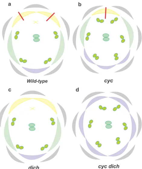
in cellular and plant growth regulation (Cubas et al., 1999; Kosugi and Ohashi, 2002). In A. majus, CYC loss of function mutants (cyc) have a strong phenotypic effect (figure 6b), with partial loss of the dorsal identity, lateral petal ventralisation and one petal and stamen gain when compared to the wild type (figure 6a). Also the remaining dorsal stamen is de-repressed resembling a lateral stamen. CYC is necessary to confer dorsal identity and this assumption is supported by the backpetals semidominant
lateral and ventral petals become mirror images of the lateral half of the dorsal petal (Luo et al. 1999).
Together with its paralogous DICH, CYC is expressed in the meristem dorsal domain. mRNA in situ hybridization showed that in the initial stage of flower development, CYC is transcribed dorsally in floral meristems. As development of the flower progresses, CYC expression becomes restricted to the dorsal petals and to the dorsal staminode (Luo et al., 1999). CYC restricts cellular proliferation in the first stages of flower development and in later stages, CYC promotes petal lobular growth and repress stamen development (Luo et al., 1996). The ability of CYC to enhance or inhibit cellular proliferation depends on CYC effect in cellular cycle genes like CYCLIN D3B and HISTONE4 (Gaudin et al., 2000). According to Gaudin et al. (2000) CYC could be acting as both promotor and repressor of the same cellular cycle regulator or binding to different co-regulators depending on the floral stage development. DICH expression is restricted to the dorsal region of the flower meristem, promoting internal asymmetry in the dorsal petals (figure 6a). DICH mutation has a much weaker phenotype compared to
cyc, resulting in the loss of asymmetry in dorsal petals (figure 6c). In cyc/dich, the
corolla is completely ventralised, with gain of one stamen and one petal (figure 6d) (Luo
et al., 1999). CYC and DICH double mutants produce radially symmetric, completely
ventralised flowers which made Luo et al. (1999) suggest that CYC and DICH could be working in an redundant way contributing in different degree to dorsal identity of A.
majus flower. Hileman and Baum (2003) suggested that CYC and DICH are the result
of a unique duplication event that occurred before the Antirrhinae species radiation. CYC and DICH functional divergence (66% amino acid identity) could have been due to the accumulation of mutations on CYC, DICH lineage or in both. The most plausible hypothesis is that CYC/DICH ancestor duplicated with both copies undergoing sub-funcionalization allowing the retention of both copies in the genome (He and Zhang, 2005).
Figure 6 – A. majus floral organ diagram showing phenotypic differences between (a) wild type and (b) cyc; (c) dich; and d) cyc/dich. Purple colour represents ventral petals, green lateral petals and yellow
dorsal petals. X represents the staminode. Grey colour depicts sepals and the red line internal petal asymmetry.
The partial ventralisation of the flower in the cyc mutant, together with the
backpetal mutant phenotype, lead to the assumption that, during flower development, CYC might be repressing the genes responsible for ventral identity.
1.3.1.2 RADIALIS
RADIALIS (RAD), like CYC and DICH, promotes dorsal identity, with rad
mutant flowers giving very similar phenotypes to cyc. Mutant flowers with loss of
lateral identity. The staminode is a bit longer than in the wild type. The number of petals and stamens is similar to wild type (Corley et al., 2005).
Figure 7 - A. majus floral organ diagram showing phenotypic differences between (a) wild type and (b) rad. Purple colour represents ventral petals, green lateral petals and yellow dorsal petals. X represents the
staminode. Grey colour depicts sepals and the red line internal petal asymmetry.
mRNA in situ hybridization demonstrated that RAD, similarly to CYC and DICH, is expressed dorsally in the flower meristem. Interestingly RAD expression is not only delayed relative to CYC and DICH, but also is absent from cyc dich indicating that RAD is genetically downstream of CYC and DICH. Accordingly, Costa et al. (2005) proved that the RAD promoter and intron contain CYC-binding sites and that RAD expression can be up-regulated by CYC in vivo.
The role of RAD in the establishment of flower dorsoventral asymmetry in A.
majus is still unclear. Similar to the cyc mutants, rad mutant flowers exhibit partial
ventralisation. This fact suggests that RAD is mediating CYC effects by antagonizing the genes responsible for ventral identity of the flower meristem.
1.3.1.3 DIVARICATA
DIVARICATA (DIV) is also a gene that has been associated with the
establishment of dorsoventral asymmetry in the A. majus flower (Galego and Almeida, 2002). In loss of function div mutants the ventral petal adopts a lateral identity. Also,
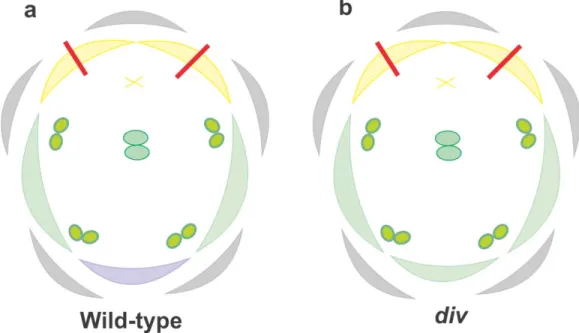
regions of the lateral petals are affected differently: the dorsal-most and central regions of the lateral petals are unaffected whereas the ventral-most region becomes more similar to the central region of the lateral petal (figure 8b) (Galego and Almeida, 2002). Dorsal petals are unaffected in div mutants and stamens develop as the wild type (figure 8a).
The lateralisation of div mutant phenotype together with the fact that the
cyc:dich:div triple mutant origins radially lateralized flowers suggest that DIV is
responsible for establishing petal ventral identity (Galego and Almeida, 2002). It has been proposed that DIV establishes ventral identity by interaction with class B floral homeotic genes in order to control downstream genes involved in differentiation and cellular proliferation (Almeida et al., 1997; Perez-Rodriguez et al., 2005).
Figure 8 - A. majus floral organ diagram showing phenotypic differences between (a) wild type and (b) div. Purple colour represents ventral petals, green lateral petals and yellow dorsal petals. X represents the
staminode. Grey colour depicts sepals and the red line internal petal asymmetry.
In the early stages of flower development, DIV is expressed in all floral organs regardless of their position along the flower dorsoventral axis. However, its phenotypical affect is only observed in the ventral petal. This suggests that the DIV gene product
increase lateral and ventral petal growth (Galego and Almeida, 2002). In later stages,
DIV transcripts are observed in the inner epidermal cell layer of the ventral and lateral
petals in the region of the petal folds. However, weaker expression is found throughout the dorsal petals. So, in later stages DIV activity results in the establishment of a clear difference between lateral and ventral petals together with the increase in lateral petal asymmetry. Together with the fact that cyc:dich double mutant has a complete ventralised phenotype it was proposed that somehow CYC and DICH antagonise DIV in the dorsal and part of lateral region of the floral meristem (Galego and Almeida, 2002). One possible explanation for this behaviour would be that RAD is mediating the antagonising action of CYC over DIV. Interestingly, RAD and DIV code for similar MYB transcription factors which might suggest competition for the same protein or DNA targets.
1.3.2 MYB family of transcription factors
MYB transcription factors are broadly distributed throughout eukaryotes (Yanhui
et al., 2006) and have been involved in essential cellular functions such as proliferation
and differentiation (Weston, 1999). Jiang et al. (2004) admit a high degree of ancestry for the MYB family in plants. The same authors propose that a MYB gene amplification occurred just before monocots and dicots divergence, which lead to the high number of MYB copies present in many plant species. Romero et al. (1998) suggested that, in plants, transcription control by the MYB family of transcription factors is particular important, being involved, among others, in circadian clock regulation (Borello et al., 1993), phosphorus deficit response (Rubio et al., 2001), secondary metabolism (Paz-Ares et al., 1987), light and hormonal signalling pathways (Ballesteros et al., 2001), seed and floral meristem development (Schmitz et al., 2002), cellular morphogenesis (Higginson et al., 2003) and cell cycle regulation (Araki et al., 2004).
A MYB protein is formed by one to four repeats of the MYB domain that are referred as R1, R2, R3 and R1/2 (from N-terminus to C-terminus of the protein). A canonical MYB domain has approximately 52 amino acid (Paz-Ares et al., 1987) with three tryptophan residues regularly spaced 18-20 amino acid apart that form an hydrophobic agglomerate that recognizes a specific DNA sequence (Ogata et al., 1995)
(figure 9). The tertiary structure of a MYB domain has three α-helix assuming a
helix-helix-turn-helix conformation. The second and third helix intercalate with the major
groove of the target DNA (Ogata et al., 1994) (figure 9 and 10).
Figure 9 - MYB transcription factor classes. Illustration showing different plant MYB protein classes,
depending on the number of adjacent MYB repeats (R). The primary and secondary structures of a typical R2R3-MYB are indicated. H, helix; T, turn; W, tryptophan; X, amino acid (X). Adapted from Dubos et
al., 2010.
MYB proteins can be divided into different classes depending on the number of adjacent MYB repeats (one, two, three or four; figure 9). The smallest class is the 4R-MYB group, whose members contain four R1/R2-like repeats (Dubos et al., 2010). The
Figure 10 – Terciary structure of a
MYB domain (helix I, II and III). The MYB protein is shown docked onto a double-stranded DNA fragment. Adapted from Stevenson et al. (2006).
class with roles, despite divergent, in cell cycle control (Ito, 2005). The third heterogeneous class comprises proteins with a single or a partial MYB repeat, collectively designated MYB-like proteins. The majority of plant MYB genes encode proteins of the R2R3-MYB class, which are thought to have evolved from an R1R2R3-MYB gene ancestor, by the loss of the sequences encoding the R1 repeat (Braun and Grotewald, 1999). However, the evolution of R1R2R3-MYB genes from R2R3-MYB genes by the gain of the R1 repeat through an ancient intragenic duplication has also been proposed (Jiang et al., 2004). R2R3-MYB transcription factors have a modular structure, with an N-terminus DNA-binding domain (the MYB domain) and a trans-activation or repression domain usually located at the C-terminus (Stracke et al., 2001).
The C-terminus domain of MYB proteins is very variable. This variability could be translated in different protein-protein or protein-DNA interactions, resulting in different transcriptional effects (Jaffé et al., 2007). There is evidence that two MYB domains in the same protein could have different functions and this situation could be correlated to the high proportion of non-conserved amino acids surrounding the DNA-binding domain or alterations in the conserved amino acids of the DNA-DNA-binding domain (Rose et al., 1999).
Proteins with MYB repeats that do not have a perfectly conserved DNA-binding domain as the observed in the canonical MYB protein (Paz-Ares et al., 1987) are called MYB-like proteins. In these MYB-like repeats one or more tryptophan residues may be replaced by another aromatic residue or by a similarly sized aliphatic residue (Corley et
al., 2005).
1.3.2.1 RAD and DIV as MYB-like transcription factors
RAD codes for a 93 amino acid protein with one single MYB-like repeat (Luo et al., 1999; Corley et al., 2005). RAD MYB-like domain has high similarity with the
MYB repeat characteristic of the MYB-like/SANT family of transcription factors (Corley et al., 2005). MYB-like/SANT transcription factors have been involved in DNA binding (Aasland et al., 1996), and some proteins in this family were also involved in the chromatin remodelling process (Boyer et al., 2005). Typically, single MYB-like/SANT proteins do not have a trans-activation domain downstream of the MYB-like
domain, which could mean that these protein could be mainly involved in transcription repression or protein-protein interactions (Boyer et al., 2005). However, RAD has an incomplete CREB domain that is shared by RAD orthologs in Arabidopsis (Baxter et al., 2007). In the animal kingdom, the CREB domain importance in transcription regulation is well established. According to Quinn (1999), the CREB constitutive activation domain interacts with the transcription factor TFIID through one or more of the TATA-binding protein-associated factors in order to control transcription. The importance of the incomplete CREB domain in RAD function is, however, still undetermined. RAD MYB-like/SANT domain is very similar to the one found in LeFSM1 (Barg et al., 2005). In tomato and Arabidopsis ectopic expression of LeFSM1 results in severe developmental alterations manifested in retarded growth, and reduced apical dominance (Barg et al., 2005). Similarly to LeFMS1, RAD is involved in the regulation of a evolutionary conserved developmental process, however, the importance of the MYB-like/SANT domain to RAD function remain unclear.
DIV is an R2R3MYB-like transcription factor with 307 amino acids (Galego and
Almeida, 2002). DIV protein is distinctively different from the typical R2R3MYB proteins not only because of the change in the conserved DNA-binding domains residues but also due to a longer linker between the R2 and R3 MYB-like domains (Galego and Almeida, 2002; Howarth and Donoghue, 2009). The two MYB-like domains of DIV have distinct functional characteristics: the first domain belongs to the MYB-like/SANT family, whereas the second belongs to like SHAQKYF class.. The second MYB-like domain of DIV has great similarity to the single MYB-MYB-like domains of the proteins CCA1 and LHY (Galego and Almeida, 2002). CCA1 and LHY are R3MYB-like transcription factors involved in DNA-binding and transcription activation (Schaffer et
al., 1998; Wang and Tobin, 1998). Accordingly, DIV might be controlling transcription
through the second MYB-like domain. The main feature of the SHAQKYF class is the highly conserved SHAQKYF motif, in the predicted third α-helix that could be essential in the transcriptional control activity (Schaffer et al., 1998).
(GATAA) described by Giuliano et al. (1988) as the binding sequence of I-box transcription factors. Similar to DIV, LeMYBI has two MYB-like domains, but only the second MYB-like repeat is involved in DNA transcriptional control (Rose et al., 1999). It is possible that the first MYB-like domain of DIV, similar to the first MYB-like repeat of LeMYB1, is not involved in transcription control, but functionally involved in protein-protein interactions as suggested for MYB-like/SANT domains by Boyer et al. (2005).
Importantly the MYB-like/SANT domain of DIV has a great similarity with the MYB-like/SANT domain of RAD, which allowed Corley et al. (2005) to infer that RAD might be resultant from an ancestral similar to DIV following a duplication event and posterior loss of the second MYB-like domain. The molecular similarity between the first MYB-like domain of DIV and the MYB-like domain of RAD suggest that DIV and RAD could be targeting the same genes or proteins (Corley et al., 2005).
1.3.3 A model for Genetic Regulation of Flower Asymmetry
The lateralised flower phenotype of cyc:dich:div triple mutant indicate that lateral identity can be considered a neutral state for petals. DIV is responsible for ventral petal identity, and DIV function might be suppressed in the dorsal and part of the lateral domain possibly due to CYC and DICH activity (figure 11) (Galego and Almeida, 2002). As well as antagonizing DIV function in the dorsal and lateral petals, CYC and DICH are responsible for dorsal petal identity (Luo et al., 1999). CYC and DICH inhibitory action over DIV appears to be partially mediated by RAD. RAD is genetically downstream of
CYC and DICH and is also required for dorsal identity (Corley et al., 2005). However, CYC and DICH can influence flower asymmetry in a RAD independent way because CYC and DICH function in the dorsal staminoide and dorsal petals lobules do not
disappear in loss of function rad mutants (Corley et al., 2005). However, it is likely that other target of CYC is responsible for dorsal petal specification because neither rad single mutants nor dich rad double mutants are radially symmetric, whereas cyc rad double mutants are (Corley et al., 2005). Nevertheless, it appears that the CYC and
DICH major effects on flower symmetry are mediated by RAD. Neither CYC or DICH,
et al., 1999; Corley et al., 2005). However, the lateral petals do not have a ventral
phenotype as expected by the ubiquitous expression of DIV (Galego and Almeida, 2002), so RAD must be influencing the lateral petals phenotype in a non-autonomous way possibly by an intercellular transport of RAD gene product or protein to the lateral domain (Corley et al., 2005), similar to the non-cell autonomous transport of DEFICIENS (DEF) and GLOBOSA (GLO) observed in Antirrhinum (Schwarz-Sommer
et al., 1992; Perbal et al., 1996).
Figure 11 – Model showing the genetic interactions underlying the establishment of zygomorphy in A. majus. CYC and DICH expression in the dorsal dominion (yellow) leads to RAD activation (brown arrow). RAD inhibits DIV activity in dorsal and lateral regions of the flower.
As previously mentioned, DIV is expressed in the entire flower meristem, however, its function is restricted to the ventral domain possibly due to RAD function. Thus, DIV could be negatively regulated by RAD through post-transcriptional mechanisms in the dorsal and lateral domains by direct or indirect competition by competing for the same DNA or protein targets (Galego and Almeida, 2002; Corley et
al., 2005). The high similarity between the MYB-like/SANT domain of RAD and the
DNA-binding and transcription activation (figure 12a). Accordingly, RAD, with no known activation domain, could therefore function as a transcriptional repressor. Moyano et al. (1996) described a similar behaviour in A. majus. The authors showed that two A. majus MYB transcription factors (AmMYB305 e AmMYB340) bind to the same gene promoter using the same cis-element having, however, distinct transcriptional activity.
Figure 12 –Mechanisms of molecular antagonism of RAD over DIV. a) RAD could bind to the DNA
cis-element of DIV; b) RAD could form a transcriptional complex with DIV altering DIV transcriptional activity; c) To be transcriptioanl activity, DIV might need a interactor partner, and in this case RAD could be sucessfully rescuing the interactor partner or DIV, the unknown partner and RAD could form a multi-protein complex that inhibits transcription.
Many proteins mediate their function through the formation of both stable or transient protein complexes (Carrol et al., 2001) and often, the same protein may interact
with various partners in response to different stimuli or at different developmental stages (Hsieh et al., 2003). Taking in consideration the fact that the MYB-like/SANT domain could be involved in the establishment of protein-protein interactions (Boyer et al., 2005), it is possible that RAD might bind directly to DIV preventing its transcriptional activity (figure 12b). It is also possible that DIV might need one or more interacting partners to control transcription (figure 12c), and in this particular case RAD might be inhibiting DIV activity by rescuing its interacting partners. A similar antagonizing pattern is observed in the determination of epidermal cell fate in Arabidopsis where the assembling of a transcriptional complex is inhibited by the antagonisation between two MYB proteins. CAPRICE (CPC), TRIPTYCHON (TRY), ENHANCER OF TRY AND CPC1 (ETC1), ETC2 and ETC3 (Simon et al., 2007) are small proteins with a single MYB domain (Simon et al., 2007; Tominaga et al., 2007), that act redundantly as negative regulators in the determination of epidermal cell fate by competition with R2R3-MYB proteins, namely GLABRA1 (GL1) and WEREWOLF (WER) (Esch et al., 2004; Kirik et al., 2004; Simon et al., 2007).
1.3.4 Genetic basis for flower asymmetry among other taxa
Flower asymmetry appears to be controlled by a group of phylogenetic related genes in different taxa suggesting a common genetic background for flower asymmetry evolution (Feng et al., 2006). Correspondence between altered expression patterns of genes and change in flower symmetry have been found in some Angiosperm taxa phylogenetically distant from A. majus, Mohavea confertiflora (Plantaginaceae) (Hileman et al. 2003); Chirita hetrotricha (Gesneriaceae) (Gao et al., 2008), Bournea (Gesnereaceae) (Zhou et al. 2008), Lotus japonica (Fabaceae) (Feng et al., 2006) and
Iberis amara (Brassicaceae) (Busch and Zachgo, 2007). There is abundant data about CYC homologous in taxa across the Asteridae and Rosids, however, there is yet little on RAD- and DIV-like homologues and their putative conserved role in the establishment of
the asymmetric dorsoventral pattern of the flower.
VmCYC were expressed in the dorsal region of the flower, becoming detected only in the
dorsal petal at late stages of development. Similarly GoRAD and GoCYC were expressed in the dorsal petals and dorsal staminoide during early to late stages of G. officinalis flower development. This is despite an apparent lack of a CYC-like binding site in the first intron of VmRAD, which is disrupted in several of the well characterized A. majus
rad mutants (Corley et al., 2005). However, according to the same authors it is unknown
whether there are CYC-like binding sites upstream of the VmRAD coding region, as have been found for A. majus RAD (Costa et al., 2005).
The ability of CYC-like genes to activate RAD-like genes appears to be not only conserved in the Veronicaceae but also in other families of the Lamiales clade. In the Gesneraceae family, work in Bournea leyophilla and Chirita heterotricha shed some light about the conservation of the genetic mechanisms that support floral dorsoventral asymmetry across the Lamiales (Zhou et al., 2008; Yang et al. 2010).
Yang et al. (2010) suggested that the floral zygomorphy in Chirita heterotricha might have been established through the co-option of the RAD-like gene into the regulatory network controlled by CYC-like genes. The authors found that an appropriate
CYC-like binding site is present in the RAD-like promoter. B. leyophylla exhibits
actinomorphic flowers at anthesis, however, its very short corolla tube with five filaments adnate to the corolla and the adaxial petals somewhat smaller than others lead Zhou et al. (2008) to suggest evolution from a zygomorphic ancestor (figure 13). The same authors have identified in B. leiophylla CYC, RAD and DIV homologues that
control flower symmetry. BlCYC1 and BlRAD are expressed in the dorsal region of the early meristem but are down regulated during the late stages, which should be responsible for the origin of the derived actinomorphy (Zhou et al., 2008). Interestingly, similarly to A. majus DIV, two DIV copies, BlDIV1 and BlDIV2 were expressed in the entire floral meristem (Zhou et al., 2008). This could mean that the regulatory mechanism that controls the dorsoventral asymmetry of the A. majus flower might be conserved in at least some families belonging to the Lamiales. However, there is little published data suggesting DIV function outside the Lamiales.
A recent study by Howarth and Donoghue (2009) provided the first phylogenetic analysis of DIV-like genes in Dipsicales. Data showed that DIV and its homologues in
Dipsicales are the result of a duplication event that occurred in parallel with CYC-like genes, their potential interacting partner. The same authors emphasised the fact that DIV and CYC maintain the same number of copies in eudicots. However, the authors were not clear whether the expression pattern of DIV is conserved in DIV-like genes and if there is correlation between expression profiles of DIV-like genes and CYC-like genes and respective shifts in flower symmetry.
Figure 13- Mature whole and dissected flowers of Bournea leiophylla. Actinomorphy was observed in
mature Bournea leiophylla flowers with nearly identical adaxial, lateral and abaxial petals and stamens. bp, abaxial (ventral) petal; bst, abaxial (ventral) stamen; dp, adaxial (dorsal) petal; dst, adaxial (dorsal) stamen; lp, lateral petal; lst, lateral stamen. Bar: 5 mm. Adapted from Zhou et al. (2008).
Outside the Asteridae, in two Fabales species, Lotus japonicus (Feng et al., 2006) and Pisum sativum (Wang et al., 2008), gene expression and functional analyses both implicate CYC-like genes in the control of bilateral flower symmetry. In these species, CYC- like gene expression is restricted to dorsal or dorsal plus lateral regions of developing flowers, similar to CYC expression in A. majus. Within the Brassicales, the expression of a CYC-like gene seems to be responsible for the bilateral petal symmetry of Iberis amara, which deviates from the radial petal symmetry in Arabidopsis (Busch and Zachgo, 2007). Also, Zhang et al. (2010) described in the Malpighales, a CYC-like gene with a conserved pattern of dorsal gene expression in two distantly related neotropical species, Byrsonima crassifolia and Janusia guaranitica.These results clearly
conserved in the core eudicots. However, in this species no data is available confirming the activation of RAD-like genes to confer dorsoventral identity to the flower.
Yang et al. (1999) described two big large groups of RAD-like genes in
Arabidopsis and Antirrhinum that were probably derived from an ancestral gene
duplication event following Rosids/Asterids divergence 112-156 million years ago. Costa et al. (2005) stated that CYC is unable to activate RAD orthologues in
Arabidopsis, however, it is unknown whether the Arabidopsis CYC-like (TCP1) can turn
on Arabidopsis RAD-like genes. Baxter et al. (2007) work revealed that RAD-like genes are not expressed dorsoventrally in Arabidopsis, suggesting that a cis or trans diversification event occurred that lead to the RAD co-option to establish dorsoventral asymmetry in the Asteridae. However, another explanation would be that Arabidopsis and its relatives, with radial, four-merous flowers, have lost a pathway that otherwise span the core eudicots (Howarth and Donoghue, 2009).
1.4 Aims of this study
The main aim of this study is to gain understanding on the molecular mechanisms underlying the antagonizing action of RAD over DIV function.
In a first approach, RAD and DIV recombinant proteins will be obtained and used in a DNA-binding assay. This assay will be useful to understand if RAD is inhibiting DIV by binding to the DNA target of DIV or by binding directly to DIV.
Recently, to understand if an interactive partner mediates the molecular antagonism between RAD and DIV, a yeast two-hybrid screening was performed using RAD as bait. Two new proteins were identified as RAD interactors, and named RAD INTERACTING PROTEIN1 (RIP1) and RAD INTERACTING PROTEIN2 (RIP2). Interestingly, both proteins interact with DIV.
The second aim of this study is to characterise the interaction of RIP1 and RIP2 with RAD and DIV and to evaluate if this newly identified proteins are responsible for the molecular antagonism between RAD and DIV. A quantitative yeast two-hybrid assay will be performed to measure the interaction strength of each pair of interactors, and the sub-cellular localisation of RAD, DIV, RIP1 and RIP2 proteins will be identified by
fluorescence microscopy, to determine whether the proteins are able to interact in the same cellular compartment.
The third objective of this study is to develop experimental procedures that will enable further characterisation of RIP1 and RIP2 and their putative role in the molecular antagonism between RAD and DIV. These assays include: the ectopic expression of RIP1 and RIP2 in Arabidopsis thaliana; the use of DNA-binding assays to determine if the RIP1 and RIP2 are able to interact with the DNA-target of DIV or to form a multi-protein complex with DIV or/and RAD; and a bimolecular fluorescent complementation assay to further characterize the interaction between RIP1, RIP2, RAD and DIV in
2. Material and Methods
2.1 Biological material
2.1.1 Plant material
2.1.1.1 Arabidopsis thalianaArabidopsis thaliana ecotype Columbia (Col-0) seeds were obtained from public
seed banks, and stored in the dark at room temperature. Seeds (20-200 per tube) were stratified by water immersion in the dark for 4ºC for 3-5 days in order to break dormancy and synchronise germination. The seeds were surfaced sterilised in a horizontal flow laminar chamber (BBH4 BRAUN Horizontal) by successive incubations with 1 mL 70% (v/v) ethanol during 5 min, 1 mL commercial 20% (v/v) bleach with 0.1% (v/v) Tween for 10 min, followed by three washes with 1 mL sterilised water and two 1 mL 100% (v/v) ethanol. Between incubations, seeds were resuspended by vortex agitation and recovered by centrifugation (10,000g during 1 min). Seeds were sown in Murashige and Skoog agar medium (MS medium, 4.209 mg L-1, 1.5% (w/v) sucrose, 1.2% (w/v) plant agar, the pH was equilibrated to 5.7 with KOH) (Murashige and Skoog, 1962) and were incubated vertically under long day conditions (16 hours light / 8 hours dark) at 20ºC in controlled environmental growth rooms, with light intensity of 40 µE m-2 s-1. Approximately 10 days after sowing, plantules were transferred to pots containing a 4:1 (v:v) mixture of turf rich soil and vermiculite. The pots were watered and covered with cling film in order to maintain humidity for up to one week after the transplant. Following this period pots were watered every day.
2.1.1.2 Nicotiana benthamiana
Material from N. benthamiana was used for the transient transformation assay.
N. benthamiana seeds were stored in the dark at room temperature. N. benthamiana
seeds (20-30) were sown in pots containing a 4:1 (v:v) mixture of turf rich soil and vermiculite. Plants were grown in a growth chamber at 22°C with 70% relative humidity under a 16 hours light/8 hours dark photoperiod for about 1–1.5 months before the transient transformation assay.
2.1.2 Bacterial material
2.1.2.1 Escherichia coli 2.1.2.1.1 XL1-Blue strainThe E. coli strain XL1-Blue (recA1 endA1 gyrA96 (nalR) thi-1 hsdR17 supE44
relA1 lac glnV44 F’[proAB+lacIqZΔM15 Tn10]) (Bullock et al., 1987) was used for vector perpetuation and cloning procedures.
2.1.2.1.2 BL21(DE3)pLysE strain
The E. coli strain BL21(DE3)pLysE (F– ompT hsdSB (rB–, mB–) dcm gal λ(DE3)
pLysE, Cmr) (Studier and Moffatt, 1986) was used for high-stringency heterologous protein expression.
2.1.2.1.3 Preparation of E. coli competent cells
E. coli cells were made competent for transformation procedures as follows: 5 ml
Luria-Bertani medium (LB medium, 10 g L-1 Bactotryptone; 5 g L-1 Yeast extract and 10 g L-1 NaCl) (Sambrook et al., 1989) was inoculated with a single colony and incubated overnight at 37ºC with vigorous shaking (200 rpm) with appropriate antibiotics. One flask containing 100 mL of LB medium was inoculated with 1 mL of the starter culture, and this was grown at 37ºC until an OD600 of about 0.25 was reached (approximately 3
hours after inoculation, depending on the strain). The cells were pelleted in four falcon tubes by centrifugation at 3,000 x g (4ºC) for 10 minutes and the supernatant was discarded. Each pellet was carefully resuspended in 8 mL 0.1 M MgCl2 (sterile and cold)
and incubated on ice during 30 min. The cells were then pelleted, as described above, and gently resuspended in 2.5 mL TG salts (75 mM CaCl2; 6 mM MgCl2; 15% (v/v)
glycerol; cold and sterile). Again cells were pelleted as described above, resuspended in 750 µL TG salts and kept on ice for 4-24 hours. 100 µL of cells were pipetted into pre-chilled eppendorfs, frozen in liquid nitrogen and stored at -80ºC.
2.1.2.2 Agrobacterium tumefaciens
The A. tumefaciens strain EHA105 was used for A. thaliana and N. benthamiana permanent and transient transformation procedure, respectively. EHA105 is a derivative of EHA101 (C58 pTiBo542; T-region::aph) (Hood et al., 1986).
2.1.2.2.1 Preparation of A. tumefaciens competent cells
A. tumefaciens EHA105 cells were made competent for transformation procedure
as follow: a single colony of EHA105 was used to inoculate 100 mL LB medium supplemented with rifampicin (100 µg mL-1) and grown overnight at 30ºC with vigorous shaking (200 rpm). When the culture reached an OD600 between0.5-0.7, the cells were
pelleted in four falcon tubes by centrifuging at 4,000 x g at 4ºC for 5 minutes. The supernatant was discarded and the pellet gently resuspended in 25 mL cold 10% (v/v) glycerol. The bacterial solution was centrifuged as described above and the supernatant discarded. The pellet was resuspended in 12.5 mL cold 10% (v/v) glycerol and then centrifuged again under the same conditions. This step was repeated using 500 µL of cold 10% (v/v) glycerol. Finally, the pellet was resuspended in 250 µL of cold 10% (v/v) glycerol. 50 µL of cells were dispensed into pre-chilled eppendorfs, frozen in liquid nitrogen and stored at -80ºC.
2.1.3 Saccharomyces cerevisiae
The Saccharomyces cerevisae strain AH109 (MATa trp1-901 leu2-3, 112 ura3-52 his3-200 gal4Δ gal80Δ LYS2::GAL1UAS-GAL1TATA-HIS3; GAL2UAS-GAL2TATA -ADE2; URA3::MEL1UAS-MEL1TATA-lacz, MEL1) was used in the Yeast-two-hybrid
assays. The yeast strain AH109 contains distinct ADE2, HIS3, lacZ, and MEL1 reporter genes, under the control of the GAL promoter, that are only expressed in the presence of the GAL4 reconstituted transcription factor.
Bacterial and S. cerevisae strains were perpetuated as glycerol stocks for long-term storage. In the former case a 500 µL aliquot from an inoculate LB culture was added to an equal volume of 80% (v/v) glycerol. In the latter case an 800 µL aliquot from a culture was added to 200 µl of 80% glycerol (v/v).
2.2
DNA Methods
2.2.1 Bacterial Transformation
2.2.1.1 E. coliAn amount of 10 ng (5-10 µL) of circular DNA was added to 100 µL of competent cells (as described in section 2.1.2.1.3) in an eppendorf tube and incubated on ice for 30 minutes. Cells were heat-shocked at 42ºC for 45 seconds, returned to ice for 2 minutes and 900 µL of LB was added to the tube. The cells were incubated at 37ºC for 60 minutes with vigorous shaking (200 rpm). A volume of 100 µL of the transformation mix was then spread onto LB-agar plates [LB medium, 1.5% (w/v) agar] containing the appropriate selection antibiotic. Plates were incubated overnight at 37ºC.
2.2.1.2 A. tumefaciens
An aliquot of thawed A. tumefaciens competent cells was added to a pre-chilled electroporation cuvette. A volume containing an amount of 10 ng was carefully added to the cuvette. An electric pulse was applied using the Gene Pulser II (Bio Rad) (field strength: 1.25 Kv mm-1; Capacitance: 25 uF; Resistance: 400 Ω; Pulse Field: 8-12 ms). The mixture was transferred to an eppendorf tube with 1 mL LB broth and incubated 3 hours at 30ºC with shaking (200 rpm). A volume of 10 µL of the transformation mix was then spread onto LB-agar plates containing the appropriate selection antibiotics. Plates were incubated overnight at 30ºC (Inoue et al., 1990).