Evaluation of cystamine-modi
fied hyaluronic acid/chitosan polyplex as
retinal gene vector
Ana V. Oliveira
a,b, Adriana Marcelo
c, Ana M. Rosa da Costa
d,e, Gabriela A. Silva
a,f,⁎
a
Centre for Biomedical Research (CBMR), University of Algarve, Faro 8005-139, Portugal
bProgram in Biomedical Sciences, University of Algarve, Faro 8005-139, Portugal c
Department of Biomedical Sciences and Medicine, University of Algarve, Faro 8005-139, Portugal
d
Department of Chemistry and Pharmacy, University of Algarve, Faro 8005-139, Portugal
e
Algarve Chemistry Research Centre (CIQA), University of Algarve, Faro 8005-139, Portugal
f
CEDOC, NOVA Medical School/Faculdade de Ciências Médicas, Universidade Nova de Lisboa, Campo Mártires da Pátria 130, 1169-056 Lisboa, Portugal
a b s t r a c t
a r t i c l e i n f o
Article history:
Received 20 February 2015 Received in revised form 16 June 2015 Accepted 25 August 2015
Available online 28 August 2015 Keywords: Gene therapy Hyaluronic acid Polymer modification Retina Biocompatibility
Purpose: A successful gene therapy approach can prevent or treat congenital and acquired diseases. However, there is still no ideal non-viral vector for gene delivery in a safe and timely manner. In this report the anionic poly-mer hyaluronic acid (HA) was investigated as a potential vector for gene therapy. Due to its intrinsic character-istics it constitutes an excellent candidate to deliver therapeutic genes, pending the modification of its surface charge.
Methods: To modify its charge, HA was modified with cystamine. Several formulations were prepared using mod-ified HA combined with sodium sulfate, sodium triphosphate, K-carrageenan and chitosan. Vectors were charac-terized with respect to size, charge, DNA load and its protection, and effect on cell viability. The better performing formulations were further evaluated in vitro for their transfection efficiency in HEK293T and ARPE-19 cells. Results: Cell viability assays showed low cytotoxicity for both polymers. Gene transfer efficiency depended on cell line and formulation, but no increased transfection efficiency was observed with the modified polymer. Conclusions: HA has great potential as a gene therapy vector, but further optimization, including incorporation of a higher percentage of positive groups in HA, is needed before its use as a gene delivery vector.
© 2015 Elsevier B.V. All rights reserved.
1. Introduction
Gene therapy is an area that has been growing rapidly and revolu-tionizing the world of research[1]. An essential requirement for the suc-cess of this technique is an efficient method for gene transfer, since DNA molecules cannot enter cells efficiently because of their large size, hy-drophilic nature and susceptibility to degradation mediated by nucle-ases[2,3]. Considering the great diversity of diseases targeted by gene therapy, it is implausible that a single gene delivery system (vector) is suitable for all applications. However, the main requirements are com-mon to all systems: vectors should transfer the genetic material to the tissue of interest and induce the proper level of therapeutic gene ex-pression with no side effects[4].
Currently, vectors used in gene therapy can be divided into two main categories: viral and non-viral. Recombinant viruses such as retrovirus, lentivirus, adenovirus, adeno-associated viruses, and herpes viruses
have been widely used for genetic material transfer[2]. Viruses have as main advantage their high transfection efficiency, however, viral vectors entail many intrinsic problems such as difficulties in production, limitations concerning repeated administrations that can lead to acute inflammatory responses, immune responses of the host to the virus and induction of mutagenesis by some viruses that integrate into the ge-nome of host cells[2]. On the other hand, non-viral vectors have a higher safety profile due to their low toxicity and low immunogenicity. Additionally, non-viral vectors have the ability to carry larger genes and present lower production costs. Despite this appealing feature from the safety standpoint, non-viral vectors have little clinical importance due to low transfer and expression of transgene[2]. However, due to prob-lems in clinical trials using viral vectors in the recent past, the interest in non-viral technologies has been renewed, particularly in the release properties of the non-viral vectors that resemble traditional drugs[4].
Non-viral vectors include synthetic or naturally occurring chemical compounds, such as lipids and cationic polymers. These can form com-plexes with the negatively charged DNA through electrostatic interac-tions that allow the therapeutic transgene across the cell membrane via endocytosis[2,3,5]. The complexes protect DNA from nuclease-mediated degradation and facilitate cell entry as well as gene transfer-ring into the nucleus[2].
Abbreviations: CS, chitosan; DNA, deoxyribonucleic acid; EDAC, 1-ethyl-3-(3-dimethylaminopropyl)carbodiimide; HA, hyaluronic acid; HASSNH2, hyaluronic
acid-co-N-cystaminyl-hyaluronamide; HASH, hyaluronic acid-co-N-cysteaminyl-hyaluronamide; MW, molecular weight; RPE, retinal-pigmented epithelia.
⁎ Corresponding author.
E-mail address:[email protected](G.A. Silva).
http://dx.doi.org/10.1016/j.msec.2015.08.047
0928-4931/© 2015 Elsevier B.V. All rights reserved.
Contents lists available atScienceDirect
Materials Science and Engineering C
j o u r n a l h o m e p a g e : w w w . e l s e v i e r . c o m / l o c a t e / m s e cous biological barriers of cells that must be overcome to achieve higher transfection rate. These barriers include the attachment of the polymer to the cell surface, then entering the cell through the cell membrane, displacement throughout the cytoplasm, escape endosomal degrada-tion and ending with the passage of the nuclear envelope and nuclear entry[3]. In order to overcome different barriers, functional groups can be introduced into polymers, such as ligands to enhance cell entry via receptor-mediated endocytosis, membrane peptides to allow endosomal release and nuclear localization signals to increase nuclear entry of the transgene[3].
In this paper, hyaluronic acid (HA) was evaluated as a possible reti-nal gene therapy vector. HA is a biocompatible, toxic, non-immunogenic, non-inflammatory anionic biopolymer that has been widely used in various biomedical applications[6]. Multiple studies on the biological function of HA have revealed that there is a strong rela-tionship between the presence of HA and the migration and prolifera-tion of cells as well as an involvement in wound healing, cell motility, angiogenesis, and extracellular matrix formation. Another important feature of HA for its use as a vector of therapeutic genes is the ability to interact with various cell receptors[6]. The negatively charged car-boxyl group of HA is responsible for the interaction with membrane re-ceptors allowing the connection with HA[7].
Despite its advantageous features, HA exists in the form of an aque-ous gel, has a short lifetime and quickly degrades after administration. In order to increase the lifetime of HA for long-term clinical applications, several strategies have been developed, particularly modifications of the polymer at the level of carboxyl and hydroxyl groups. These new polymers, although they have different physico-chemical properties compared to unmodified HA, they maintain essential biological proper-ties that allow their use in non-viral therapies[6]. Among possible chemical modifications to perform, the more relevant for gene therapy is the inclusion of amine groups[6]. A higher amount of amine groups may lead to greater DNA loads and increase the efficiency of transfection and transgene expression. In this study, HA was modified with cysta-mine through a coupling reaction with the carboxyl groups of HA. This modification with cystamine not only adds amine groups to the poly-mer but it also contains a disulfide bond that can be cleaved in the pres-ence of intercellular glutathione, thereby promoting a more rapid release of genetic material[8,9].
In this work we compare the transfection efficiency between modi-fied and unmodimodi-fied HA using two types of cells: human embryonic kid-ney (HEK 293T) cells and retinal pigment epithelium cells (ARPE-19). The former are a commonly used cell line in transfection studies, while the latter are a model of retinal pigmented epithelial (RPE) cells, our target in the retina, due to their important role in the support of the retinal homeostasis and involvement in several retinal diseases[8]. 2. Materials and methods
2.1. Materials
Chitosan (MW of 80 kDa) with a degree of de-acetylation of 83% was purchased from Polysciences, Inc., USA. Hyaluronic acid, with MWs of 132 and 214 was purchased from Lifecore Biomedical. All other reagents were analytical grade and used without further purification.
2.2. Plasmid constructs and cell lines
A plasmid expressing enhanced greenfluorescent protein driven by the cytomegalovirus promoter (kindly provided by Jean Bennett, Uni-versity of Pennsylvania, USA) was amplified in Top 10 bacteria and
HEK 293T cells (kindly provided by Guilherme Ferreira, University of Al-garve, Portugal) and a human retinal pigment epithelial cell line (ARPE-19, kindly provided by Francisco Ambrósio, University of Coimbra). All cell culture reagents were purchased from Sigma-Aldrich® (St. Louis, MO/USA).
2.3. Methods
2.3.1. Modification of hyaluronic acid with cystamine (HASSNH2)
The modification reaction was performed in an adaptation of what was previously described elsewhere and depicted inFig. 1 [10–12]. Briefly, 500 mg of hyaluronic acid was dissolved in 100 ml of distilled H2O, a one and a half molar excess (relative to the carboxylic acid groups
in HA) of 1-ethyl-3-(3-dimethylaminopropyl)carbodiimide (EDAC, 0.378 g, 1.974 mmol), and three molar excess of cystamine (0.889 g, 3.947 mmol) were added to the solution. The mixture was stirred at room temperature for 72 h. The reaction mixture was dialyzed against 4 g/l NaCl (MW cut off 2000) for 6 h and then against distilled H2O for
24 h. Thefinal product – HASSNH2– was lyophilized for 3 days and
stored at room temperature until use. 2.3.2. Determination of the thiolation extent
In order to determine the extent of the modification, the thiol groups were quantified by the Ellman's test. Firstly, 50 mg of HASSNH2was
dis-solved in distilled H2O and treated with 30 molar excess of dithiothreitol
(DTT, 0.418 g, 2.712 mmol) in order to expose the thiol groups. The mix-ture was stirred under N2atmosphere for 24 h, at room temperature.
The reaction mixture was dialyzed against 4 g/l NaCl (MW cut off 2000) for 6 h and then against distilled H2O for 24 h. Thefinal product
HASH was lyophilized for 2 days and stored at room temperature until use. Secondly, for the Ellman's test, 3.8 mg of HASH was dissolved in 1 ml of 0.1 M Tris buffer, pH 8.0. For each measurement, a tube contain-ing 100μl of Ellman's reagent (10 mM of 5,5′-dithio-bis-(2-nitrobenzoic acid) in 0.1 M Tris buffer), 1800μl of 0.1 M Tris buffer and 100 μl of sam-ple was prepared, incubated at room temperature for 15 min and then the absorbance at 412 nm was measured[13].
2.3.3. Potentiometric titration of HASSNH2
In order to characterize the protonation behavior of the modified polymer, a titration was performed[14]. Polymer solutions, HA and HASSNH2, were prepared with a concentration of total titratable
carbox-yl group concentration of 10 mM in a volume of 10 ml. The initial pH was adjusted to 2–3 by adding 2.00 M HCl prior to the dropwise addi-tion of the titrant 0.08 M NaOH to the polymer soluaddi-tions under constant stirring. Potentiometric measurements were made using a pH measure-ment electrode (Orion 9157BN) connected to an Orion pH meter (Ther-mo Orion 4 Star pH-ISE Benchtop; Ther(Ther-moFisher Scientific (Waltham, MA USA).
2.3.4. Polyplex preparation
Polyplexes were prepared as previously described by our lab[8,15]. Vector formulations were prepared at various ratios by adding anionic solutions to the HASSNH2solution, as indicated inDiagram 1. Briefly,
polymer solutions (1 mg/ml in MilliQ H2O, pH 5.5) and sodium sulfate
solutions were separately heated to 55 °C. Equal volumes of both solu-tions were quickly mixed together, vortexed for 30 s, placed on ice and stored at 4 °C. Alternatively, HA and other anionic species (TPP andκ-carrageenan) were tested by combining them to the sodium sul-fate solutions. Different weight ratios were tested keeping constant the HASSNH2amount (250μg) and varying the HA amount. In order to use
equal volumes of both solutions, HA was diluted in sodium sulfate and then mixed with the HASSNH2solution, as described above.
Vectors were prepared based on mixtures with chitosan (CS), origi-nating CSHA or CSHASSNH2vectors. The CS:HA or CS:HASSNH2weight
ratios 5:1 and 7:1 were chosen based on previous studies[15], keeping constant the CS amount (250μg). In order to use equal volumes of both solutions, HA or HASSNH2was diluted in 25 mM sodium sulfate and
then mixed with the CS solution, as described above.
To prepare polyplexes loaded with DNA at a NH3+:PO4−ratio of 15:1,
250μg of CS and 26.5 μg of DNA, were used. DNA was mixed with the sodium sulfate solution and this solution mixed with the CS solution, as described above. The resulting polyplex formulations are described inDiagram 2.
2.3.5. Polyplex characterization
Size analysis was performed by dynamic light scattering (DLS) with a detection angle of 173°, and zeta potential (ZP) measured by laser Doppler velocimetry (Zetasizer Nano ZS, Malvern instruments, UK). All measurements were performed in ddH2O at 25 °C. The
polydispersi-ty index (PdI) was calculated based on DLS measurements using the Zetasizer Nano Series software version 7.11.
The complexation and integrity of the plasmids (DNA) in the polyplexes were assessed by gel electrophoresis. Free DNA and polyplexes were incubated with 1 unit of DNAse I (Sigma-Aldrich®, USA) for 15 min at 37 °C. The reaction was stopped by the addition of 1μl of a 50 mM EDTA solution and heating at 70 °C for 10 min. Complex-ation and integrity of the DNA were analyzed by agarose gel electropho-resis in 1% (W/V) agarose in TAE buffer with GreenSafe Premium (NZYTech, Portugal). Gels were subjected to a 90 mV voltage for 1 h
and further visualized under UV light (AlphaImager®, Alpha Innotech, USA).
Polyplex stability was also evaluated regarding physiological tem-perature (37 °C) and pH (7.4). Briefly, polyplexes were incubated in equal volumes of either PBS or DMEM (with 10% FBS [fetal bovine serum]) at 37 °C for 1 to 3 days. Polyplex stability, evaluated by DNA re-tention, was performed as described in the previous section.
To evaluate the cleavage of disulfide bonds incorporated into the modified polymer, polyplexes were incubated with glutathione and DTT. These assays were performed as described previously[8,9]. Briefly, a 0.4 M glutathione reduced-form (Sigma) stock solution was prepared in 0.1 M potassium phosphate buffer (pH 7.5), with 2 mM EDTA, pH 7.5 (potassium phosphate buffer with EDTA from here on referred to as PB). Polyplexes, prepared as previously described, were incubated for 24 h at 37 °C in PBS in the presence of 20 mM glutathione, 2 units/ml of gluta-thione reductase (GlutR; Sigma), and 0.5 mM of nicotinamide adenine dinucleotide phosphate (NADPH, Sigma). Polyplexes were incubated for 6 h at 37 °C with different concentrations of DTT (10 and 100 mM) in sodium acetate at 10 mM. Samples were then analyzed by agarose gel electrophoresis as described in the previous section.
2.3.6. In vitro assays
Cells were cultured at 37 °C, under a 5% CO2atmosphere. Different
culture media were used, according to each cell's specifications: HEK293T in Dulbecco's Modified Eagle's Medium (DMEM) and ARPE-19 in DMEM mixture with F-12 HAM; both supplemented with 10% of fetal bovine serum, 1% penicillin/streptomycin and 1% glutamine.
An MTT assay was performed to evaluate the cytotoxicity of the polymers. Cells were seeded at a density of 1.5 × 10⁴cells/well in
48-wellflat bottom tissue culture plates and the assay was carried out as
Diagram 1. Tested vector formulations with different anionic agents and different weight ratios, nd—not determined. Fig. 1. Schematic representation of modification reaction and conditions.
described previously[8]. Cells were incubated in culture medium con-taining different amounts of polymer (from 0.01 up to 0.1 mg/ml) for 24 and 72 h. As positive and negative controls of cell viability, cells cul-tured in standard cell culture conditions and cells incubated with a latex extract in culture medium (1.5 cm2/ml) were used, respectively.
Absor-bance was measured using a microplate reader (Tecan Infinite 2000, USA), at 570 and 630 nm, for cell viability/formazan formation and background, respectively. Cell viability was calculated as follows: Cell viability (%) = (ODsample) / (ODcontrol) × 100, where ODcontrol and ODsample are cells not challenged and challenged by polyplexes, respectively. Each value was averaged from triplicates and each experi-ment was carried out thrice.
For the transfection studies, cells were seeded at 2 × 10⁵cells/well in
6-well tissue culture plates 24 h prior to the transfection. FuGENE® HD (Promega, USA) was used as positive transfection control according to the manufacturer's instructions. Polyplexes were added to cells at a ratio of 1μg of DNA per well and further incubated for 5 h in serum-free media. Then, media was replaced by complete media and transfec-tion efficiency was evaluated quantitatively 72 h post-transfection by flow cytometry by scoring GFP-positive cells (FACScalibur, BD Biosci-ences, USA) using FL-1H, green channel. A total of 5 × 10⁴events were counted for each sample. The percentage of positive events corresponds
to the gated events minus the non-transfected cells. Transfection results were also evaluated qualitatively byfluorescence microscopy (Axiovert 40 CFL, Zeiss).
2.3.7. Statistical analysis
Results presented are mean ± standard deviation of at least three in-dependent experiments. Statistical analysis was performed with GraphPad Prism version 6.04 software. Data were subjected to analysis of variance and multiple comparisons tests using a confidence interval of 95%.
3. Results
3.1. Characterization of the modified polymer: degree of modification Thefirst step in the characterization of the polymer was to deter-mine the extent of modification by quantification of the added thiol groups. After treatment with an excess of DTT it was possible to quantify the thiol groups present in the polymer through Ellman's test. The aver-age percentaver-age of thiol substitution to the total carboxylic acid groups of HA was 9.88% ± 0.34.
Diagram 2. Polyplex formulations with chitosan and hyaluronic acid. All polyplexes containing DNA were prepared at a N:P ratio of 15:1. Formulation names used in subsequent studies are shown in bold.
Fig. 2. Potentiometric titration curves of HA and HASSNH2in water. The initial polymer solutions were prepared to contain afixed amount of protonable amine groups (0.100 mmol) in a
total initial solution volume of 10.0 ml and the pH of the solution was adjusted to 2.00–3.00 with 2.00 N HCl. The solution was then titrated using 0.0800 N NaOH (the x-axis label of the plot denotes the total volume of added NaOH inμl).
The behavior of HASSNH2as a function of pH was then evaluated by
titration, where the carboxylic group (COO−) was considered to be the protonatable group. Comparing the unmodified and modified titration curves it is possible to observe differences in the equivalence points present confirming the success of the modification reaction (Fig. 2).
3.2. Physical characterization of HASSNH2polyplexes
Several conditions were tested in order to produce vectors formula-tions with adequate characteristics. Firstly, formulaformula-tions based on HASSNH2and different concentrations of anionic agents were tested
(Na2SO4, TPP, HA andκ-carrageenan) but did not yield polyplexes
with the appropriate size (b 500 nm) or polydispersity (b0.5, data not shown). Then, formulations with the cationic polymer chitosan and the modified polymer or HA were examined, yielding positive results as described in Fig. 3. Only formulations CSHASSNH25N and
CSHASSNH27N presented size and polydispersity above average.
Re-garding polyplexes prepared in water (CSHA5H, CSHA7H, CSHASSNH25H and CSHASSNH27H) these presented much higher zeta
potential values with approximately a 2-fold increase in the surface charge.
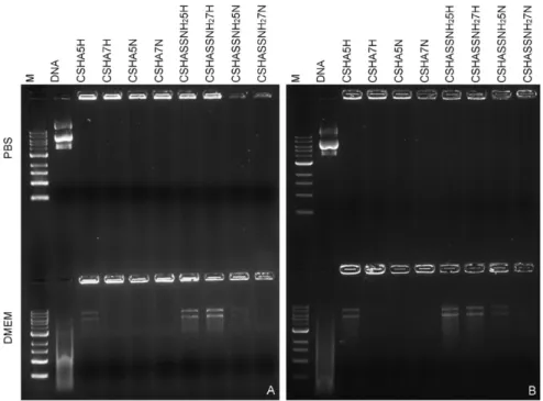
Next, polyplexes were evaluated regarding their DNA complexing capacity and nuclease protection. Polyplexes were capable of efficient DNA complexion and protection against DNAse-induced degradation, as verified by agarose gel retardation assays (Fig. 4). Also, polyplexes remained stable and did not release DNA in detectable amounts, even after incubation for several days at physiological conditions. However, upon incubation in media with serum in some of the formulations it is possible to observe some DNA release (Fig. 5) indicating a less stable polyplex. Nevertheless it is noteworthy that the DNA released by the polyplexes is still undamaged whereas the free DNA used as control is clearly degraded indicating the polyplexes did protect the DNA.
In order to evaluate the effect of a reductive environment on polyplex DNA release, polyplexes were incubated in the presence of glu-tathione and DTT and DNA release was subsequently analyzed by gel
retardation assays. No detectable amount of DNA was released from the polyplexes in either condition, as observed inFig. 6.
3.3. In vitro studies: cytotoxicity and transfection
The cytotoxicity evaluation was performed by an MTT assay to study possible changes in the cytocompatibility of the modified polymer by comparison with the unmodified HA. The results are presented in
Fig. 7, and show no differences both for tested concentrations and polymers.
After verifying that there were no cytotoxicity associated with the polymers, the polyplexes were evaluated regarding their transfection efficiency in two cell lines, HEK293T and ARPE-19, the first are a com-monly used cell line in transfection studies, while the latter are a model of retinal pigment epithelial (RPE) cells, our target in the retina. In ARPE-19 cells no differences between different formulations were found whereas in HEK293T there was statistically lower transfection ef-ficiency when HASSNH2polyplexes were prepared in sodium sulfate
(Fig. 8). Nonetheless, polyplexes prepared in water, with both HA and HASSNH2, had similar transfection results (Figs. 8 and 9).
4. Discussion
4.1. HA was successfully modified with cystamine
Hyaluronic acid was modified by reaction of the carboxylic groups of HA with the amine groups of cystamine dihydrochloride using EDAC ac-tivation. Later, a portion of the modified polymer was treated using an excess amount of the DTT to cleave the disulfide bonds. The resulting product (HASH) was purified by dialysis and isolated after freeze-drying. The average percentage of thiol substitution (approximately 10%) was determined by Ellman's test. This percentage of substitution is in agreement with values in the literature since it is considered that a partial modification (10–20%) is desirable to achieve polyplexes with well-defined sizes[10].
Fig. 3. Size, polydispersity and zeta potential of polyplexes. Statistical differences and their significance are indicated by the star (*) symbol, with ***P b 0.001, ****P b 0.0001, ns — not significant.
Fig. 4. DNA complexation and Dnase I protection in polyplexes were analyzed by 1% agarose gel electrophoresis and DNA visualized with GreenSafe Premium after incubation with Dnase I for 15 min at 37 °C (+) for formulations A) in H2O and B) in Na2SO4.
Fig. 5. DNA complexation in polyplexes was analyzed by 1% agarose gel electrophoresis and DNA visualized with GreenSafe Premium after incubation with PBS or DMEM with 10% FBS for A) 3 and B) 7 days at 37 °C.
Fig. 6. DNA retention by polyplexes in reductive conditions: A) 24 h incubation in the presence of glutathione and glutathione reductase and B) 6 h incubation in the presence of dithio-threitol (DTT); polyplexes in lanes: 1—CSHA5N, 2—CSHA5H, 3—CSHA7N, 4—CSHA7H, 5—CSHASSNH25N, 6—CSHASSNH25H, 7—CSHASSNH27N, 8—CSHASSNH27H.
The presence of two equivalence points in the potentiometric titra-tion curve of HASSNH2further confirmed the success of the
modifica-tion reacmodifica-tion and indicated the pH range more adequate for polyplex preparation. At pH values much below thefirst equivalence point the carboxyl groups are predominantly deprotonated. This may hinder the preparation of polyplexes because in this case carboxyl groups may in-teract with amine groups in the polymer, leaving very few amine groups available to interact with DNA phosphate groups. On the other hand, at pH values exceeding the second equivalence point all amine groups are deprotonated and there are fewer positive charges available to interact with DNA phosphate groups, establishing less electrostatic interactions. Thus it is preferable to prepare polyplexes at pH values between 6 and 9, which is also desirable for later in vitro and in vivo experiments. 4.2. HASSNH2does not produce polyplexes with anionic agents
Initially nanoparticles were evaluated and characterized with re-spect to their size and polydispersion. Several formulations with HASSNH2and different concentrations of anionic agents were tested
(Na2SO4, TPP, HA and κ-carrageenan) with no satisfactory results.
HASSNH2/DNA polyplexes were also tested at various N:P ratios but
complete complexation was not achieved (data not shown). The modi-fied polymer was unable to generate polyplexes by itself or with the ad-dition of other anionic agents, probably due to insufficient electrostatic interactions. This may be related to the degree of substitution of the modified polymer that seems to have a low number of positive charges available to interact. In order to increase the number of positive charges and foster electrostatic interactions, chitosan was added to the formula-tion yielding the results described in the next secformula-tion.
4.3. CSHA and CSHASSNH2polyplexes: a comparison
Previous studies in our lab had shown positive results in terms of transfection with formulations based on chitosan and hyaluronic acid
[15]. In this study we wanted to evaluate and compare the performance of polyplexes prepared with normal unmodified hyaluronic acid and polyplexes prepared with HASSNH2. Different formulations were
pre-pared and thefirst step in their evaluation was to characterize them Fig. 7. Cytotoxicity in HEK293 and ARPE-19 cell line, given as percentage of viable cells after 24 or 72 h incubation with polymer at increasing concentrations (0.01, 0.05 and 0.1 mg/ml per well). Un-challenged cells and cells incubated with latex extracts were used as positive and negative controls of cellular viability.
Fig. 8. Transfection efficiency 72 h post-transfection in HEK293T and ARPE-19 cells as percentage of GFP positive cells. Statistical differences and their significance are indicated by the star (*) symbol, with *Pb 0.05, ****P b 0.001, ns — not significant.
for their size, polydispersity, and zeta potential (Fig. 3). Polyplexes were compared through a statistical multiple comparison analysis. Among the various comparisons, those with higher significance for the inter-pretation of the results were comparisons 1) between formulations with different solvents but the same polymer and ratio; 2) between for-mulations with different ratios while maintaining the same polymer; and 3) between formulations with different polymers but keeping the same ratio and solvent. Given the comparisons described above, in
Fig. 3are represented all comparisons with a significant statistical differ-ence. All other comparisons show no statistical differdiffer-ence.
The different formulations yielded polyplexes with similar sizes ex-cept for CSHASSNH25N and CSHASSNH27N. These two formulations
also presented high polydispersity and a slightly lower surface charge (given by the zeta potential values). The zeta potential can be used to predict the stability of the dispersion so this reduced surface charge might have caused aggregation of the polyplexes hence the large size and polydispersity[16]. Since chitosan is a polycation, a positive zeta potential was expected, which occurred in all cases. However, statistical differences were observed between polyplexes with the same polymer and ratio but different solvents (Na2SO4and H2O). This may be due to
the presence of the negative charges from sulfate anions, which reduce the overall surface charge.
4.4. Polymer modification did not affect cytotoxicity
The cell viability assay main objective was to evaluate the possible cytotoxic effect of the polymers in the cells after DNA release and to as-sess if the modification changed the polymer cytotoxicity profile. HA is known as a biocompatible polymer and studies have shown that it is safe for concentrations up to 1–2% (W/V)[17,18]. Comparing the results obtained for the HA and HASSNH2polymers, no statistical differences
were observed in all four tested situations. This is possibly due to the
fact that the difference in composition of the two polymers is small, since only 10% modification was achieved, and this did not sufficiently alter the toxicity profile of the polymer.
However, after 24 h of incubation HA and HASSNH2showed a
reduc-tion of cell viability in HEK293 cells for all tested concentrareduc-tions when compared to the control. This was not observed for the other tested con-ditions and this could be related to the fact that at 24 h the cells underwent acute exposure while at 72 h it resembles a chronic expo-sure. The fact that the cell viability had decreased slightly in some cases may also be related to the fact that the polymer solutions have not been extensively purified before addition to cells, and so the ob-served cytotoxicity may be due to residual impurities of the modified polymer. Nevertheless, at concentrations to be used in future in vitro and in vivo transfection studies, no cytotoxicity was observed.
4.5. Polyplex stability at physiological conditions varies with temperature and presence of serum
All polyplexes were capable of efficient DNA complexion and protec-tion against DNAse-induced degradaprotec-tion when exposed to DNase I (Fig. 4). This is a critical aspect for all non-viral vectors, since a successful gene therapy vector needs to protect its load until it reaches the target and only unload it at the appropriate time[19,20]. Our stability assay re-sults shown that all polyplexes remained stable up to 7 days at physio-logical temperature and pH (polyplexes incubated with PBS). However, when polyplexes were incubated in DMEM with serum some DNA re-lease was observed, particularly in polyplexes with the modified poly-mer. It is hypothesized that serum components establish electrostatic interactions with the polyplexes, destabilizing them, and causing DNA release. The fact that polyplexes prepared with the modified polymer appear less stable can actually be advantageous since several studies Fig. 9. Fluorescence images of transfected cells 72 h post-transfection. Non-transfected cells and cells transfected with FuGENE HD were used as negative control (C−) and positive control (C +), respectively. Cells transfected with different formulations: A) CSHA5H, B) CSHA5N, C) CSHASSNH25H, D) CSHASSNH25N, E) CSHA7H, F) CSHA7N, G) CSHASSNH25H,
have shown a negative correlation between polyplex stability and trans-fection efficiency[15,21–23].
4.6. Transfection efficiency is formulation-dependent
One of the main objectives of this study was to evaluate if the mod-ification of HA would affect the stability of the polyplexes and conse-quently their transfection efficacy. In our study, different formulations of CSHASSNH2polyplexes showed very similar transfection efficiencies.
Analyzing the results obtained for ARPE-19 and HEK293 cells, the latter had much higher transfection efficiency in all formulations due to their higher permissibility to transfection, associated with their higher mitot-ic rate. Despite the higher transfection values there were only differ-ences in the transfection efficiency between CSHA and CSHASSNH2
polyplexes prepared in sodium sulfate. These polyplexes (CSHASSNH25N and CSHASSNH27N) were expected to have a reduced
efficacy since their size was too large for an efficient and timely cellular internalization and this was indeed confirmed.
Moreover, less stable formulations, with visible DNA release in the stability assay, displayed higher transfection values as expected, based on the aforementioned relation between polyplex stability and transfec-tion efficiency.
Polyplex charge is a key parameter due to its association with toxic-ity and transfection efficiency. Other studies have shown an association between surface charge and increased transfection efficiency as well as a decrease in toxicity[24,25]. Also, cellular uptake is one of the major obstacles for a successful gene delivery strategy and polyplexes are thought to enter the cell via endocytosis in a process that may be depen-dent on polyplex charge[26]. A higher surface charge can facilitate elec-trostatic interactions with the negatively charged proteoglycans in cellular membranes and hence promote polyplex internalization[27]. Our results show a positive correlation between polyplex surface charge and transfection efficiency that is in agreement with the literature. Polyplexes prepared in water displayed nearly a 2-fold increase in sur-face charge that correlated with higher transfection values, irrespective of the polymer used. This data strongly indicates that the preparation method used for polyplex formation is as important as the polymer used.
5. Conclusions
This work had as main objective the characterization of a novel poly-mer, HASSNH2, for subsequent use as a retinal gene therapy vector.
After confirming the success of the modification reaction and determin-ing its extent (~ 10%), several strategies were tested in order to yield polyplexes with adequate characteristics. The resulting polyplexes were capable of effective DNA complexation though their stability at physiological conditions varied. Transfection studies using HEK293 and ARPE-19 cells showed relatively modest transfection efficiency where no differences were observed between polyplexes with modified or unmodified HA. Higher transfection values correlated with lower polyplex stability as well as higher surface charge.
In the future, higher degrees of modification will be pursued by in-troducing changes in the reaction conditions, in order to modulate the stability and DNA release from the polyplexes, to allow further improve-ment of the transfection efficiency of these gene therapy vectors. Acknowledgments
The authors acknowledge thefinancial support of Fundação para a Ciência e Tecnologia (PTDC/SAU-BEB/098475/2008 to Gabriela A. Silva, SFRH/BD/70318/2010 individual fellowship to Ana V. Oliveira, IBB/LA under the project PEst-OE/EQB/LA0023/2013, and
PEst-OE_QUI_UI4023_2011) and the Marie Curie Reintegration Grant (PIRG-GA-2009-249314 to Gabriela A. Silva) under the FP7 program.
References
[1] P.D. Robbins, S.C. Ghivizzani, Viral vectors for gene therapy, Phamacological Therapy 80 (1998) 35–47.
[2] M.S. Al-Dosari, X. Gao, Nonviral gene delivery: principle, limitations, and recent progress, AAPS J. 11 (2009) 671–681.
[3] I.A. Khalil, Uptake pathways and subsequent intracellular trafficking in nonviral gene delivery, Pharmacol. Rev. 58 (2006) 32–45.
[4] D.C. Gorecki, "Dressed-up" naked plasmids: emerging vectors for non-viral gene therapy, Discovery Medicine 6 (2006) 191–197.
[5] I.M. Verma, N. Somia, Gene therapy— promises, problems and prospects, Nature 389 (1997) 4.
[6] E.J. Oh, K. Park, K.S. Kim, J. Kim, J.-A. Yang, J.-H. Kong, et al., Target specific and long-acting delivery of protein, peptide, and nucleotide therapeutics using hyaluronic acid derivatives, J. Control. Release 141 (2010) 2–12.
[7] R.J. Peach, D. Hollenbaugh, I. Stamenkovic, A. Aruffo, Identification of hyaluronic acid binding sites in the extracellular domain of CD44, J. Cell Biol. 122 (1993) 257–264.
[8] A.V. Oliveira, A.P. Silva, D.B. Bitoque, G.A. Silva, A.M. Rosa da Costa, Transfection ef-ficiency of chitosan and thiolated chitosan in retinal pigment epithelium cells: a comparative study, Journal of Pharmacy & Bioallied Sciences 5 (2013) 111–118.
[9] C. Pichon, E. LeCam, B. Guérin, D. Coulaud, E. Delain, P. Midoux, Poly[Lys-(AEDTP)]: a cationic polymer that allows dissociation of pDNA/cationic polymer complexes in a reductive medium and enhances polyfection, Bioconjug. Chem. 13 (2002) 76–82.
[10] R. Jin, L.S. Moreira Teixeira, A. Krouwels, P.J. Dijkstra, C.A. van Blitterswijk, M. Karperien, et al., Synthesis and characterization of hyaluronic acid–poly(ethylene glycol) hydrogels via Michael addition: an injectable biomaterial for cartilage repair, Acta Biomater. 6 (2010) 1968–1977.
[11] H. Lee, S.H. Choi, T.G. Park, Direct visualization of hyaluronic acid polymer chain by self-assembled one-dimensional array of gold nanoparticles, Macromolecules 39 (2006) 23–25.
[12]H. Lee, H. Mok, S. Lee, Y.-K. Oh, T.G. Park, Target-specific intracellular delivery of siRNA using degradable hyaluronic acid nanogels, J. Control. Release 119 (2007) 245–252.
[13] G.L. Ellman, K.D. Courtney, J. Valentino Andres, R.M. Featherstone, A new and rapid colorimetric determination of acetylcholinesterase activity, Biochemical Pharmaco-logical 7 (1961) 88–95.
[14] D.B. Bitoque, S. Simao, A.V. Oliveira, S. Machado, M.R. Duran, E. Lopes, et al., Efficien-cy of RAFT-synthesized PDMAEMA in gene transfer to the retina, Journal of Tissue Engineering and Regenerative Medicine (2014)http://dx.doi.org/10.1002/term. 1909(in press).
[15] Oliveira AV, Bitoque DB, Silva GA. Combining hyaluronic acid with chitosan en-hances gene delivery. J. Nanomater. 2014;2014:9.
[16] Nobbmann U.Protein sizing by light scattering, molecular weight and polydisper-sity. In: Instruments M, editor.http://www.malvern.co.uk/proteins.
[17] D.G. Boeckel, R.S. Shinkai, M.L. Grossi, E.R. Teixeira, In vitro evaluation of cytotoxicity of hyaluronic acid as an extracellular matrix on OFCOL II cells by the MTT assay, Oral Surgery, Oral Medicine, Oral Pathology and Oral Radiology 117 (2012) e423-8.
[18] Becker LC, Bergfeld WF, Belsito DV, Klaassen CD, Jr JGM, Shank RC, et al. Final report of the safety assessment of hyaluronic acid, potassium hyaluronate, and sodium hyaluronate. Int. J. Toxicol. 2009;28:5–67.
[19] S. Danielsen, S. Strand, D.C. de Lange, B.T. Stokke, Glycosaminoglycan destabilization of DNA–chitosan polyplexes for gene delivery depends on chitosan chain length and GAG properties, Biochim. Biophys. Acta 2005 (1721) 44–54.
[20] M. Koping-Hoggard, K.M. Varum, M. Issa, S. Danielsen, B.E. Christensen, B.T. Stokke, et al., Improved chitosan-mediated gene delivery based on easily dissociated chito-san polyplexes of highly defined chitochito-san oligomers, Gene Ther. 11 (2004) 1441–1452.
[21] X. Zhao, S.-B. Yu, F.-L. Wu, Z.-B. Mao, C.-L. Yu, Transfection of primary chondrocytes using chitosan-pEGFP nanoparticles, J. Control. Release 112 (2006) 223–228.
[22] T. Kiang, J. Wen, H.W. Lim, K.W. Leong, The effect of the degree of chitosan deacetylation on the efficiency of gene transfection, Biomaterials 25 (2004) 5293–5301.
[23] N. Duceppe, M. Tabrizian, Factors influencing the transfection efficiency of ultra low molecular weight chitosan/hyaluronic acid nanoparticles, Biomaterials 30 (2009) 2625–2631.
[24] P. Erbacher, S. Zou, T. Bettinger, A.M. Steffan, J.S. Remy, Chitosan-based vector/DNA complexes for gene delivery: biophysical characteristics and transfection ability, Pharm. Res. 15 (1998) 1332–1339.
[25]C. Perez, A. Sanchez, D. Putnam, D. Ting, R. Langer, M.J. Alonso, Poly(lactic acid)– poly(ethylene glycol) nanoparticles as new carriers for the delivery of plasmid DNA, J. Control. Release 75 (2001) 211–224.
[26]T. Ishii, Y. Okahata, T. Sato, Mechanism of cell transfection with plasmid/chitosan complexes, Biochimica et Biophysica Acta (BBA) 1514 (2001) 51–64.
[27] M. Agirre, J. Zarate, G. Puras, E. Ojeda, J.L. Pedraz, Improving transfection efficiency of ultrapure oligochitosan/DNA polyplexes by medium acidification, Drug Delivery 22 (2015) 100–110.