Hélder Ricardo Cardoso Pereira
outubro de 2013
Genotoxic, phytotoxic and protective
effects of Portuguese propolis
UMinho|20 13 Hélder Ricar do Car doso P er eira Genoto xic, ph ytoto
xic and protective ef
fects of P
or
tuguese propolis
Universidade do Minho
Escola de Ciências
Hélder Ricardo Cardoso Pereira
outubro de 2013
Dissertação de Mestrado
Mestrado de Biologia Molecular, Biotecnologia e
Bioempreendedorismo em Plantas
Genotoxic, phytotoxic and protective
effects of Portuguese propolis
Universidade do Minho
Escola de Ciências
Trabalho realizado sob a orientação da
Professora Doutora Ana Cunha
e do
Professor Doutor Rui Oliveira
e da
iv
Acknowledgments/Agradecimentos
My academic studies were only possible with the unwavering support of my family. Their love and caring in both the finest and darkest hours of the past few years were an unsurmountable contribution that lead me to this very moment. I am most thankful.
To my supervisors Ana Cunha, Rui Oliveira and Cristina Aguiar, I thank them for all their patience, availability and understanding that helped me to learn countless invaluable lessons throughout the course of this work.
I would also like to thank my colleagues in the laboratory, Ana Sofia Freitas, Juliana, Walid and Rita Marques for their friendship, help and company.
Finally, I would like to extend the same courtesy to all the staff of the Department of Biology for all assistance in the realization of this work, especially to Cristina Ribeiro for the great help in cytometry procedures.
v
Abstract
Propolis is a mixture produced by bees (Apis mellifera L.) from various plant sources with a diverse chemical composition including many bioactive phenolic compounds and terpenes characteristic of plant secondary metabolism. The bees use propolis mainly as a sealant for cracks in the beehive, but it has been used in human folk medicine for several millennia. A renewed interest in its study emerged in recent years due to the increasing popularity of natural products in foods, beverages and medicines.
Propolis extracts have been associated with varied biological activities, like antioxidant, antimicrobial and anti-inflammatory among others, and indicated for the treatment of several pathologies such as cancer or neurodegenerative diseases. Portuguese propolis, usually regarded as a by-product without any value in apiaries, remains insufficiently studied, hence the need for a better characterization of its properties.
In this study Portuguese propolis samples from different apiaries and different collection dates were selected and ethanolic and n-hexane extracts were prepared and tested using a yeast (Saccharomyces cerevisiae) and in vitro flax (Linum usitatissimum) cultures as models. Viability assays with yeast cells and plant growth analysis were performed to assess extracts toxicity. The yeast model was used to investigate mechanisms of cytotoxicity by the mitochondrial membrane potential-targeting fluorochrome rhodamine 123 approach and to evaluate genotoxicity by the comet assay. The in vitro plant model was used to evaluate extracts effects at the multicellular developmental level as well as in the photosynthetic function by pulse amplitude modulated fluorometry. Both photoinhibitiory and photoprotective potential against oxidative damage of the extracts were investigated using plants grown under high light intensities. Extracts antioxidant properties were also studied in yeast by flow cytometry using the redox-sensitive fluorochrome dichlorofluorescein diacetate.
Our results suggest that ethanolic extracts from Pereiro and Póvoa do Varzim apiaries are among the most toxic for both yeast and plants. They decrease yeast viability and mitochondrial membrane potential and dramatically affect early plant development inhibiting particularly root growth, photosynthetic efficiency and increasing non-photochemical quenching in a dose-dependent manner. Significant genotoxicity was found only in ethanolic extracts from Pereiro collected in 2010, also one of the most toxic in both models tested. However, some ethanolic extracts were also able to revert oxidative-induced damage. They reduced intracellular oxidation induced by hydrogen peroxide in yeast, and greatly recovered the total chlorophyll content reduced by high-light-induced photooxidative stress. Here, also n-hexane extracts were effective.
Globally our results are in line with the antioxidant properties revealed by propolis worldwide but also underline the strong toxicity in different cellular models suggesting that eukaryotic universal mechanisms/structures may be the most affected, possibly mediated by the production of reactive oxygen species. These effects are promising for different applications namely in food industry as preservative, agro-chemical as bioherbicide or pesticide and in pharmaceutical industry as a source of new drugs.
vii
Resumo
O própolis é uma mistura produzida pelas abelhas (Apis mellifera L.) a partir de várias plantas, possuindo uma constituição química diversa que inclui numerosos compostos fenólicos e terpenos bioativos característicos do metabolismo secundário vegetal. O própolis é usado pelas abelhas como selante para fendas na colmeia, mas tem sido também usado na medicina humana há vários milénios. Um interesse renovado no seu estudo surgiu, em tempos recentes, devido à popularidade dos produtos naturais em alimentos, bebidas e medicamentos.
Os extratos de própolis estão associados a atividades biológicas variadas: antioxidante, antimicrobiana, anti-inflamatória e indicados para aplicação na terapia de várias patologias tais como cancro ou doenças neuro-degenerativas. O própolis português, frequentemente considerado um sub-produto sem valor na apicultura nacional, permaneceu até aos nossos dias insuficientemente estudado, daí a necessidade de realizar uma melhor caracterização das suas atividades.
Para estudar os extratos portugueses de própolis foram escolhidas várias amostras de vários apiários e períodos de coleta, que foram testadas numa levedura (Saccharomyces cerevisiae) e na planta do linho in vitro (Linum usitatissimum), utilizados como modelos biológicos. Ensaios de viabilidade com levedura e análise do crescimento do linho foram realizados para avaliar a toxicidade. O modelo levedura foi usado para investigar mecanismos de citoxicidade com o fluorocromo rodamina 123, que tem como alvo a membrana mitocondrial interna, e a genotoxicidade com o ensaio cometa. O modelo vegetal in vitro foi usado para avaliar os efeitos dos extratos ao nível do desenvolvimento multicelular, bem como na função fotossintética por fluorometria. Tanto o potencial fotoinibitório como fotoprotetor dos extratos contra danos oxidativos foram investigados com plantas expostas a intensidades luminosas elevadas. Novamente em levedura, realizou-se citometria de fluxo para aferir a atividade antioxidante com o fluorocromo fluoresceína diacetato e os efeitos ao nível mitocondrial com rodamina 123.
Os resultados obtidos sugerem que os extratos etanólicos de Pereiro e Póvoa são os mais tóxicos quer em levedura quer em linho. Diminuem a viabilidade celular e potencial de membrana mitocondrial em levedura, e afetam dramaticamente o desenvolvimento das plantas, inibindo o crescimento radicular, eficiência fotossintética e aumentando o quenching não fotoquímico. A genotoxicidade do extrato etanólico de Pereiro 2010 foi confirmada por ensaio cometa. Contudo, alguns extratos etanólicos também reverteram danos oxidativos. Reduziram a oxidação intracelular em levedura e recuperaram o teor de clorofilas reduzido por stresse fotooxidativo. Também, aqui os extratos de n-hexano foram eficazes.
Globalmente, os resultados estão em linha com as propriedades antioxidantes reportadas para o própolis, mas também sublinham a forte toxicidade em diferentes modelos, o que sugere que mecanismos universais dos eucariontes possam ser o alvo, possivelmente através da produção de espécies reativas de oxigénio. Estes efeitos são promissores para diferentes aplicações nomeadamente na indústria alimentar como conservante, agroquímica como bioherbicida ou pesticida, ou farmacêutica como fonte de novas substâncias.
ix
Index
Introduction ... 2
1. Propolis: nature, composition and biological activities ... 2
1.1 Nature and composition of propolis ... 2
1.2 Biological activities of propolis ... 3
2. Oxidative stress: sources of reactive oxygen species, damage and repair ... 6
2.1 Reactive oxygen species formation and scavenging mechanisms ... 6
2.2 DNA integrity, toxicity and repair ... 8
3. Plant secondary metabolites and protection against environmental challenges ... 8
3.1 Coping with high light stress conditions in the chloroplast: a tough “iron-arm” ... 8
3.2 Plants chemical wealth: defence against abiotic stress and communication in war and peace .. 11
3.3 Propolis as a blend of allelochemicals ... 13
4. Assessing effects on photosynthesis by pulse amplitude modulated fluorometry ... 14
5. Assessing cellular damage and protection against oxidative stress ... 15
5.1 DNA damage assessment by comet assay ... 15
5.2 Flow cytometry as a mean to assess cell damage and protection in yeast ... 16
6. Biological models to study propolis biological activities ... 16
6.1 Yeast as a model of genotoxicity ... 17
6.2 In vitro cultures of Linum usitatissimum as a plant versatile multicellular platform ... 18
7. Objectives and scope of this work ... 18
Materials and Methods ... 20
1. Preparation of propolis extracts ... 20
2. Establishment of in vitro cultures of flax (Linum usitatissimum L.) ... 21
3. Plant growth and root microscopy analyses ... 21
4. Photochemical efficiency of PSII by PAM fluorometry ... 22
5. Chlorophylls and carotenoids quantification ... 23
6. Yeast strain, culture media and growth conditions ... 23
7. Yeast viability assays by drop test ... 24
8. Yeast intracellular oxidation state assessment by flow cytometry ... 24
9. Yeast mitochondrial membrane potential assessment by flow cytometry ... 25
10. DNA damage assessment by comet assay... 25
x
Results ... 28
1. Effects of propolis extracts on yeast cell growth and viability ... 28
2. Propolis extracts effects on yeast DNA integrity ... 29
3. Effects of propolis extracts on intracellular oxidation status of yeast cells ... 30
4. Effects of propolis extracts on mitochondrial function and membrane potential ... 33
5. Propolis extracts effects on plant growth and photosynthetic activity... 34
6. Effects of propolis extracts in plants exposed to light induced photooxidative damage ... 37
Discussion ... 48
1. Propolis ethanolic extracts are cytotoxic decreasing yeast cell growth and viability ... 49
2. Propolis extracts from Pereiro samples are genotoxic inducing DNA damage on yeast ... 49
3. Propolis extracts promote mitochondrial damage in yeasts induced by reactive oxygen species ... 50
4. Propolis ethanolic extracts have antioxidant activity in yeast cells decreasing the basal intracellular oxidation status but also the high oxidation levels induced by hydrogen peroxide ... 50
5. Propolis extracts can inhibit plant growth and photosynthesis ... 51
Final Remarks and Future Perspectives ... 57
References: ... 59
2
Introduction
1. Propolis: nature, composition and biological activities
1.1 Nature and composition of propolis
Propolis is a natural mixture produced by bees (Apis mellifera L.) from various plants’ exudates, being abundant in resins and waxes with a myriad of compounds such as flavonoids. Characteristically, it is hard and brittle when cold but soft, pliable, and very sticky when warm, being also known as bee glue. This product is employed mainly as a sealant material on the beehive but also as an antiseptic. The very origin of the word propolis stems from the Greek language words pro – which means for or in defence of – and polis – city, in this particular case the beehive (Sforcin, 2007).
Besides this natural usage by bees, propolis has been used as medicine for over several millennia - with records dating from the ancient civilisations of Egyptians, Arabs or Greeks -, but in recent years a renewed scientific interest emerged with many studies focusing its biological activities, mainly envisaging pharmacological but also other applications, partially due to the fact that the majority of its components are considered safe substances for human ingestion, as most of its components are natural constituents of food (Grange and Davey, 1990; Lofty, 2006; Sforcin, 2007; Falcão et al., 2010). For instance, given its known antibacterial and antioxidant activities, the use of propolis as a food preserver or as a food supplement to improve human health has already been tested (Banskota et al., 2002; Moreira et al., 2008; Fokt et al., 2010).
Propolis’ compounds originate from three main sources: plant exudates, bee's metabolic secretions and other materials added during propolis elaboration (Marcucci, 1995). Bees mix resins with their own salivary secretion, which contains ß-glucosidases that hydrolyse glycosyl flavonoids into flavonoid aglycones (Pereira et al., 2002). Propolis composition is extensive and diverse, varying geographically with the flora from which bees collect the raw materials, the time period when such collections are made and also with the different behavioural patterns exhibited by different bee communities. In the Northern Hemisphere, bees collect in the end of spring, during the summer and in the beginning of autumn, while in some countries of the Southern Hemisphere such as Brazil the collection is made throughout the year (Bankova et al., 1998). In Europe, poplar (Populus sp.) buds are the main source for the bees, hence the “poplar type” propolis.
3
Hundreds of compounds have been identified in propolis worldwide and range from polyphenols, phenolic aldehydes, sesquiterpenes, quinones, coumarins, amino acids and steroids to inorganic compounds. Propolis from temperate regions is rich in galangin, chrysin, pinocembrin, caffeic acid, ferulic acid and cinnamic acid (Marcucci, 1995; Falcão et al., 2010). Other compounds such as isosakuranetin or kaempferide are characteristic of “poplar type” samples, but not typically present in large proportions (Marcucci, 1995; Park et al., 2002; Falcão et al., 2010). Portuguese propolis contains the most common compounds of the temperate zones already mentioned, but also several other phenolic components such as methylated, sterified or hydroxylated derivatives of already described flavonoids, rare forms of pinocembrin and p-coumaric ester derivative dimers (Falcão et al., 2010). Generally, flavonoids from the resins and other phenolic compounds constitute approximately 50% of the mixture, while beeswax, pollen, and others represent, respectively, 30, 10 and 5% (Grange and Davey, 1990; Lofty, 2006; Sforcin et al., 2007; Falcão et al., 2010).
1.2 Biological activities of propolis
Due to the very complex chemical composition of propolis samples, different solvents must be used to extract and isolate the fractions/compounds before testing for biological activities. Propolis extracts, also known as balsams, are usually obtained with ethanol, methanol, dimethyl sulfoxide (DMSO), n-hexane, glycerol or even water, though many non-standard solvents can be used as well (Marcucci, 1995; Cunha et al., 2004; Najafi et al., 2007; Fokt et al., 2010).
Antibacterial activity of propolis extracts has been reported against a wide range of Gram-positive bacterial strains of cocci or rods like Streptococcus or Bacillus, but only limited activity against Gram-negative bacilli (Grange and Davey, 1990; Mirzoeva et al., 1997; Menezes et al. 1997). Growth inhibition of Staphylococcus aureus or Escherichia coli was described, albeit less significant for the latter. Antimicrobial activity varies depending on the type of propolis, the dosage and the solvents used (Lofty, 2006). Ethanolic extracts were found to be effective against anaerobic Gram-positive bacteria (Mirzoeva et al., 1997), being this activity attributed to their polyphenol content. Propolis could inhibit bacterial growth by preventing cell division, disorganizing the cytoplasmic membrane and cell wall, and by inhibiting protein synthesis. What is interesting is that this plethora of actions does not match a single classic antibiotic mode of action (Grange and Davey, 1990; Takasi et al., 1994; Lofty, 2006), foretelling a wide range of new antimicrobial molecules to unveil.
4
Virucidal activity of propolis extracts on several DNA and RNA viruses such as Herpes simplex type 1 and 2 (enveloped), poliovirus type 2 (non-enveloped), adenovirus type 2 (non-enveloped) were demonstrated in vitro, although the latter was less susceptible than herpes and polioviruses. This virucidal activity has been attributed to the high content in flavonoids like galangin and chrysin (Amoros et al., 1992).
Propolis has also demonstrated antifungal activities with effectiveness on Candida yeast strains, particularly on Candida albicans (Ota et al., 2001; Lofty, 2006). Other species proved also to be susceptible, as Penicillium notatum or Aspergillus flavus growth was inhibited with concentrations of propolis extracts of 15 to 30 mg/ml (Pepeljnajak, 1982). Regarding antiprotozoal activity, it was reported that ethanolic and DMSO extracts of propolis were active against Trypanosoma cruzi (Pepeljnjak, 1982; Higashi and de Castro, 1995; Lofty, 2006).
Ethanolic extracts of propolis could also exhibit anti-inflammatory activity, which might be promising to treat diseases caused by chronic inflammation. Caffeic acid phenethyl ester (CAPE), a component of propolis, was shown to have this property. This phenolic compound is a potent inhibitor of early and late events of T-cell activation and their immune response in inflammatory processes (Park and Kahng, 1999; Lofty, 2006).
Extracts from Brazilian propolis samples have shown cytotoxicity and the ability to inhibit the proliferation of human malignant tumour cells, possibly due to the compound artepillin C (3,5-diprenyl-4-hydroxycinnamic acid) (Kimoto et al., 1998). The cytotoxic effects of artepillin C were most noticeable in carcinoma and malignant melanoma by apoptosis, abortive mitosis and massive necrosis, as suggested by histological observations. Besides tumour growth suppression, the immune system was activated, with increased numbers of helper T cells. Propolis and artepillin C also appear to inhibit lipid peroxidation (Kimoto et al., 1998, 2001). Besides artepillin C, other cinnamic acid derivatives, such as baccharin or drupanin, a common and abundant component of bee propolis, induce tumour cell death with less genotoxicity to haemopoetic cells than normal anticancer drugs (Lofty, 2006). Portuguese propolis extracts have also shown antitumoral activities (Carvalho, 2013). There is some evidence that propolis could block tumour angiogenesis. CAPE, already mentioned for its anti-inflammatory properties, could also be effective in tumour angiogenesis inhibition as was demonstrated by studies with chick embryo chorioallantoic membrane as an animal model (Song et al., 2002).
Propolis has also been described as a protective agent of liver, heart and brain (Irmak et al., 2003; Fuliang et al., 2005; Lofty, 2006). Regarding liver protection, effectiveness was assessed with rat hepatocytes pre-treated with propolis extract and on which hepatotoxicity was induced by acetaminophen, yielding a decrease in mortality and severity of hepatocyte necrosis (Seo et al., 2003).
5
It was also shown that propolis is able to inhibit the action of the enzyme hyaluronidase, which degrades hyaluronic acid, a major constituent of the extracellular matrix that contributes to cell proliferation (Starr and Engleberg, 2006). This enzyme has a role in infection by some pathogens like Staphylococcus aureus, being produced by the pathogen to obtain hyaluronic acid that is used as carbon source (Starr and Engleberg, 2006; Moreira et al., 2008).
Several human pathologies result from an increased level of reactive oxygen species (ROS) and propolis extracts as well as some of its compounds have already been reported to have a role in reducing ROS levels and thus on the related disorder symptoms (Ilhan et al., 1999; Lofty, 2006). For instance, oxygen-derived free radicals have been implicated in the pathogenesis of cerebral injury after ischaemia – a restriction of blood supply to tissues that causes oxygen deprivation. The subsequent restoration of circulation can cause additional damage on ischaemic tissues as the sudden supply of oxygen leads to the formation of many free radicals, and CAPE, which also possess antioxidant properties and can act as free radical scavenger, demonstrated relevant activity in alleviating the symptoms of this disease (Lofty, 2006). Also, cardiomyopathy is the consequence of oxidative stress through the action of free radicals, when in the presence of the cancer treatment drug doxorubicin. The effects of propolis on doxorubicin-induced cardiomyopathy were studied in rats by intraperitoneal administration and it was observed that propolis had a protective effect towards the cardiac muscle comparable to that of rutin, a well-know cardioprotective flavonoid (Lofty, 2006).
6
2. Oxidative stress: sources of reactive oxygen species, damage and repair
2.1 Reactive oxygen species formation and scavenging mechanisms
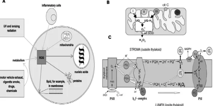
Reactive oxygen species (ROS) are formed through the reduction of molecular oxygen to water by the transfer of four electrons, a process that all aerobic organisms perform during the accumulation of energy through the electron transport chain or metabolism of exogenous compounds. Many environmental factors induce ROS formation in cells (Fig. 1A). The mitochondria (Fig. 1B) and chloroplast (Fig. 1C) are the main sources of ROS in eukaryotic cells, due to the electron transport chain of oxidative phosphorylation and of photosynthesis, respectively. Superoxide anion (O2-), hydrogen peroxide (H2O2
)
and hydroxyl radicals (.OH) are examples of common and noxious ROS that affect cellular components like nucleic acids, proteins and lipids, inflicting severe damage to cells and to the organism. These ROS are known causes of degenerative diseases such as cancer, epilepsy or neurodegenerative diseases and integral part of the biological ageing process (Harman, 1991, 2006; Malinska, 2010; Mubarakshina and Ivanov, 2010).C
Figure 1. Potential sources and targets for reactive oxygen species in animal cells. A) Enviromental factors and
intracellular targets of ROS; B) Oxidative phosphorylation on mitochondria; C) Electron transport chain on chloroplast (Cooke and Evans, 2013; Novo and Parola, 2005; Mubarakshina and Ivanov, 2010).
A B
7
The presence of intracellular H2O2 can oxidise cysteine and methionine residues of iron-sulfur proteins, leading to the formation of free radicals by the Fenton reaction (Fig. 2) catalysed by transition metals such as iron or copper.
𝑀𝑛++ 𝐻
2𝑂2 → 𝑀(𝑛+1)++ 𝑂̇𝐻 + 𝑂𝐻−
The resulting oxidised metals can be re-reduced by molecular oxygen free radical on the Haber-Weiss reaction so that these metals can be reused once again in the Fenton reaction. The conjugation of hydrogen peroxide, molecular oxygen radicals and transition metals make for a very dangerous predicament for the cells. The formation of ROS like superoxide or singlet oxygen in the mitochondria and in the chloroplast are well-documented facts (Novo and Parola, 2005; Malinska, 2010). Singlet oxygen particularly is produced in the photosystem II of the chloroplast (Pospíšil, 2012). Enzymes such as catalases or peroxidases catalyse the scavenging of hydrogen peroxide, being one important line of defence on living organisms. Cells need to maintain a reducing intracellular state, in spite of a largely oxidising extracellular environment, allowing them to perform many functions such as the proper folding of proteins (Smirnoff, 2005; Drakulic et al., 2005).
In a state of homeostasis, ROS production is counter-balanced by enzymatic processes, as above mentioned, and antioxidant compounds present in the cells. These molecules are in balance, so it must be accepted that a “normal” level of ROS always occurs causing minimal damage. In the event of a loss of homeostasis caused by increased free radical production or failure of antioxidant defences, it is said that cells are under oxidative stress and subject to cellular damage (Izawa et al., 1995; Munné-Bosh and Alegre, 2003; Smirnoff, 2005; Collins, 2009).
While ROS molecules are undoubtedly harmful in high concentrations, they are also needed at low concentrations as inter- and intracellular signalling molecules. The ROS signal transduction network is an evolutionary conserved pathway on all aerobic organisms. Molecules such as singlet oxygen, superoxide and hydrogen peroxide act as signal transductors to control a large array of biological processes ranging from the regulation of development and growth to responses to biotic and/or abiotic stimuli. In plants, ROS signaling was shown to be involved in processes of seed after-ripening, lignification, root hair formation, closure of stomata or programmed cell death. The key to this phenomenon seems to be the cells ability to maintain a low steady-state level of ROS molecules, while allowing for its accumulation in specific subcellular locations
Figure 2. The generic reaction mechanism as first described by Fenton, on which transition metals convert hydrogen
8
that act as signals (Izawa et al., 1995; Munné-Bosh and Alegre, 2003; Smirnoff, 2005; Collins, 2009; Suzuki et al., 2011).
2.2 DNA integrity, toxicity and repair
DNA integrity is essential for the viable and normal function of organisms. Numerous endogenous and exogenous agents such as ROS molecules produced in the electron transport chain in the mitochondria and chloroplasts, ionizing radiation, exogenous toxic compounds or inflammation compromise that integrity and are sources of genotoxicity. A popular hypothesis, albeit not unanimously accepted, is that oxidative stress is directly connected to the ageing process (Harman, 1991). This postulate assumes that our cells accumulate damage in its constituent biomolecules slowly over time causing organs to deteriorate over the years, in an unavoidable obsolescence. DNA can be damaged through single and double-stranded breaks, base and sugar replacements, apurinic/apyrimidinic lesions or DNA-proteins binding (Collins, 2009). Cells have some defensive mechanisms, including superoxide dismutase, catalase, several peroxidases and antioxidants such as ascorbate, tocopherol, uric acid, β-carotene and glutathione that allow for the elimination of ROS deleterious effects (Izawa et al., 1995).
3. Plant secondary metabolites and protection against environmental challenges
3.1 Coping with high light stress conditions in the chloroplast: a tough “iron-arm”
Plants are exposed to a wide array of environmental stress conditions, ranging from low water availability, temperature fluctuations, nutrient deprivation and high light exposures. These stresses lead up to an imbalance between the amount of reactive oxygen species and the antioxidant defences (Smirnoff, 1993; Pastori and Foyer, 2002; Xiong et al., 2002).
The photosynthethic pathway allows the plant to gather sunlight and generate enough chemical energy to proceed with overall thermodynamically unfavourable reactions of the Calvin cycle, involved in sugar synthesis from CO2. Molecular diatomic oxygen is a by-product of photosynthesis that diffuses passively between cells and leaves source organs through stomata. However, when exposed to light, the O2
9
concentrations may reach high levels and an imbalance between the electron transfer flow and the recycling rate of photochemical products in Calvin cycle renders the chloroplasts particularly susceptible to oxidative damage by ROS formation (Smirnoff, 2005). In addition, exposure to higher than normal light intensities may lead to higher intracellular amounts of ROS due to increased rates of molecular oxygen photoreduction in photosystem II but also to and increased flux of H2O2 in peroxisomes via photorespiration (Niyogi, 1999; Mittler, 2002; Heldt, 2005; Smirnoff, 2005; Pospíšil, 2012).
Plants have several mechanisms to cope with this variable oxidative challenge conditions and maintain homeostasis, but when the rate of light absorption far exceeds the capacity of their photosynthetic apparatus, this frequently leads to the repression of photosynthesis in a process known as photoinhibition.
Indeed, while ROS are needed at low concentrations for cell signaling, at higher concentrations they cause severe damage at several organization levels of the plant cells such as the chloroplasts. Apart from the enzymatic (superoxide dismutase, catalase and peroxidases) and non-enzymatic antioxidant compounds already mentioned above, the plant cells have other mechanisms that also protect the chloroplast from oxidative damage like the xanthophyll cycle (XC) and photorespiration (Doke, 1997; Munné-Bosh and Alegre,. 2003). Carotenoids, tocopherol, ascorbate and glutathione help maintaining the integrity of the photosynthetic membranes under oxidative stress (Havaux, 1998; Smirnoff and Wheeler, 2000; Munné-Bosch and Alegre, 2003). Tocopherol and β-carotene have also been shown to act in singlet oxygen scavenging in lipid membranes and in photosystem II protection (Munné-Bosch and Alegre, 2003).
The de-epoxidised carotenoids of the XC zeaxanthin and antheraxanthin coupled with a low thylakoid lumen pH, which results from high light conditions, and a minor light harvesting complex protein named PsbS, can act to dissipate the excessive electron energy from photosynthesis as heat, in a process known as non-photochemical quenching (NPQ). The XC consists on the interconversion of violaxanthin, antheraxanthin and zeaxanthin during the period of high light exposure when the violaxanthin accumulated on leaves starts to be converted into the other two (Fig.3) (Young et al. 1997; Smirnoff, 2005; Baker, 2008).
10
Fig. 3 Interconversion of xanthophyll cycle plant carotenoids, and the mechanism of heat dissipation by
non-photochemical quenching (Baker, 2008).
A major cause for photoinhibition is the overexcitation of photosystem II reaction centre, which leads chlorophyll molecules to attain a triplet state (3Chl.) and resulting in the formation of singlet oxygen (1O
2) (Fig.4). The triplet excitation energy of some chlorophyls at specific sites of the light harvesting complex can be effectively quenched by lutein, another carotenoid, but others are not and in the presence of molecular oxygen contribute to singlet oxygen formation. Some carotenoids are able to revert the triplet state of chlorophyll and the singlet state of oxygen to their ground fundamental state, forming a triplet carotenoid that dissipates its energy as heat via non-photochemical quenching.
11
Plants have other mechanisms to cope with light stress besides scavenging, dissipating and repairing. The avoidance of excessive photosynthetic-dependent ROS production and thence oxidative damage is a way to escape stress effects. The well-known chloroplast movement observed under high light conditions, which consists of the relocation of chloroplast from the cell surface to the side cell walls parallel to sunbeams (Heldt, 2005), is an important example.
3.2 Plants chemical wealth: defence against abiotic stress and communication in war and peace
Terpenes, or isoprenoids, are a major class of secondary metabolites in plants. Terpenes are highly diverse, both in functions and activities as well as in their chemical structures, but they share the same method of sequential assembly from a couple building blocks, each of which consists of a branched five-carbon atoms chain. The two building blocks are the interconvertible isopentenyl pyrophosphate (IPP) and
B
A
A
A
Figure 4. Singlet oxygen generation in the light harvesting complex (A) and the reaction center of PSII (B) (Pospíšil,
12
dimethylallyl pyrophosphate (DMAPP), which are condensed together in a sequential way through enzymes called prenyltransferases. The major families of terpenes are mono-, di-, tri- and sesquiterpenes. All the carotenoids are in fact diterpenes, synthesised from building blocks of the precursor molecule geranyl-geranyl pyrophosphate (GGPP) and the sterols like cholesterol are triterpenes (Humphrey and Beale, 2006; Crozier et al., 2006).
Limonoid triterpenes have biological activities against insects and are used to develop commercial insecticides (Isman et al., 1997). Terpenes are constituents of many plant species essential oils with reported antimicrobial activities (Delaquis et al., 2001). There are also terpenes with anti-cancer potential, for instance well know anti-cancer drug, paclitaxel, is a terpene metabolite (Humphrey and Beale, 2006).
In plants, the phenylpropanoid metabolism constitutes a major pathway of secondary metabolism leading to the synthesis of phenolic compounds. These compounds are a wide group of molecules characterised for possessing at least one aromatic ring with one or more hydroxyl groups attached. Phenolic compounds can be classified in two groups, flavonoids or non-flavonoids. Flavonoids are a family of diverse, over 9000 compounds, which comprise a large portion of the secondary metabolism of plants. The main classes of flavonoids include the flavones, flavonols, isoflavones, chalcones, coumarins and anthocyanidins. In plants, flavonoids could have diverse roles such as UV protection, pigmentation, in the stimulation of nitrogen fixing and disease resistance. Flavonoids can act as singlet oxygen quenchers and although mainly distributed on the leaf surface and epidermal cells, they are also present in chloroplasts (Crozier et al., 2006; Hernández et al., 2008). The phenylpropanoid pathway is frequently induced by stress conditions of a wide range, such as high light radiation, high temperature or pathogens. Many phenolic compounds of the plant secondary metabolism such as caffeic acid, kaempferol or apigenin have reported antioxidant activities (Zheng and Wang, 2001). The production of ROS is also upregulated at periods of stress and phenylpropanoids such as coumarins, flavonoids, phenolic acids or stilbenes have been for a long time associated with several stress related function, most remarkably protection against photoinhibition and scavenging of ROS (Young et al., 1997; Li et al., 2000; Smirnoff, 2005; Crozier et al., 2006). Some flavonoids like galangin and 7-hydroxyflavanone can act also as pro-oxidants besides their superoxide scavenging activities (Dewick, 2002).
13 3.3 Propolis as a blend of allelochemicals
Plant allelochemicals, plant secondary metabolites with communication roles between species, include an array of compounds such as phenols, terpenes, alkaloids, quinones, saponines, tannins, fatty acids or peptides (Crozier et al., 2006). Allelopathy, an important ecological phenomenon, consists on the production and release of allelochemicals by certain plant species that have effects on other plants species physiology and development or even in other type of organisms. Some plants produce phenolics compounds such as rutin or chlorogenic acid that are toxic to certain insects’ larvae that predate on plants foliage (Isman and Duffey, 1982, Medeiros, 1990; Delachiave et al., 1999). Many plant phenolic compounds like p-hydroxyacetophenone, p-hydroxybenzoic acid, catechol and protocatechuic acid have allelopathic effects against mycorrhizal fungi such as Laccaria laccata and Cenococcum graniforme (Pellissier, 1993). Also, many plant species used as source of raw materials by bees to produce propolis have reported biological activities and allelopathic effects. Propolis extracts from Bulgaria, whose plant source is Populus nigra similar to Portuguese propolis display antimicrobial activities against fungi and bacteria (Salomão et al., 2004). It is known, for instance, that Brazilian propolis is made from exudates of Baccharis dracunculifolia, a plant with recognised allelopathic potential against other plant species (Gusman, 2008).
Plant allelochemicals are likely present in the composition of propolis, and considering the biological activities attributed to secondary metabolites like alkaloids, phenolic compounds and terpenes, namely the antioxidant potential, it is reasonable to hypothesize a protective effect of propolis against induced oxidative stress on living cells. Indeed, some phenolic compounds present in propolis extracts are known to play in vivo the role in maintaining homeostasis and counter oxidative stress damage, thus explaining the antioxidant properties of those propolis samples (Kasai, 2002; Humphrey and Beale, 2006; Collins, 2009).
As referred before, many plant secondary metabolites like phenylpropanoids or terpenes have toxic activities that protect the plant against pathogenic microorganisms or herbivores. These compounds can act as natural pesticides, which in some plants can account for 10% of its dried biomass. In response to microbial infections plants synthesise substances called phytoalexins, which comprise many of the above-discussed chemical classes such as flavonoids, isoprenoids or stilbenes. Many of these phytoalexins like psoralen, xanthotoxin or bergaptol have demonstrated antibiotic activity against a broad spectrum of pathogenic fungi and bacteria. Psoralens in particular have phototoxic effects, that is, their toxicity is activated by UV light exposure (Pathak and Fitzpatrick, 1992; Manderfeld et al., 1997; Hendt, 2005).
14
Although there is a huge amount of accumulated knowledge about phytochemicals and related bioactivities, about the wide infochemical web related to species-species communication at the ecological level, and many studies focusing on propolis composition and bioactivities, we believe that there are many interesting biological and other properties still to uncover. Two examples are propolis potential genotoxicity and genoprotection and phytotoxicity. To address these topics two powerful techniques were used in this work, and in this sense some detailed information will be provided in the next two sections.
4. Assessing effects on photosynthesis by pulse amplitude modulated fluorometry
Light energy absorbed by the leaves, and ultimately by the photoreceptors of photosystems I and II, remain only very transiently in the excited pigments. There are three main competitive pathways by which this excitation energy will decay: most of it is relayed to the electron transport chains (the photochemical pathway or photochemical quenching), some can be dissipated as heat (the non-photochemical quenching) or emitted back as fluorescence, as light causes the transient closure of some PS reaction centres and hence limits the photochemical pathway (White and Critchley, 1999; Baker, 2008). The proportion of energy that is channelled by each of the pathways will depend on the light conditions, light adaptation status of the plant and stress conditions that may affect different aspects of the photosynthetic machinery.
The pulse amplitude modulated (PAM) fluorometry is a very useful method to study the effect of different factors - environmental, biotic, abiotic, extracts, compounds - on photosynthesis. While being rapid and very sensitive, it can be also non-intrusive and used on intact leaves as well as isolated chloroplasts or subchloroplast particles (White and Critchley, 1999; Schreiber et al., 1995), but virtually in all photoautotrophic organisms or samples. Concerning only plant photosynthesis, this technique has been widely used to evaluate the effects of many types of stresses, like water deficit in crop species (Carvalho et al., 2011) or light stress (Dixon and Paiva, 1995; Hutin et al., 2003), but also to study non-foliar systems (Breia et al., 2013).
The PAM technique is based on the analysis of chlorophyll fluorescence yields of photosystem II (PSII) under different experimental controlled conditions, and makes use of short light saturation pulses (typically less than 1 s at several thousand µmol of photons m-2 s-1) to transiently drive a very high proportion of PSII reaction centres to closure (virtually all), with QA (the primary quinone electron acceptor of PSII) at its most reduced state, thus unabling the photochemical pathway (Baker, 2008; Papagergiou and Govindjee, 2004)
15
what will result in maximum fluorescence yield. This method allows the determination of many important photochemical and non-photochemical parameters, and hence to take conclusions about the photosynthesis status. The most commonly used parameters are the maximum quantum yield of PSII (Fv/Fm), measured in dark-adapted samples, and the effective quantum yield of PSII (ΦII) and non-photochemical quenching (NPQ) in samples under light conditions. Fv/Fm represents the intrinsic or maximum quantum yield of photosystem II (PSII) measured in dark-adapted samples; the effective quantum yield (ΦII) of PSII represents the efficiency by which the absorbed energy is actually channeled to photochemistry and measured under light-adapted samples. The non-photochemical quenching (NPQ) represents the absorbed light energy that is dissipated by other processes (like in the form of heat) than photosynthesis.
Considering all this characteristics, this technique will be used to assess the effects of propolis extracts added to the culture medium in the photochemical and non-photochemical capacities of in vitro grown plantlets of flax.
Carotenoids have essential roles in photosynthesis as they contribute to light harvesting and are associated with the photosystem II reaction centre (Smirnoff, 2005). Carotenoids can contribute to photoprotection and act as antioxidants by quenching singlet oxygen and also by reacting with superoxide and other free radicals. If carotenoid synthesis is somehow inhibited or the degradation accelerated, the chloroplast undergoes rapid photo-oxidative damage. Zeaxanthin is a carotenoid noteworthy for its involvement in the non-photochemical quenching of excitation energy from PSII, in which the excess energy is transferred to zeaxanthin and freed as heat (Smirnoff, 2005; Havaux and Nyogi, 1999; Barry et al., 1990).
5. Assessing cellular damage and protection against oxidative stress
5.1 DNA damage assessment by comet assay
To assess DNA damage in individual cells a technique such as single-cell gel electrophoresis can be employed. Single-cell gel electrophoresis, also known as the comet assay or microgel electrophoresis, is a widely used method for assessing damage of DNA, hence its usefulness for genotoxicity tests or DNA damage and repair studies amongst many others. Östling and Johanson reported this assay in 1984 as a technique for the direct visualisation of DNA damage in individual cells. Cells are embedded in an agarose microgel, lysed, electrophoresed and stained with an appropriate DNA binding fluorochrome. The electric current makes the negatively charged supercoiled DNA molecules migrate and the relaxation imposed by single strand breaks or
16
fragmentation by double strand breaks lead to higher displacement of DNA towards the anode. The cells with the staining dye and a clear one-directional path of leaked DNA outside resemble a comet and its tail and thus prompted the naming of the assay. The average tail length is taken as a measure for genotoxic damage. The original Östling and Johanson method, performed under neutral conditions, had limitations related with the sensitivity for single-stranded DNA breaks on supercoils only detecting double-strand breaks, but the assay was easily adapted by Singh et al. in 1988 to more stringent alkaline lysing conditions so that it also allows for the detection of the single-strand DNA breaks, by relaxing and unwinding the supercoils (Fairbairn et al., 1995; Menke et al., 2000; Collins, 2009).
The comet assay is nowadays ubiquitous in genotoxicity testing. It is simple and easy to perform and allows for rapid and visual assessment of DNA damage in individual cells (Dhawan et al., 2009).
5.2 Flow cytometry as a mean to assess cell damage and protection in yeast
Flow cytometry is a laser-based technique that allows for the detection and counting of cells on suspension by using several fluorescent labels, the fluorochromes. Flow cytometry techniques offer several advantages over traditional culture-based techniques to quantify cells, the latter being often time consuming and not suited for non-culturable microorganisms (Veal et al. 2000).
Perhaps the most attractive proposition of flow cytometry cell measurement is the ability to obtain real-time in vivo information about the microorganisms. The two fluorochromes used in this work (fluorescein diacetate and rhodamine 123) for cell staining will allow for both the quantification of propolis-induced damage and to assess protective effects on yeast cells (Veal et al., 2000) after an imposed oxidative challenge.
6. Biological models to study propolis biological activities
To evaluate a range of biological effects of propolis extracts at the developmental, physiological, cellular, and DNA level, two different eukaryotic models were used: the unicellular yeast Saccharomyces cerevisiae and the plant species Linum usitatissimum (flax). The use of different models allows comparing responses and also identifying specific effects and more transversally fundamental modes of action.
17 6.1 Yeast as a model of genotoxicity
The potential genotoxicity of a drug candidate such as propolis extracts or compounds can be assessed using a simple organism like yeast as a model. Saccharomyces cerevisiae is a prime example of a unicellular eukaryote that shares the essential cellular pathways with even the higher multicellular eukaryotes (Seioghe and Wolfe, 1999; Liu et al., 2008) and thus is widely used to study complex physiological and molecular processes on metazoan cells.
Major benefits of S. cerevisiae include the rapid growth and tractability, being cheap and simple to maintain in culture. Adding to that, the availability of the full genome sequence of S. cerevisiae makes this organism a very interesting proposition for various fields of biology (Pabla et al., 2006; Grzelak et al., 2006), namely in the investigation of drug effects on particular molecular, metabolic or other cellular mechanisms.
Yeast cells adapt their growth and development depending on the nutrients available. Yeast can grow on a variety of compounds as carbon sources such as glucose, fructose, sucrose, raffinose or trehalose. However, yeast cells have a preference for glucose or fructose over all other mono-, di- or trisaccharides, and prefer fermentable carbon sources like all the aforementioned over nonfermentable compounds like ethanol, glycerol or acetate. These nonfermentable energy sources are not usable in anaerobic processes, only being catabolised by oxidative phosphorylation in the mitochondria. This conditional nutrient preference is regulated by several key enzymes in glycolysis and gluconeogenesis, in which glucose act as the repressor of the transcription of genes needed for less favourable energy sources catabolism. This repression by glucose is the basis of the Crabtree effect, allowing for the distinction between species that aerobically perform fermentation, such as Saccharomyces cerevisiae, from those that do not, like Kluyveromyces sp. (Broach, 2012). In this work the yeast model will be used to assess propolis effects on cytotoxicity, DNA damage and protection and also on cell redox status.
18
6.2 In vitro cultures of Linum usitatissimum as a plant versatile multicellular platform
Linum usitatissimum L., commonly known as flax, is a widely used, easily to cultivate angiosperm. This species is cultivated worldwide for the production of oil and fibre (Chakravarty and Srivastava, 1996; Millam et al., 2005). Its high regeneration rate in vitro, short life cycle and small genome size are key attributes for researchers.
In vitro cultures of flax have been well established in the late 1990s (Cunha and Fernandes-Ferreira, 1996, 1999) and recently this plant system was successfully used in the evaluation of the effects of propolis extracts on different physiological and early plant developmental aspects (Pereira, 2008; Paulo, 2009; Oliveira, 2011; Amorim, 2011; Apresentação, 2012; Carvalho, 2012). Major benefits of this biological plant platform include its versatility – a wide variety of types of cultures (suspensions, calli, shoot and root cultures, plantlets) allowing to evaluate the effects of extracts and compounds at different organizational levels (from sub-cellular to plant developmental); sensitivity; the robustness and consistency of responses; as well as the ease and economy of culture maintenance. In this work, this model will be used to assess propolis effects at the plant developmental level and also in the photosynthetic function, evaluating both the inhibitory properties and protective effects against oxidative stress induced by high light.
7. Objectives and scope of this work
There is a renewed interest in natural products for their potential applications on several industries (pharmaceutical, cosmetic, agrochemical), which have intensifiedprospection efforts for active substances. Given the concerns often raised by the possible toxicity of chemically synthesised active compounds, persistence in the soils and effects on non-target organisms in the case of pesticides and herbicides, the generalized acquisition of resistance by the target organisms, as well as the often difficult and expensive process that is to synthesise the complex molecules that most of the bioactive metabolites are, there is a need to study sources of natural bioactive compounds. Also, the narrow range of chemical motifs and correspondent molecular target sites of the currently available herbicides, responsible for the increasing number of weed resistances (Duke, 2011), demand an urgent response.
19
Bee propolis is a well-known by-product of the beehive with a rich and complex chemical composition and many reported bioactivities and therefore it appears to constitute an excellent natural raw material to address these concerns for finding cheap, natural and safe substances. The polyphenols such as the flavonoids are often associated with biological activities namely antioxidant, antimicrobial, anti-inflammatory or anti-tumour (Mirzoeva et al., 1997; Kimoto et al., 1998; Park and Kahng, 1999; Lofty, 2006; Moreira et al., 2008). In this context the present work was devised and thought to be of value by studying propolis activities in two different kinds of organisms: yeast and plant, choosing Saccharomyces cerevisiae as a model species and in vitro cultures of Linum usitatissimum as a model plant platform, respectively.
The aim of this work is to investigate the potential genotoxic effects of Portuguese propolis, as well as the antioxidant potential and toxicity it may pose for plants and yeast. The main objectives for this work were therefore obtaining propolis extracts that can be sources of safe natural compounds with antioxidant activity; extracts that can inhibit plant growth and photosynthesis with potential for the development of new herbicides, and extracts that cause inhibition of yeast growth, DNA damage and protection.
To do so in yeast, drop test viability assays, comet assay and flow cytometry with Rhodamine 123 and fluorescein diacetate were carried on. On the plant front, in vitro culture medium incorporated with the selected propolis extracts were made to analyse effects on seedling growth and photochemical parameters by non-invasive PAM-fluorometry. Also, under photoinhibitory light conditions, photosynthetic pigments and photochemical efficiency were assessed to determine photo-protective effects from photooxidative stress induced by excess light.
20
Materials and Methods
1. Preparation of propolis extracts
Portuguese propolis samples were collected during the summer of 2012 from apiaries in Gerês (Minho region) and Pereiro (Douro region). Each sample was extracted both with ethanol (Carlo Erba, analytical grade) and n-hexane (Merck, analytical grade). Approximately 15 g of each sample was incubated with 100 mL of solvent in an Erlenmeyer flask, under agitation (100 rpm), at 25 ºC, in the dark. The solutions were filtered (Macherey-Nagel fast flow filter paper) using a Büchner funnel and Kitasato system coupled to a vacuum pump. The solid residues were further incubated with 80 mL and a third time with 50 mL of the respective solvent. The three filtrates obtained were mixed and the solvent separated in a rotary evaporator (Büchi Rotavapor RE 121) under low pressure, at 50 rpm and 35 ºC (Büchi 461 water bath).
The propolis extracts obtained were named P12.EE and G12.EE for the Pereiro (P) and Gerês (G) ethanolic extracts (EE), and P12.HE and G12.HE for the Pereiro and Gerês n-hexane extracts (HE), respectively (Table I). Table I also includes other propolis samples/extracts obtained in previous investigations (Paulo, 2009; Oliveira, 2011; Amorim, 2011; Carvalho, 2012, da Apresentação, 2012) that were also tested in this work.
The extracts were stored in the dark at 4 ºC until use. In subsequent experiments working solutions were freshly prepared at the necessary concentrations by diluting the extracts in the appropriate solvent (ethanol or n-hexane).
Table I: Designations of propolis extracts used in this work. The designations adopted took into consideration sample provenance, year of propolis collection and the solvent used for extraction, respectively in this order. Location, year / Solvents Pereiro
2010 Pereiro 2012 Póvoa 2009 Côa 2010 Gerês 2011 Gerês 2012
Ethanol P10.EE P12.EE PV09.EE C10.EE G11.EE G12.EE
21
2. Establishment of in vitro cultures of flax (Linum usitatissimum L.)
To test the effect of propolis extracts on plant development and photochemistry in vitro cultures of flax (Linum usitatissimum) were used. MS (Murashige and Skoog, 1962) basal medium (Duchefa) was prepared supplemented with 2% (w/v) sucrose (Fischer Scientific), and the pH adjusted to 5.8 prior to the addition of agar (VWR, Prolabo) (0.8% w/v). Volumes of 20 mL were dispensed into glass culture flasks with transparent polypropylene caps, autoclaved (121 ºC, 20 min) and kept warm (ca 50 ºC) for ulterior incorporation of propolis extracts. Working dilutions of propolis extracts were prepared at the concentrations needed for each experiment (ranging from 25 to 400 mg/mL), and 50 µl were added to each culture flask medium, by dropping and gently stirring, in a laminar flow chamber. Controls were prepared adding 50 µl of the correspondent solvent. Five replicates (flasks) per treatment were used.
Two independent in vitro plant cultures were established for this project: a first culture where all the extracts were tested at the concentration of 200 mg/mL diluted in 20 mL MS medium (0.5 mg/mL final concentration), and cultured under an average regular light intensity of 40 µmol m-2 s-1 (NL) – the screening experiment -, and a second culture where only selected extracts were tested at a range of concentrations and cultures grown under high light intensity (102 µmol m-2 s-1) (HL) to evaluate photoinhibition and photoprotective capacities. The concentrations tested were the following: 25, 50, 100 and 200 mg/mL extract concentrations for ethanolic extracts and 50, 100, 200 and 400 mg/mL for the less active n-hexane extracts.
Flax seeds (kindly provided by the Banco Português de Germoplasma Vegetal, INIAV) were sterilized by immersion in 70% (v/v) ethanol for 2 min, followed by sodium hypochlorite (1.5% active chlorine) for 10 min and then washed thoroughly several times with deionized water. Seven seeds were plated per flask. The cultures were maintained in an acclimatized room at 25 ºC, under a photoperiodic regime of 16 h and a mean light intensity of 40 µmol m-2 s-1 (OSRAM L35W/77).
3. Plant growth and root microscopy analyses
The effects of propolis extracts in the early development of in vitro grown plantlets were analysed. Two weeks after seeding, flax plantlets were taken from the flasks with care and the main root, hypocotyl and epicotyl lengths were measured.
22
Particular morphological trait identified in the root apical region were examined by microscopy. The 2-3 mm apical segments of selected roots were excised, mounted in water and observed with a DM5000B fluorescence microscope equipped with an ebq100 light source (Leica).
4. Photochemical efficiency of PSII by PAM fluorometry
To evaluate the effects of propolis extracts in in vivo photosynthesis of plantlets, the technique of chlorophyll fluorescence analysis by pulse amplitude modulated fluorometry (PAM) was selected and a PAM-210 fluorometer device (Heinz Walz GmbH, 1997), controlled via the PAMWin software, was used. The emitter-detector unit consists of the following essential components: measuring light LED with short-pass filter (<690 nm), peak wavelength circa 650 nm; actinic LED, unfiltered, peak wavelength ca. 665 nm; far-red LED, long-pass filter (>710 nm), peak wavelength ca. 730 nm; PIN photodiode and dichroic filter, reflecting fluorescence at 90º towards the detector.
The photochemical parameter Fv/Fm, which represents the intrinsic or maximum quantum yield of photosystem II (PSII) measured in dark-adapted samples, which is calculated by the following formula
𝐹𝑣 𝐹𝑚 =
𝐹𝑚− 𝐹0
𝐹𝑚 , and the effective quantum yield (II) of PSII, which represents the efficiency by which
the absorbed energy is actually channelled to photochemistry and measured under light-adapted samples, were determined in independent well-developed leaves of two-week-old plantlets. The effective quantum yield was calculated by the following formula:
ΦII = 𝐹′𝐹𝑚−𝐹′𝑚 , where F’m represents the maximum fluorescence emitted by the sample under actinic light exposure after a short and intense saturation pulse (SP) and F the variable fluorescence emission before the application of the SP. The non-photochemical quenching (NPQ), a measure of the absorbed light energy that is dissipated by other processes than photosynthesis, is measured by the following equation:
𝑁𝑃𝑄 = (𝐹𝑚−𝐹′𝑚)
𝐹′𝑚 (White and Critchley, 1999). Fv represents the variable fluorescence emission on the dark-adapted sample at a specific moment and Fm is the maximum fluorescence emitted when the PSII reactions centres are closed by a short saturation pulse.
Before the PAM experiments each flask was adapted to dark conditions for 20 min. Leaves were cut and placed individually on the magnetic support and an operational minimum fluorescence (F0) above 0.150 was guaranteed. The maximum fluorescence (Fm) was registered following a short saturation pulse (800 ms,
23 3500 µmol m-2 s-1) and the F
v/Fm parameter was computed. Leaves were then exposed for 5 min to actinic
light at 66 µmol m-2 s-1 for normal light (NL) samples and at 102 µmol m-2 s-1 for the high light (HL) samples, after which another SP was emitted to obtain maximum fluorescence under light adapted conditions (F’m) and to calculate ΦII.
5. Chlorophylls and carotenoids quantification
Leaves physiologically analogous to those used for PAM experiments were selected, placed in pre-weighted Eppendorf tubes and total fresh weight was determined using a high precision scale (Mettler H54AR). Photosynthetic pigments were extracted adding 1 mL acetone (80% v/v) per tube and incubating in the dark, at 4 ºC, for 24 h. Absorbance was read at 663.2, 646.8 and 470 nm, and the concentration in chlorophyll a, b and carotenoids of the cetonic solutions was calculated according to Lichtenthaler (1987; Table II).
Table II: Equations for the determination of chlorophyll a and b and carotenoids concentrations in solutions (µg/mL), when using acetone 80% (v/v) as solvent (Lichtenthaler, 1987).
Pigments Equations
Chlorophyll a = 12.25 × 𝐴663.2 − 2.79 × 𝐴646.8 Chlorophyll b = 21.50 × 𝐴646.8 − 5.10 × 𝐴663.2 Carotenoids = 1000 × 𝐴470 − 1.82 × 𝐶ℎ𝑙. 𝑎 − 85.02 × 𝐶ℎ𝑙. 𝑏
198
6. Yeast strain, culture media and growth conditions
The yeast Saccharomyces cerevisiae, haploid strain BY4741 (genotype: MATa; his3Δ 1; leu2Δ 0; met15Δ 0; ura3Δ 0) (Brachmann et al., 1998) was used in this work. Cell cultures were grown in liquid YPD medium [1% w/v yeast extract (Panreac), 1% w/v peptone (Becton, Dickinson and Company) and 2% w/v glucose], in Erlenmeyers with 1:5 ratio of culture to flask volume, in an orbital shaker at 30 ºC and 200 rpm, or in YPethanol (1% w/v yeast extract, 2% w/v peptone and 1% ethanol with 1:10 ratio of culture volume and
24
flask volume. Spectrophotometric measurements at 600 nm were taken to monitor culture growth for all experiments performed.
7. Yeast viability assays by drop test
In order to evaluate the antimicrobial activity of propolis extracts, a screening was developed to assess yeast viability when co-incubated with the extracts. The extracts tested were P10, P12, G11, G12, PV09, both ethanolic (EE) and n-hexane (HE), and C10.EE (Table I). A pre-inoculum of the selected yeast strain was inoculated in 5 mL YPD medium and incubated overnight at 30 ºC, 200 rpm in an orbital shaker. The culture was then diluted in fresh medium to OD600 0.1 and incubated again at 30 ºC, 200 rpm, until exponential phase (OD600 0.4 to 0.8). For each assay condition, a 5 mL volume of the exponential phase culture was transferred to glass test tubes, followed by the addition of the specific treatment and the mixture was incubated at 30 ºC, 200 rpm. Aliquots were taken at incubation times of 0, 30, 60 and 90 min and sequentially diluted from 10-1 to 10-4. A small volume (7.5 µL droplets) of each dilution was spotted on dried agar YPD plates (YPD with 2% w/v agar). The Petri dishes were left to dry at room temperature, incubated at 30 ºC and photographed 48 h later.
The selected treatments –propolis extracts with concentrations ranging from 10 µg/mL to 500 µg/mL and controls (absolute ethanol or n-hexane) – were added to the glass test tubes and incubated as above described. The tested extract concentrations were: 50, 200 and 500 µg/mL for P10.EE; 200 and 500 µg/mL for P12.EE and P12.HE; 10, 50, 75, 125, 200 and 500 µg/mL for PV09.EE; 500 µg/mL for PV09.HE; 50 and 500 µg/mL for C10.EE; 50 and 500 µg/mL for G11.EE and G11.HE; and 200 and 500 µg/mL for G12.EE and G12.HE.
8. Yeast intracellular oxidation state assessment by flow cytometry
Flow cytometry was performed to assess cell redox status by using dichlorofluorescein diacetate (H2DCFDA; Sigma) as fluorochrome. An overnight-grown pre-inoculum (incubation at 30 ºC, 200 rpm) of the selected yeast strain was diluted to obtain a 10 mL culture at OD600 0.1. The culture was then incubated under the same conditions until the exponential phase. Cells were washed twice (with centrifugations at 17608 xg, 2
25
min) with PBS (137 mM NaCl, 2.7 mM KCl, 4.3 mM Na2HPO4, 1.47 mM KH2PO4, pH 7.4), diluted to OD600 0.02 and 500 µL was taken for auto-fluorescence measurement. The fluorochrome was then added to the cells (0.05 mM final concentration) and incubation was performed in an orbital shaker for 1 h in the dark. Cells were washed in the same volume of PBS and the suspension distributed by 1 mL aliquots for the treatments. Treatments were made with propolis extracts (25, 50 and 100 µg/mL P10.EE and P12.EE; and 12.5, 25 and 50 µg/mL PV09) and controls were included (negative with absolute ethanol and positive with 0.01M H2O2) before incubation for 20 min at 200 rpm, 30 ºC. Each sample was analysed by flow cytometry in a Beckam Coulter Epics® XL cytometer equipped with an argon-ion laser emitting a 488 nm beam at 15 mW and the data was analysed with the “Flowing 2” software (Beckam Coulter, 2010).
9. Yeast mitochondrial membrane potential assessment by flow cytometry
A pre-inoculum was prepared with a colony of the yeast strain in 5 mL YPethanol and was incubated at 30 ºC, 200 rpm overnight. The suspension was diluted to OD600 0.2 with fresh medium followed by incubation under the same conditions until the exponential phase. Cells were washed (with centrifugations at 17608 xg, 2 min) with deionized sterilized H2O2, diluted to OD600 0.02 and distributed by aliquots for the treatments. Treatments were made with propolis extracts (50, 100 and 200 µg/mL P10.EE and P12.EE; and 50, 100 and 150 µg/mL PV09.EE) and a negative control (absolute ethanol) was included. Mixtures were incubated at 30 ºC, 200 rpm for 60 min and rhodamine 123 (final concentration 50 mM) was added to each tube with subsequent incubation for 10 min at room temperature. Each sample was then analysed by flow cytometry in a Beckam Coulter Epics® XL cytometer as mentioned above in order to detect mitochondrial membrane potential variations.
10. DNA damage assessment by comet assay
A 5 mL YPD pre-inoculum was prepared with a colony of the yeast strain and was incubated overnight at 3 ºC, 200 rpm. The suspension was diluted to OD600 0.1 with fresh medium and was incubated under the same conditions until the exponential growth phase (OD600 ~0.4).
26
Cells were harvested by centrifugation at 17608 xg, 2 min, and washed twice with the same volume of deionised water at 4 ºC. Subsequently cells were incubated with lyticase buffer [200 U/mL lyticase, 500 μL S buffer 2x (2 M sorbitol, 50 mM KH2PO4, pH 6.5), 300 μL deionized H2O and 50 mM β-mercaptoethanol (Sigma Aldrich) for 40 min at 30 ºC, 200 rpm to obtain spheroplasts. Spheroplasts were washed twice with S buffer (1 M sorbitol, 25 mM KH2PO4, pH 6.5), ressuspended and distributed by 100 µL aliquots. Propolis extracts were added (25, 50, 100 and 200 µg/mL P10.EE), controls were also included (absolute ethanol and 0.01 M H2O2) and the mixtures were incubated for 20 min at 30 ºC, 200 rpm. The spheroplasts of each treatment were harvested by centrifugation, washed once with S buffer and the final pellet was ressuspended in 60 µL low-melting agarose (1.5% w/v in S buffer) at 35 ºC. The mixture was spread onto glass slides previously coated with normal-melting agarose (0.5% w/v in deionized H2O) and covered with cover slips. The slides were placed on ice in order to solidify the agarose and cover slips were removed. The slides were submerged for 20 min in lysing buffer [300 mM NaOH, 5 M NaCl, 0.5 M ethylenediamine tetraacetic acid (EDTA), 0.1 M Tris-HCl, 0.05% w/v lauroylsarcosine [Sigma Aldrich], pH 10], followed by 20 min in electrophoresis buffer (300 mM NaOH, 0.5 M EDTA, 0.1 M Tris-HCl, pH 10). Electrophoresis was performed with this buffer at 0.7 V/cm, 10 min at 4 ºC. The microgels were neutralised with neutralisation buffer (10 mM Tris-HCl buffer, pH 7.4) for 10 min and fixed for 10 min with ethanol 76% (v/v) followed by 10 min with ethanol 96% (v/v). The glass slides were dried at room temperature and stored at 4 ºC. The microgels were stained in GelRed (3x, Biotium) for visualization in a fluorescence microscope. Tail length was measured with the CometScore software.
11. Statistical analyses
Data from the various experiments were analysed with the Prism v.5 (GraphPad, software, Inc.) statistical software. Data from flax plant growth measurements were analysed running one-way ANOVA followed by Tukey post-hoc test for multiple comparisons. The distribution's normality was assumed and homogeneity of variances tested with Bartlet’s test. The data was then plotted as column or line graphs as mean values with 2 standard deviation (SD) bars. To also analyse the factor “year of collection”, a two-way ANOVA with Bonferroni post-hoc test was performed with results obtained with both Pereiro extracts (P10 and P12). The pigment quantification experiments statistical data and difference between conditions can be found on the Annex section. For the maximum quantum efficiency of PSII (Fv/Fm) parameter, and to meet homogeneity of variances assumption, a data transformation was employed as follows:
27
From the transformed data, a one-way ANOVA followed by a Tukey test was performed. In each comet assay experiment, at least 20 random comets were analysed to calculate the mean. Comet assay data are presented as the mean ± standard deviation of the means obtained in three independent experiments. Data were analysed with one-way ANOVA with Kruskal-Wallis test and Dunn’s multiple comparison post-hoc tests.