outubro de 2013
Ana Margarida Arantes Pereira
Development of a voltammetric aptasensor
for the detection of proteins with biomedical
relevance
UMinho|20 13 Ana Mar garida Ar ant es P er eir a De velopment of a volt amme tric ap tasensor for t he de tection of pro teins wit h biomedical rele vanceDissertação de Mestrado
Mestrado Integrado em Engenharia Biomédica
Ramo de Engenharia Clínica
Trabalho realizado sob a orientação da
Doutora Lígia Rodrigues
e do
Mestre Sofia Meirinho
outubro de 2013
Ana Margarida Arantes Pereira
Development of a voltammetric aptasensor
for the detection of proteins with biomedical
relevance
II
DECLARAÇÃO
NOME: Ana Margarida Arantes Pereira
ENDEREÇO ELECTRÓNICO: [email protected] CARTÃO DE CIDADÃO: 13577652
TÍTULO DA DISSERTAÇÃO DE MESTRADO:
Development of a voltammetric aptasensor for the detection of proteins with biomedical relevance Desenvolvimento de um aptasensor voltamétrico para a deteção de proteínas com relevância biomédica
ORIENTADOR:
Professora Doutora Lígia Rodrigues Mestre Sofia Meirinho
ANO DE CONCLUSÃO:2013
MESTRADO INTEGRADO EM Engenharia Biomédica – Ramo Engenharia Clínica
É AUTORIZADA A REPRODUÇÃO INTEGRAL DESTA DISSERTAÇÃO APENAS PARA EFEITOS DE INVESTIGAÇÃO, MEDIANTE DECLARAÇÃO ESCRITA DO INTERESSADO, QUE A TAL SE COMPROMETE.
Universidade do Minho, ___/ ___ /______ Assinatura:
III
Acknowledgements
In this section, I would like to thank many people who, in several different ways, contributed to the completion of this thesis.
To my advisor, Lígia Rodrigues, for the support, availability and understanding demonstrated throughout the period of this thesis, along which there had been countless difficulties, however never doubting about my commitment.
To my co-advisor, Sofia Meirinho, for the patience shown during these months of joint work in the lab.
Also to all researchers from the Molecular Biology Platform, Department of Biological Engineering (DEB), for all the support they gave me in the lab, even when setbacks occurred, predisposing themselves to give me useful advices, suggestions and recommendations. Thank you Andrea for your fellowship during your lab-rotation, also to Maria José, Graça, Leonel, Carla and Franklin and to my friend Fatinha Pinto, for all their support.
To my family, To those who were always beside me at the worst moments, that comforted and transmitted me the best feelings and wisdoms. A sincere thank you, without you it would not be possible to believe that I could go forward and end this challenge the best way. Thank you again and excuse me because I recognize that it was not always easy to stand by me in several of these moments.
A very special thanks to my parents and sister, especially to my mother Fátima, despite the time of emotional fragility that went through, you always believed in my capacities and above all you gave me motivation to finish the final stage of my academic career, so crucial for my future.
I also want to dedicate this work to my dear grandfather, who was an example of struggle and fight; he would feel very proud of me. I love you Grandpa.
IV
Abstract
Translating biomedical knowledge of biomarkers into clinically relevant devices that could be used as diagnostic or monitoring tools for disease management using effective analytical techniques is extremely important and still remains a challenge. However, the existing analytical methods for real-time protein detection in homogeneous solutions are limited.
In this research, molecular aptamers were combined with fluorescence techniques and electrochemical sensors to provide an easy and efficient method to detect proteins. In a first step, two types of high-affinity thrombin-binding aptamers (TBA1 and TBA2) and OPN-R3, an aptamer that has been reported to bind specifically to human osteopontin, were labeled with 6-FAM and used as molecular recognition probes to conduct ELISA experiments with human thrombin, human osteopontin and interferences such as bovine osteopontin and bovine serum albumin. Through non-linear fitting it was found a dissociation constant value of 1.820 nM and 0.867 nM for TBA1 and TBA2, respectively, and 5.65 nM for OPN-R3. This constant, Kd is commonly used to describe
the affinity between a ligand and a protein i.e. how tightly a ligand binds to a particular protein. An Electrophoretic Mobility Shift Assay (EMSA) was also conducted to validate the formation of the aptamer:protein complexes, however the results obtained were inconclusive, possibly due to the low protein concentrations used.
The ultimate goal of this work was to develop a voltammetric aptasensor for the detection of thrombin, using the aptamer as the detection probe and [Fe(CN)6]4-/3 as the electrochemical active
redox solution. Appropriate aptamer sequences (TBA1 and TBA2) were designed to enable binding to thrombin, and also to include a biotin molecule in the 5’-end for the immobilization step. The aptamers were immobilized through the avidin-biotin methodology on screen-printed electrodes adequate for electrochemical detection. Thrombin detection was studied using cyclic voltammetry. The aptasensor presented a linear response for thrombin concentrations in the range between 0.5 nM and 50 nM, and a detection limit of 0.025 nM. Furthermore, this aptasensor was found to be specific for thrombin. The results gathered in this thesis are promising, suggesting that aptasensors constitute an alternative approach for the detection of proteins with biomedical relevance.
V
Resumo
O desenvolvimento de dispositivos que constituam ferramentas de diagnóstico e
monitorização de doenças, fazendo recurso ao conhecimento existente sobre biomarcadores e usando técnicas analíticas eficazes, é actualmente uma área de grande interesse e relevância. No entanto, as técnicas analíticas disponíveis para a deteção de proteínas em tempo real e em soluções homogéneas são ainda limitadas.
Neste trabalho de investigação, os aptámeros moleculares foram combinados com técnicas de fluorescência e sensores eletroquímicos para desenvolver uma abordagem simples e eficiente para a deteção de proteínas. Numa primeira etapa, dois tipos de aptámeros de elevada afinidade de ligação à trombina (TBA1 e TBA2) e o aptámero OPN-R3, que foi reportado por se ligar especificamente a osteopontina humana, foram marcados com 6-FAM e usados como sondas de reconhecimento molecular para conduzir ensaios ELISA com trombina e osteopontina humana e interferentes como osteopontina bovina e albumina de soro bovina. Através do ajuste não linear dos dados foi possível determinar um valor para a constante de dissociação de 1.820 nM e 0.867 nM para TBA1 e TBA2, respectivamente, e 5.65 nM para o OPN-R3. Esta constante, Kd é usada para
descrever a afinidade entre um aptámero e uma proteína, ou seja, saber quão forte é a ligação entre o aptámero e a respectiva proteína. Adicionalmente, foram realizados ensaios de mobilidade electroforética, a fim de validar a formação do complexo aptámero:proteína, no entanto os resultados obtidos foram inconclusivos, possivelmente devido às baixas concentrações de proteína usadas.
O principal objetivo deste trabalho foi desenvolver um aptasensor voltamétrico para a deteção de trombina, usando o aptámero como sonda de detecção e [Fe(CN)6]4-/3 como solução
redox electroquímica. Foram desenhadas sequências de aptámeros marcados com uma molécula de biotina na sua extremidade 5’ para auxiliar o passo de imobilização. Estas sequências: TBA1 e TBA2 ligam especificamente à trombina. Os aptámeros foram imobilizados usando a metodologia da avidina-biotina em eléctrodos desenhados especificamente para a deteção electroquímica. A detecção da trombina foi estudada por voltametria cíclica. O aptasensor apresenta uma resposta linear para concentrações de trombina na gama entre 0.5 nM e 50 nM, e um limite de detecção de 0.025 nM. Adicionalmente, verificou-se que o aptasensor é específico para a trombina. Os resultados desta tese são bastante promissores, sugerindo que os aptasensores podem constituir uma abordagem alternativa para a deteção de proteínas com relevância biomédica.
VI
List of contents
Acknowledgements ... III Abstract ... IV Resumo ... V List of contents ... VI List of figures ... VIII List of tables ... XI Abbreviations ... XI1. Introduction ... 1
1.1. Biosensors ... 5
1.1.1. Biorecognition element - Aptamers ... 6
1.1.2. Transducer ... 11 1.2. Electrochemical detection ... 13 1.2.1. Voltammetry ... 15 1.3. Immobilization methods ... 16 1.4. Immobilization techniques ... 17 1.4.1. Physical adsorption ... 17 1.4.2. Covalent immobilization ... 18 1.4.3. Biocoatings/Bioaffinity ... 18
1.5. Design Strategies for Aptasensors ... 20
1.5.1. Sandwich Assay (SA) ... 21
1.5.2. Label-Free Aptamer Sensor -Using Electrochemical Impedance Spectroscopy (EIS)21 1.5.3. Aptamer Conformational Changes ... 21
1.5.4. Target-Induced Displacement Mode (TID) ... 22
1.5.5. Other electrochemical strategies... 22
1.6. Biosensor parameter criteria ... 26
2. Materials and methods ... 28
2.1. Fluorescence anisotropy assays ... 28
2.1.1. Materials and chemicals ... 28
VII
2.1.3. Fluorescence anisotropic measurements and dissociation constant (Kd) data fitting
31
2.2. Electrophoretic mobility shift assays (EMSA) ... 32
2.2.1. Reagents ... 33
2.2.2. Equipment ... 33
2.2.3. EMSA Analysis for Binding Complexes ... 33
2.3. Immobilization Assay on Screen-Printed Electrode ... 35
2.3.1. Apparatus ... 35
2.3.2. Reagents and solutions ... 35
2.3.3. Aptamer immobilization onto the gold electrode surface by avidin-biotin methodology 36 2.3.4. Experimental detection of Thrombin ... 37
2.3.5. Electrode sensor-surface regeneration ... 37
3. Results and discussion ... 38
3.1. Binding Assays ... 38
3.1.1. Fluorescence: Effect of the Labeling Strategy. Predictions & Observations. ... 38
3.2. Development, optimization and validation of an aptasensor for thrombin ... 48
3.2.1. Optimization of experimental conditions for aptasensor ... 49
3.2.2. Design of an electrochemical aptasensor ... 50
3.2.3. Electrochemical detection of thrombin using the aptasensors TBA1-biotin and TBA2-biotin ... 52
3.2.4. Determination of the SPE linear response range for the detection of thrombin ... 55
4. Conclusion ... 57
5. Future work: Improving and refining the techniques ... 58
VIII
List of figures
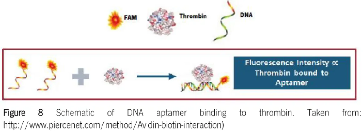
Figure 1 llustration of a biosensor functioning process. ... 5 Figure 2 Isolation of aptamers using the SELEX procedure... 7 Figure 3 Schematic representation of the G-quartet of (a) TBA1 and (b) TBA2 (by Mfold web server) ...9 Figure 4 Screen-printed electrode with its diagram. Ref., reference electrode; Aux., auxiliary electrode; and Work, working electrode. ... 14 Figure 5 Illustration of Avidin-biotin interaction. Each Avidin molecule can bind four biotinylated aptamers. 19 Figure 6 The schematic representation of protein strategies for electrochemical biosensors: (a) Sandwich assay using aptamers combining various signal amplification methods: enzyme, nanoparticle and carbon nanotubes means of measuring the concentration of bound target via impedance spectroscopy; (b) Impedimetric aptasensor: the binding of a target to the aptamer reduces the electron transfer (et) of a small redox mediator diffusing to the electrode surface and the increase the interfacial resistance, which provides a means of measuring the concentration of bound target via impedance spectroscopy; (c) Aptamer Conformational Changes: in the absence of target, the aptamer is largely unfolded, allowing for frequent collisions between the terminal redox moiety and the electrode. After target binding, the aptamer folds, enhancing electron transfer and producing a signal-on aptasensor; (d) Displacement Assays: target molecules in a sample displace labeled-target molecules previously bound to the sensor surface. ... 20 Figure 7 Signal-off (A) and signal-on (B) approaches using an aptamer labeled with a redox active molecule. ... 22 Figure 8 Schematic of DNA aptamer binding to thrombin. ... 28 Figure 9 Scheme of the 96-dark well plate for the fluorescence measurements ... 30 Figure 10 Electrophoretic mobility shift assay representation. The gel shift assay consists of three key steps: binding reactions, electrophoresis and probe detection. ... 32 Figure 11 a) A schematic diagram of a TBA aptamer immobilized on the working electrode region of a screen-printed electrode chip. b) Scheme of signal off system through interference in the electron transfer. ... 36 Figure 12 Electrochemical sensing of thrombin using thrombin binding aptamer (TBA) functionalized gold electrode. ... 37 Figure 13 Single binding assay for OPN-R3FAM and hOPN. The aptamer concentration used was 20 nM, the binding reaction time was 30 min and the fluorescence was recorded at 518 nm ... 39 Figure 14 Plot 20nM OPN-R3FAM fluorescence responses at 528nm versus protein concentrations (bovine osteopontin, bOPN, bovine serum albumin, BSA, human blood thrombin, Thr and human osteopontin, rhOPN) in the standard phosphate buffer ... 40
IX
Figure 15 Fluorescence binding studies for TBA1FAM (20 nM) with concentrations from 0 to 376.31 nM of hThr, after 30 minutes incubation ... 42 Figure 16 Fluorescence binding studies for TBA2FAM (20 nM) with concentrations from 0 to 376.31 nM of hThr, after 30 minutes incubation ... 42 Figure 17 Plot 20nM TBA1FAM fluorescence responses at 528nm versus protein concentrations (bovine osteopontin, bOPN, bovine serum albumin, BSA, human blood thrombin, Thr and human osteopontin, rhOPN) in the standard phosphate binding buffer, after 30 minutes incubation... 43 Figure 18 Plot 20nM TBA2FAM fluorescence responses at 528nm versus protein concentrations (bovine osteopontin, bOPN, bovine serum albumin, BSA, human blood thrombin, Thr and human osteopontin, rhOPN) in the standard phosphate binding buffer, after 30 minutes incubation... 43 Figure 19 Electrophoretic Mobility Shift Assay (EMSA) using different concentrations of a) TBA1FAM and b) TBA2FAM under the conditions described in Materials and Methods section. Binding reaction mixtures were applied on a 12 % non-denaturing gel containing 5X TB TBE buffer. The mobility of free aptamers, stained by SybrGreen Safe®, was detected using the Chemidoc System. ... 46 Figure 20 Electrophoretic Mobility Shift Assay (EMSA) of a) TBA1FAM and b) TBA2FAM under increasing concentrations of human thrombin, collected from ELISA assays. Binding reaction mixtures were applied on a 12% non-denaturing gel containing 5X TBE buffer. The mobility of free aptamers, stained by SybrGreen Safe®, was detected using the Chemidoc System. ... 47 Figure 21 Electrophoretic Mobility Shift Assay (EMSA) of a) TBA1FAM and b) TBA2FAM under increasingly concentrations of interferents, harnessed from ELISA assays. Binding reactions were applied on a 12% non-denaturing gel containing 5X TBE buffer. The mobility of free aptamers, stained by SybrGreen Safe®, was detected using the Chemidoc System. ... 48 Figure 22 Cyclic voltammetry scan rate optimization results (0,01; 0,05; 0,1 and 1 v/s) using a [Fe(CN)6]−3/−4 redox probe ... 49 Figure 23 Cyclic voltammograms for a thrombin TBA2 aptasensor regarding the [Fe(CN)6]−3/−4 redox probe: SPE control (voltammogram to the electrode itself), After DSP (voltammogram after DSP modification), After Avidin (voltammogram after Avidin incubation, After BSA (voltammogram after BSA blockage), TBA2-biotin immobilized (voltammogram after aptamer immobilization) ... 51 Figure 24 Electrochemical analysis of human thrombin hThr using modified TBA1-biotin-aptamer - immobilized on screen-printed electrode chip: a considerable current drop occurred by the treatment of hThr in a range of 0.025 nM–50nM from cyclic voltammetry; scan rate: 0.05V/s with a step potential of 0.5V to -0.6V/s, using 5mM [Fe(CN)6]4-/3- in 100mM KCl prepared in PBS, pH 7.4 ... 52 Figure 25 Electrochemical analysis of human thrombin (hThr) using modified (0.5 μM) TBA1-biotin-aptamer - immobilized on screen-printed electrode chip: a considerable current drop was occurred by the treatment of hThr in a range of 0.05 nM and 50nM from cyclic voltammetry; scan rate: 0.05V/s with a step potential of -0.5V to -0.6V/s, using 5mM [Fe(CN)6]4-/3- in 100mM KCl prepared in PBS, pH 7.4 ... 53
X
Figure 26 Electrochemical analysis of human thrombin (hThr) using modified TBA2-biotin-aptamer - immobilized on screen-printed electrode chip: a considerable current drop was occurred by the treatment of hThr in a range of 0.025 nM–50nM from cyclic voltammetry; scan rate: 0.05V/s with a step potential of -0.5V to -0.6V/s, using 5mM [Fe(CN)6]4-/3- in 100mM KCl prepared in PBS, pH 7.4 ... 54 Figure 27 Thrombin concentration versus oxidation peak. The concentration of the aptamer employed during sensor fabrication was 1 μM. The incubation time was 30 minutes at RT ... 55
XI
List of tables
Table 1 List of biomarkers tested. ... 2 Table 2 Assays reported from different authors in which TBA has been used. ... 10 Table 3 Types and sensing mechanisms involved in different biosensor. ... 11 Table 4 The comparison of the representative electrochemical aptasensor using different detection schemes. ... 23 Table 5 List of the DNA and RNA sequences used in the experiments ... 29 Table 6 Solutions for preparing resolving gels for NATIVE-PAGE gels ... 34 Table 7 Dissociation constants (Kd) for TBA1 and TBA2 with different protein targets for two different incubation times ... 44
XII
Abbreviations
CA15-3 Biomarker analyzed in breast cancer patients
CEA Carcinoembryonic antigen
CA-125 Elevated cancer antigen
RT-PCR Reverse transcription – polymerase chain reaction
CA 19-9 Biomarker analyzed in pancreatic and gastrointestinal cancer patients AFP Biomarker analyzed in testis, ovary and liver cancer
MIF Biomarker analyzed in mammary tumors
OPN Osteopontin
Thr Thrombin
PSA Prostate-specific antigen
NCBI National Centre for Biotechnology Information
BC Breast Cancer
IUPAC International Union of Pure and Applied Chemistry
RNA Ribonucleic acid
DNA Deoxyribonucleic acid
IL-1 Interleukin-1
PCR Polymerase chain reaction
SELEX Systematic Evolution of Ligands by Exponential enrichment FDA Food and Drug Administration
AMD Macular Degeneration Treatment VEGF Vascular Endothelial growth factor TBA 1 Thrombin binding aptamer 1 TBA 2 Thrombin binding aptamer 2 R3-OPN Osteopontin-binding RNA sequence
ELISA Enzyme-Immunosorbent Assay FA Fluorescence Anisotropic
EMSA Electrophoretic Mobility Shift Assays ISFETS Ion-sensitive field-effect transistors
SAM Self- Assembled Monolayers
LBL Layer- by- Layer
Fc- PEI Ferrocene-appended poly(ethyleneimine)
CNT`s Carbon Nanotubes
SPE Screen- printed electrodes
Kd Dissociation Constant
EDC / NHS 1-ethyl-3-[3-(dimethylamino)propyl] carbodiimide sulfosuccinimide QCM Quartz crystal microbalance
SA Sandwich Assay
EIS Electrochemical impedance spectroscopy TID Target- Induced Displacement Mode
QD`s Quantum Dots
NP`s Nanoparticles
LOD Limit of Detection
PBS Phosphate Buffer Saline
MB Methylene Blue
GDH Glucose dehydrogenase
MG Methylene green
Fc Ferrocene
XIII
RT Room temperature
FAM Fluorescein
FT Total fluorescence
FAPTNB Fluorescence of the aptamer not bound FAPTBOUND Fluorescence of the aptamer bound
FCOMPLEX Fluorescence of the complex
APS Ammonium persulfate
TEMED (N,N,N′,N′-Tetramethylethylenediamine
TBE Tris-borate-EDTA
DSP 3,3`-dithiopropionic acid-di(N-succinimidylester
DMSO Dimethyl sulfoxide
BSA Bovine serum albumin
CV Cyclic voltammetry
rhOPN Human recombinant osteopontin bOPN Bovine serum osteopontin
1
1. Introduction
Current demands regarding human health have encouraged the development of an increasing number of clinical tests. As a result, there is a growing requirement to develop more sensitive, reliable, time-efficient and inexpensive methods of analysis (Hong, Li, & Li, 2012). Despite the outstanding progresses in the field of biomedicine, substantial challenges remain in translating biomedical knowledge on disease markers into clinically relevant devices that could be used as diagnostic or monitoring tools for disease management (X. Y. Wang, Gao, Lu, He, & Yin, 2013). Progress in the development of consistent malignancy markers is imminent due to the advances in genomics and bioinformatics fields that enabled the analysis of great amounts of data through the use of less complex procedures involving less wet-lab analysis, and also with a lower requirement for qualified and experienced technical staff to conduct the analysis (Opstal-van Winden et al., 2011; Rodrigues, Teixeira, Schmitt, Paulsson, & Lindmark-Mänsson, 2007). The adoption of high-throughput methods led to the discovery of many new biomarkers that have been reported for prognostic and predictive purposes. However, out of these only a few have made their way into clinical routine mainly due to the lack of sufficient validation (Bohunicky & Mousa, 2010).
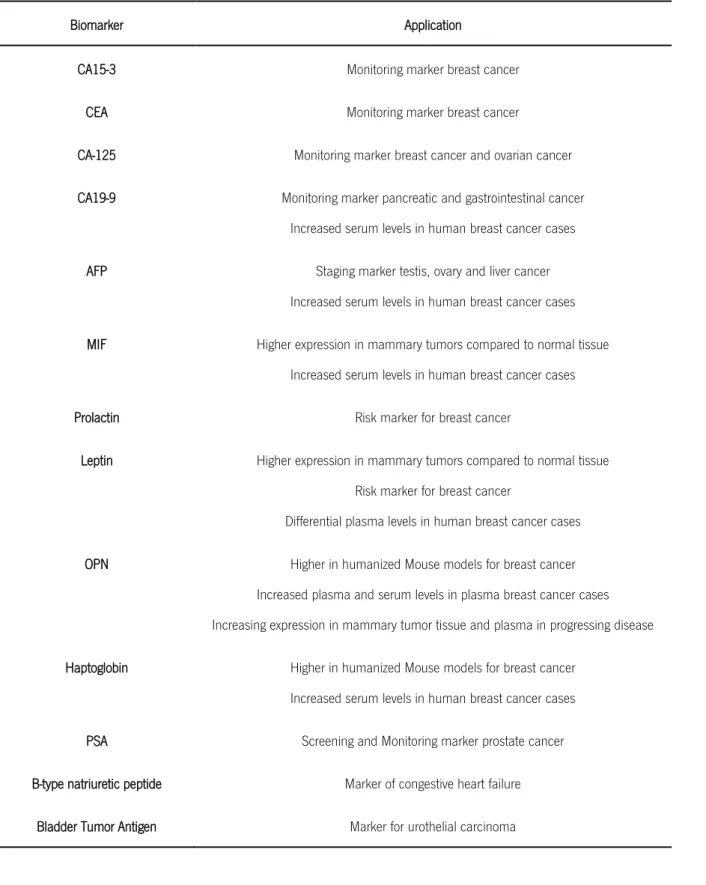
A biomarker is an indicator of a biological state or condition which, besides its value for diagnosis purposes, can be helpful to evaluate the body reaction to a given treatment (Bohunicky & Mousa, 2010; J. Wang, Chen, Jiang, Li, & Wang, 2013). The use of biomarkers to anticipate the outlines of the disease has been an emerging issue, especially in the case of cancer for which several treatments have been quite successful in the last few years. Biomarkers can also provide information on the mechanism underlying the initiation of a disease, and ultimately they constitute a powerful tool to precisely define the disease states, as well as to define appropriate treatments even for early stages of diseases (J. Wang et al., 2013). Some of the biomarkers that undergone rigorous testing are summarized in Table 1.
2
Table 1 List of biomarkers tested. Adapted from: Polanski & Anderson, 2006; Tothill, 2009; Opstal-van Winden et al., 2011 and Bohunicky & Mousa, 2010
Biomarker Application
CA15-3 Monitoring marker breast cancer
CEA Monitoring marker breast cancer
CA-125 Monitoring marker breast cancer and ovarian cancer
CA19-9 Monitoring marker pancreatic and gastrointestinal cancer
Increased serum levels in human breast cancer cases
AFP Staging marker testis, ovary and liver cancer
Increased serum levels in human breast cancer cases
MIF Higher expression in mammary tumors compared to normal tissue
Increased serum levels in human breast cancer cases
Prolactin Risk marker for breast cancer
Leptin Higher expression in mammary tumors compared to normal tissue
Risk marker for breast cancer
Differential plasma levels in human breast cancer cases
OPN Higher in humanized Mouse models for breast cancer
Increased plasma and serum levels in plasma breast cancer cases Increasing expression in mammary tumor tissue and plasma in progressing disease
Haptoglobin Higher in humanized Mouse models for breast cancer
Increased serum levels in human breast cancer cases
PSA Screening and Monitoring marker prostate cancer
B-type natriuretic peptide Marker of congestive heart failure
Bladder Tumor Antigen Marker for urothelial carcinoma
The discovery of new biomarkers is a time-consuming and costly task, since it requires the systematic separation and identification of biological molecules from complex body fluids (e.g. urine, blood) or tissues (Bohunicky & Mousa, 2010; Medley, Bamrungsap, Tan, & Smith, 2011;
3
Phillips, Xu, Xia, Fan, & Tan, 2009). Biomarkers that have been recognized as reliable for disease diagnosis and prognosis are listed in several reference cancer databases (e.g. National Centre for Biotechnology Information (NCBI), Cancer Research United Kingdom) and these have been approved by the FDA (Food and Drug Administration) (Opstal-van Winden et al., 2011; Tothill, 2009). Moreover, guidelines have also been developed describing the best approach to report the discovery of novel tumor markers, in order to guarantee that such studies are clear and easily understood in the context of their conclusions (McShane et al., 2005).
Biomarkers can either be present inside the cancer cells or be extracellular. Besides, they can also be of various molecular origins, including DNA (i.e. specific mutation, translocation, amplification, and loss of heterozygosity), RNA, or protein (i.e. hormone, antibody, oncogene or tumor suppressor)(Bohunicky & Mousa, 2010). Proteins that are selectively overexpressed as the result of cancer cells growth have been selected as potential biomarkers for cancer diagnosis and/or prognosis. Thus, these proteins can provide the basis for screening techniques, treatment options (Medley et al., 2011; Tothill, 2009), metastasis evaluation, and for determining the response to pharmacologic intervention (Sadana & Sadana, 2011).
Several proteins in the blood were indeed found to be related with the presence of breast cancer (J. Li et al., 2005; Lord et al., 2007; Mathelin, Cromer, Wendling, Tomasetto, & Rio, 2006). Based on the hypothesis that these proteins can be useful biomarkers for the early detection of cancer, great efforts have been developed to discover such proteins that are overexpressed in patient’s body fluids (Misek & Kim, 2011).
During the past decade, emerging evidence has refined the value of osteopontin (OPN) as a potential biomarker. OPN is an extracellular matrix protein with adhesive properties, that possesses a thrombin cleavage site and a cell attachment sequence found in all body fluids as plasma and serum (D. X. Cao et al., 2012; Fisher, Torchia, Fohr, Young, & Fedarko, 2001; Higashikawa, Eboshida, & Yokosaki, 2007; Ke et al., 2011; Macrì et al., 2009). Current research showed that OPN is often overexpressed in human cancers and contributes to regulate tumor growth and progression (Rangaswami, Bulbule, & Kundu, 2006; Rittling & Chambers, 2004; Servais et al., 2011). Increased OPN expression is one of the characteristics often associated with metastatic cancer cells (Kadkol et al., 2006; Rudland et al., 2002; P. Y Wai & Kuo, 2004), and it has been suggested as a main player in the progression and metastasis of a variety of cancers, including breast, liver, prostate and lung (Shojaei et al., 2012; P. Y. Wai & Kuo, 2008; Weber, Lett, & Haubein, 2010). Beausoleil and co-workers (2011) demonstrated that the changes in the OPN
4
plasma levels after therapy and over time could be used to monitor the clinical outcome (Beausoleil, Schulze, Goodale, Postenka, & Allan, 2011).
Several recent studies describe the multiple and complex mechanisms in which OPN is involved and suggest it’s potential as a biomarker for ovarian (J.-h. Kim et al., 2002; Matsuura, Suzuki, & Saito, 2010), bladder urothelial carcinoma (Park et al., 2012), gastric and liver (D. X. Cao et al., 2012), and for breast cancer (BC) (Mirza et al., 2008).
As mentioned, OPN contains two integrin-binding sites and a thrombin cleavage domain located in close proximity to each other. When OPN is cleaved at these domains by thrombin, it is separated into two approximately equivalent sized pieces, and since thrombin is also often overexpressed on the surface of cancer cells, the tumor microenvironment favors the activation of thrombin, and therefore the OPN cleavage (Beausoleil et al., 2011).
Human thrombin, highly specific serine protease, is also a biomarker that plays an important role on procoagulant and anticoagulant functions. This protein plays multiple roles in endothelial and smooth muscle cell functions, as well as coagulation and hemostasis (P. Wang et al., 2011). It is activated by the proteolytic cleavage of its precursor molecule pro-thrombin generation factor Xa (W. Z. Xie et al., 2005). Thrombin converts fibrinogen to insoluble fibrin that forms the fibrin gel, which is responsible either for some physiological plugs or for the formation of pathological thrombus (Neundlinger et al., 2011). The concentration of thrombin in blood varies considerably and it can be almost absent in the blood of healthy individuals. Some studies refer that its concentration in blood is normally around 0.01 nM (Frense et al., 2013). However, thrombin can reach low-micromolar concentrations during the coagulation process, and low levels of thrombin can even be generated in the early hemostatic process (Sosic, Meneghello, Cretaio, & Gatto, 2011).
In addition to its central role in coagulation, thrombin has been reported to induce mitogenesis and differentiation in cancer cells (W. Z. Xie et al., 2005); to regulate microvascular permeability (Guttridge, 1997); and also to be involved in blood coagulation, incrustation, inflammation and pulmonary metastasis (Chiu & Huang, 2009; Lin et al., 2011). Due to its crucial involvement in both thrombosis and hemostasis, thrombin is a major target for anticoagulation and cardiovascular disease therapy (Paborsky, N. McCurd, C. Griffin, J. Toole, & L. K. Leung, 1993).
Thrombotic disorders and their common clinical phenotypes of ischemic stroke, and venous thromboembolism cause substantial health care expenditures, morbidity, and mortality worldwide. The in vivo detection of thrombin, for example using aptamers, could be a promising method to prevent and/or treat these diseases (P. Wang et al., 2011).
5
1.1.
Biosensors
Currently, there is a high demand for convenient methodologies that allow detecting and measuring the levels of specific proteins in biological and environmental samples. In general, their detection, identification and quantification using conventional techniques, such as molecular assays and microbial culture-based tests, can be very complex, expensive and time consuming (Hong et al., 2012).
A biosensor, as per definition of IUPAC, is an integrated receptor-transducer device, which is capable of providing bio-recognition processes into measurable signals via a physicochemical transducer, with electronic and optical techniques as two major transducers (Shiping Song, Xu, & Fan, 2006; Beate Strehlitz, Nikolaus, & Stoltenburg, 2008). The use of biosensors brings about a combination of advantages. First, biosensors are highly sensitive, mainly because biomolecules often possess high affinity towards their targets. Second, biological recognition is usually very selective, thus it often leads to selective biosensors. Third, arising due to the development of modern electronic industry, it has been relatively easy to develop inexpensive, integrated and ready-to-use biosensor devices. These biological sensors certainly improve the ability to detect molecules or pathogens, besides being of particularly usefulness for small clinics and even for point-of care analysis (K. M. Song, Lee, & Ban, 2012; Shiping Song et al., 2006).
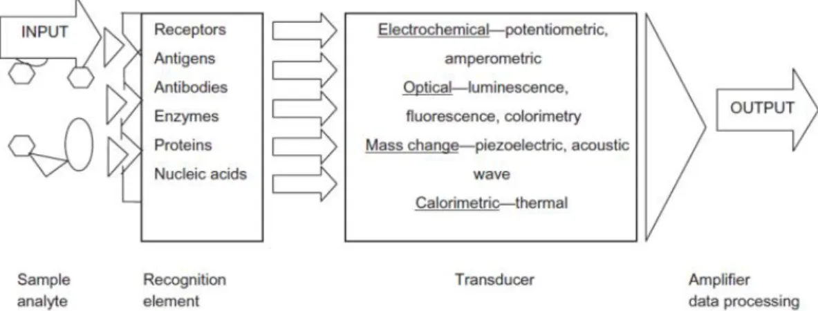
The core design of a biosensor mainly includes three components namely the probe-target recognition (that detects the presence or concentration of biological molecules or biological structures), the signal transduction (which translates the biochemical interaction into a quantifiable physical signal) and the physical readout (Bohunicky & Mousa, 2010; D'Orazio, 2003; J. O. Lee et al., 2008), as illustrated in Figure 1.
6
1.1.1. Biorecognition element - Aptamers
The biorecognition element is a critical key component of any biosensor. Fortunately, due to advances in technology and synthetic chemistry, many biosensor recognition elements used today are synthesized in the laboratory for improved stability and reproducibility of the biosensor function. Examples of recognition elements include receptor proteins, antigens, antibodies, enzymes, and nucleic acids (specifically aptamers) (Bohunicky & Mousa, 2010).
Aptamers (from the Latin word “aptus” meaning to fit and the Greek word “meros” meaning particle or piece) (Mairal et al., 2008; Mayer, 2009) are artificial nucleic acid ligands which can be selected from combinatorial libraries of synthetic nucleic acids, possessing motifs that recognize their targets (Hong et al., 2012). These molecules are 20–80 base pair long single-stranded ribonucleic acid (RNA) or deoxyribonucleic acid (DNA) oligonucleotides which are folded into distinct three-dimensional conformations (Kanwar, Roy, & Kanwar, 2011). Binding occurs because of their specific and complex three-dimensional shape characterized by loops, hairpins, stems, bulges, pseudoknots, triplexes, or quadruplexes. The structure compatibility results in the aptamer-target binding by stacking of aromatic rings, van der Waals and electrostatic interactions, and hydrogen bonding, or from a mixture of these effects (B. Strehlitz, Reinemann, Linkorn, & Stoltenburg, 2011).
Aptamers are similar to antibodies concerning their binding affinities; however they possess unique features, which turn them into potential alternatives to the antibodies. Firstly, the aptamers are generated using in vitro settings contrarily to the antibodies that are generated in vivo (Jayasena, 1999; J. H. Lee, Yigit, Mazumdar, & Lu, 2010). In vitro selection allows oligomers to be screened against molecules that have weak immunogenicity or high toxicity (Jayasena, 1999; J. H. Lee et al., 2010; Tombelli, Minunni, Luzi, & Mascini, 2005). In addition, non-physiological and/or harsh conditions can be applied while selecting aptamers (Jayasena, 1999; J. H. Lee et al., 2010; Tombelli et al., 2005). For instance, the thermal denaturation of nucleic acids is reversible, as exhibited in polymerase chain reaction (PCR), whereas antibodies or proteins such as interleukin-1 receptor (IL-1) are permanently denatured at temperatures higher than 53.5 °C (Remmele et al., 2005). Secondly, since the method for the synthesis and purification of oligonucleotides is well developed, the batch-to-batch difference of aptamer products is minimal compared with that of antibodies (Jayasena, 1999; J. H. Lee et al., 2010; Tombelli et al., 2005). Also, the aptamers can be chemically modified with a variety of functional groups and/or fluorophores without loss of binding functionality (J. H. Lee et al., 2007). Besides, as mentioned above, aptamers are structurally versatile because they have basic stem-loop arrangements that form proper
three-7
dimensional structures. These structures facilitate the formation of a complex with the target molecule. Thus, aptamers have high affinities to their targets, with dissociation constants at the low picomolar level, comparable to or better than antibodies (Gopinath, Awazu, & Fujimaki, 2012).
Furthermore, aptamer-based ligands may exhibit prominent advantages that include site-specific labeling, structure-controlled design and sequence-dependent amplification, which makes them an ideal molecular recognition tool for the development of biosensors (Zhang, Huang, Jiang, Shen, & Yu, 2007). In this context, besides the proliferated uses of aptamers instead of antibodies in established immunoassay techniques, the development of aptamer sensors with unique response strategies for homogeneous assays has been a subject of intensive research (Cho, Lee, & Ellington, 2009).
In addition, aptamers can be easily labeled for their use in diagnostics (Ulrich & Wrenger, 2009). As the use of aptamers has been extended from basic biology of cellular processes and gene regulation to therapeutic and diagnostic applications, many patented aptamers are currently being tested in clinical trials and have been recently reviewed (Kanwar et al., 2011). One of the major challenges in this field is the development of general methods to convert the highly specific molecular recognition between aptamers and their targets into detectable signals. Conjugation of aptamers with labels, nanoparticles, enzymes, among others, can be an ideal way to overcome this hurdle (Y. S. Kim, Lee, & Gu, 2008).
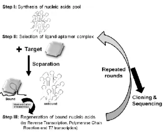
Systematic Evolution of Ligands by Exponential enrichment (SELEX) methodology is a combinatorial chemical procedure, commonly known as “in vitro selection” that allows the identification of nucleic acid sequences with unique properties from a random pool of sequences (an aptamer library), which were described above as aptamers (Figure 2).
8
Since then, the SELEX methodology has been used to identify aptamers with high-affinity and specificity against hundreds of targets, becoming an important research tool especially in the discovery of new drugs and disease targets, clinical diagnosis, therapy and in food inspection. There have been attempts to search for aptamers that are specific to targets involved in various diseases, such as cancer and viral infection (K. M. Song et al., 2012). Developed aptamers have been studied primarily for applications as diagnostic or therapeutic tools. In 2004, the Food and Drug Administration (FDA) approved the first RNA aptamer (Macugen, Pegaptanib) for human use in age-related macular degeneration treatment (AMD) targeting the vascular endothelial growth factor (VEGF) (Bunka & Stockley, 2006; Djordjevic, 2007; K.-T. Guo, Paul, Schichor, Ziemer, & Wendel, 2008). SELEX was used in many of these cases to identify dsDNA sequences that are the strongest (consensus) binders to the protein of interest (Djordjevic, 2007).
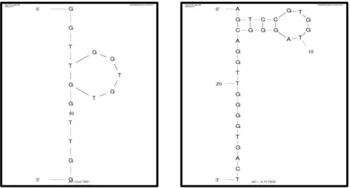
The choice between DNA or RNA aptamers depends essentially on practical considerations, and in part on the final application envisaged. In terms of a wide conformational diversity, RNA aptamers are naturally more flexible than the DNA molecules, but the RNA is more susceptible to degradation by nucleases which restricts their use in the presence of biological fluids. In terms of stability, using chemical modifications of the nucleotides, DNA aptamers are considered naturally more robust and display high levels of stability for biological applications than RNA aptamers. (Kulbachinskiy, 2007). Several strategies have been proposed to overcome the issue of RNA aptamers degradation, including their modification with 2′-aminopyrimidine, 2′-fluoropyrimidine or 2′-O-methyl nucleotides to increase their stability (Burmeister et al., 2005; Chiu & Huang, 2009; Feng et al., 2008; Keefe & Cload, 2008; Mayer, 2009; Stoltenburg, Reinemann, & Strehlitz, 2007). Virtually, aptamers can be raised against any target. This feature makes them interesting molecules to recognize for example proteins that have been previously identified as biomarkers. One of the most well-known examples of in vitro selection of DNA oligonucleotides for targeting a specific protein is the thrombin-binding aptamer (TBA). This thrombin aptamer has been extensively studied and characterized, thus enabling the monitoring of thrombin levels in plasma or blood (Diculescu, Chiorcea-Paquim, Eritja, & Oliveira-Brett, 2010) It has been shown that in the presence of alkali metals, TBA forms an antiparallel quadruplex consisting of two G-quartets connected by two TT loops and one TGT loop. (Bock, 1992). In particular, the thrombin binding aptamer 1 (TBA1) and 2 (TBA2) consist of two G-quartet conformations that selectively bind to specific and different epitopes of human -thrombin (Tasset, Kubik, & Steiner, 1997 ) (Figure 3). TBA1 is a 15-mer DNA aptamer which binds exosite I of thrombin (Fibrinogen Binding Site), with a dissociation constant (Kd) of approximately 26 nM (Bock, 1992; Paborsky et al., 1993; Tasset et al., 1997 ). TBA2 is a 29-mer
9
DNA aptamer binding to exosite II of thrombin (Heparin Binding Domain) with subnanomolar affinity (Sosic et al., 2011). This distinct recognition pattern allows their use in tandem, since a ternary complex could possibly be formed by simultaneous recognition of thrombin with different formats and detection methods have been demonstrated by few groups (Bang, Cho, & Kim, 2005; D. W. Huang, Niu, Qin, Ruan, & Zeng, 2010; A.-E. Radi, Sánchez, Baldrich, & O`Sullivan, 2005; Zhao et al., 2011)
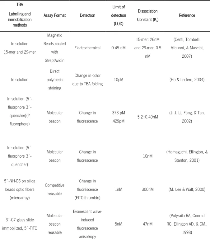
Some studies have reported the use of TBA to detect thrombin as summarized in Table 2. In most cases, it appears that the methodology adopted has been strongly influenced by the authors’ background, usually taking advantage of ELISA or nucleic acid-related protocols, as described below in Table 2. Discrepancies in values may also arise from the fact that each aptamer has a unique structure for target binding (E. Baldrich, Restrepo, & O'Sullivan, 2004).
Figure 3 Schematic representation of the G-quartet of (a) TBA1 and (b) TBA2 (by Mfold web server)
10
Table 2 Assays reported from different authors in which TBA has been used. Adapted from Baldrich, E. et al., 2004 and Centi, S. et al., 2007
TBA Labelling and immobilization
methods
Assay Format Detection
Limit of detection (LOD) Dissociation Constant (Kd) Reference In solution 15-mer and 29-mer
Magnetic Beads coated with StreptAvidin Electrochemical 0.45 nM 15-mer: 26nM and 29-mer: 0.5 nM (Centi, Tombelli, Minunni, & Mascini,
2007) In solution Direct polymeric staining Change in color
due to TBA folding 10pM (Ho & Leclerc, 2004)
In solution (5`-fluorphore 3`-quencher)(2 fluorophore) Molecular beacon Change in fluorescence 373 pM 429pM 5.2±0.49nM
(J. J. Li, Fang, & Tan, 2002) In solution (5`-fluorphore 3`-quencher) Molecular beacon Change in fluorescence 10nM
(Hamaguchi, Ellington, & Stanton, 2001)
5`-NH-C6 on silica beads optic fibers
(microarray) Competitive reusable Change in fluorescence (FITC-thrombin)
1nM 300nM (M. Lee & Walt, 2000)
3`-C7 glass slide immobilized, 5`-FITC Molecular beacon reusable Evanescent wave-induced fluorescence anisotropy 5nM 47nM
(Potyrailo RA, Conrad RC, Ellington AD, & GM.,
1998)
An OPN-directed RNA aptamer, OPN-R3, was recently isolated through SELEX technology. Binding studies with human OPN, were conducted using the OPN-R3 aptamer (Kd value of 18 ± 0,2
nmol/l) and an unspecific competitor RNA aptamer. It was found that human OPN binds to OPN-R3 in a specific manner (Zhiyong Mi et al., 2009). Besides, further research indicated that RNA aptamer binding to OPN blocks its interaction with cancer cell surface receptors to significantly
11
inhibit adhesion, migration and invasion in vitro, thus inhibiting local progression and distant metastases (Mi, Guo, & Kuo, 2009).
1.1.2. Transducer
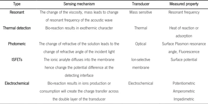
Regarding the physical transducer, it is the part of the device that converts the recognition signal events into electrical (often digital) signals – biosensors can be categorized in various types, including resonant, photometric, thermal detection, ion-sensitive field-effect transistors (ISFETs), and electrochemical sensors (Tothill, 2009; Y. Zhou, Chiu, & Liang, 2012). Table 3 summarizes the sensing mechanism of each type of biosensor.
Table 3 Types and sensing mechanisms involved in different biosensor. Adapted from: Tothill, 2009 and Zhou, Y. et al., 2012
Type Sensing mechanism Transducer Measured property
Resonant The change of the viscosity, mass leads to change
of resonant frequency of the acoustic wave
Mass sensitive Resonant frequency
Thermal detection Bio-reaction results in exothermic character Thermal Heat of reaction or
adsorption Photomeric The change of refractive of the solution leads to the
change of refractive angle of the incident light
Optical Surface Plasmon resonance
angle, Fluorescence
ISFETs The ionic analyte diffuses into the membrane
hence change the potential difference at the detecting interface
Ion-selective membrane
Surface potential
Electrochemical Bio-reaction results in ions production or
consumption will create the charge transfer across the double layer of the transducer
Electrochemical Potentiometric
Amperometric Impedimetric
1.1.2.1. Techniques for modification of surfaces: Self-assembled monolayers (SAM) and Layer-by layer (LBL)
Self-assembled monolayers (SAM) and Layer-by-Layer (LBL) are methodologies that lead to the deposition of organic compounds on the biosensor surfaces. Among the commonly used substrates, gold presents some benefits for many electrochemical measurements. The gold surface can be functionalized and the type of chemistry selected is dependent on the type of terminal functional group linked to the aptamer (amine, thiol or biotin termini) (Velasco-Garcia & Missailidis, 2009). In addition, gold surfaces can be functionalized with SAM or LBL.
12
SAM provides molecular level control over the density and position of assembled molecules. SAM is capable of packing different types of molecules in an orderly fashion at the molecular level, which generates a multifunctional surface for multitasks. This layer of biological/chemical molecules is advantageous due to its simplicity of preparation, high sensitivity, and few limitations in the detection range of an analyte, and most importantly, the versatility of the modification that no other organic materials could match (Y. Zhou et al., 2012). As a substrate for monolayers assembly, gold presents interesting features: it is air-stable and commercially available from several sources as films and particles; it binds thiols with a high affinity (Balamurugan, Obubuafo, Soper, & Spivak, 2008), and the films are stable in complex liquid media containing target biomolecules. The monolayer can be designed to prevent non-specific adsorption of aptamers to the gold surface and it plays an important role in applications that require long-term storage stability (Kuralay, Campuzano, & Wang, 2012).
On the other hand, the layer-by-layer (LBL) approach is a convenient technique for the bottom-up assembly of multi-layered polymer films, because it allows the deposition of oppositely charged polyelectrolytes onto solid substrates (Du et al., 2010). The nature of the assembly process leads to precise, nanoscale control of film thickness and composition through the appropriate choice of the components, the number of layers, and the order of their deposition (Sultan, Walsh, Monreal, & DeRosa, 2009; J. Wang, 2006; S. Xie & Walton, 2010). This method is highly versatile since several polyelectrolytes can be chosen. Biological macromolecules such as polypeptides/proteins, polysaccharides, nucleic acids and even viruses can serve as building blocks for these multilayer films. Sultan et al. (2009) reported that when embedded in or attached to LBL polyelectrolyte matrix, aptamers could maintain their affinity and specificity for the cognate target. The LBL technique has been reported for protein detection using self-assembled multilayers with ferrocene-appended poly(ethyleneimine) (Fc-PEI), carbon nanotubes (CNTs), and aptamers (Du et al., 2010).
Compared with SAM, these LBL multi-layers with three dimensional structure can bring in, not only more probes to produce an amplified signal, but also more molecular recognition elements to improve the sensitivity of the detection, making the stable membrane property of the multi-layer widely used in biosensors (Du et al., 2010).
13
1.2. Electrochemical detection
Electrochemical transducers are the most widely used in platforms for patient diagnosis since they are portable, simple, easy to use, low cost, minimal simple-to-operate, robust power requirements and independence of sample turbidity and in most cases disposable (A.-E. Radi, 2011; F. Wei, Lillehoj, & Ho, 2010). The electrochemical instruments used with the biosensors have been miniaturized to small pocket size devices which make them applicable for “alternative-site” testing, emergency-room screening, bedside monitoring or home self-testing (Tothill, 2009; J. Wang, 2006).
The name electrochemical biosensor is applied to a molecular sensing device which intimately couples a biological recognition element to an electrode transducer, in which the purpose of the transducer is to convert the phenomenon into a suitable and readable electrical signal.
Potentiometric and amperometric biosensors are the two most common types of electrochemical biosensors. Potentiometric biosensors use ion-selective electrodes to detect an electrical response in the molecular recognition element (Han, Liang, & Zhou, 2010). A true potentiometric aptasensor has been recently reported. This device was based on poly(phenothiazine) conducting polymers electropolymerized on a glassy carbon electrode. Avidin-modified polymer surfaces obtained by direct electrostatic precipitation have then been used to immobilize biotinylated anti-thrombin DNA aptamers. Measurement of the difference in the potential of the sensor enabled the potentiometric thrombin detection in a concentration ranging from 10-9 to
10-6 M (Evtugyn, Porfireva, Hianik, Cheburova, & Budnikov, 2008).
Amperometric transducers measure the current that is produced when a potential is generated between two electrodes. Oxidation or reduction reactions produce a current, which can then be measured (J. Wang, 2006). Amperometric-based biosensors for cancer detection, using sequence-specific DNA as the recognition element, have an enormous potential in the diagnosis field (Ikebukuro, Kiyohara, & Sode, 2005). Other examples include the work by Ikebukuro and co-workers (2004) that developed the first electrochemical aptasensor with an amperometric sandwich-based biosensor sandwich-based on glucose dehydrogenase-labeled signaling aptamers. The GlucoWatch™ is a glucose sensor that was developed to enable non-invasive, continuous measurement of blood glucose levels in diabetic patients. This device works like a wristwatch and takes blood glucose readings through the skin via reverse ion-transfer, a process whereby a small electrical signal brings glucose to the skin surface so that it can be measured.
14
Electrochemical aptasensors have been recently developed for multiplexed protein measurements. In fact, since most cancer diseases are associated with the presence of more than one tumor marker, developing an effective aptasensor for simultaneous measurement of co-existing tumor markers may be valuable to improve the detection accuracy and to deliver more precise information on diagnosis, prognosis and treatment (Zhao(a) et al., 2012).
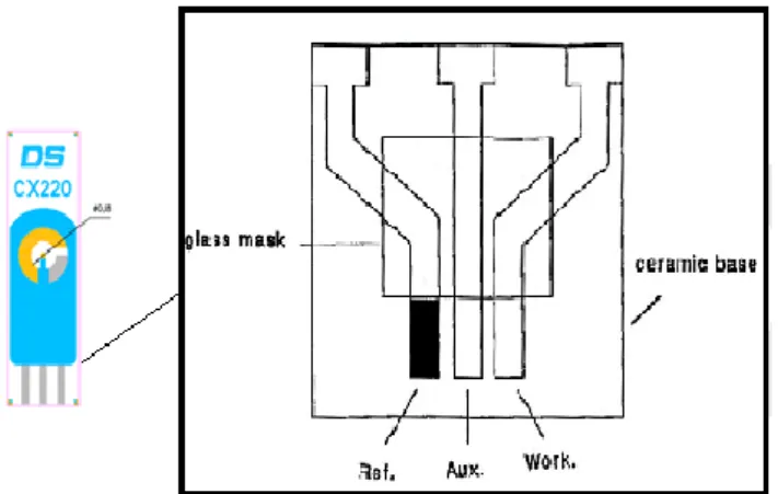
Electrochemical sensors are part of an electrochemical cell that often consists of three electrodes. A distinctive three electrode electrochemical cell consists of a working (or indicator) electrode of a chemically stable solid, conductive material, such as gold; a reference electrode and an auxiliary electrode (Ronkainen, Halsall, & Heineman, 2010) as shown in Figure 4. The working electrode serves as the transduction element in the biochemical reaction, while the counter electrode establishes the connection with the electrolytic solution so that a current can be applied to the working electrode (Grieshaber, MacKenzie, Voros, & Reimhult, 2008; Beate Strehlitz et al., 2008). Electroactive reporters such as methylene blue (MB), ferrocene, ferrocene-bearing polymers, ruthenium complexes and [Fe(CN)6]4−/3− are
commonly used for signal transduction in electrochemical aptamer-based sensors. These reporters can be covalently conjugated to the aptamer itself, conjugated to a complementary oligonucleotide or indirectly linked to aptamers (Cho et al., 2009). Screen- printed electrodes (SPEs) have been used in several electrochemical DNA sensors due to their straightforward fabrication, high uniformity and material versatility (F. Wei et al., 2010). Therefore, SPEs can offer an inexpensive solution with the additional advantage of flexibility in the biosensor shapes (D. Wei, Bailey, Andrew, & Ryhanen, 2009). Screen-printed technology has been reported as an attractive method for mass production of sensors (Alonso-Lomillo, Dominguez-Renedo, Matos, & Arcos-Martinez, 2009; Tothill, 2009).
Figure 4 Screen-printed electrode with its diagram. Ref., reference electrode; Aux., auxiliary electrode; and Work, working electrode. Adapted from Ronkainen et al., 2008
15
1.2.1. Voltammetry
Voltammetry provides an electroanalytical method for gathering information on one or more analytes by measuring the current as a function of the potential in electrochemical aptasensors. Several types of experiments may be performed to collect information from voltammetry including cyclic voltammetry, square-wave voltammetry, and stripping voltammetry to name a few common techniques (Feng et al., 2008; Ikebukuro et al., 2005; K. M. Song et al., 2012; Beate Strehlitz et al., 2008; F. Wei et al., 2010; D. Xu et al., 2005).
Since there are many ways to vary a potential, there will also exist many forms of voltammetry, including polarography (DC Voltage), linear sweep, differential staircase, normal pulse, reverse pulse, differential pulse, among others. However, cyclic voltammetry is one of the most generally used forms and it is useful to obtain information about the redox potential and electrochemical reaction rates (e.g. the chemical rate constant) of analyte solutions.
The voltage is measured between the reference and the working electrodes, while the current is measured between the working and the counter electrode. The measurements are plotted as current versus voltage, also known as a voltammogram. The shape of the voltammogram for a specific target depends not only on the scan rate and the electrode surface, which is different after each adsorption step, but can also depend on the catalyst concentration (Grieshaber et al., 2008). Moreover, the detection can be either “signal on” or “signal off”, depending on how the redox reporter is shielded from the electrode, resulting in maximum or minimum peaks, respectively on the voltammograms (Cho et al., 2009; Shiping Song, Wang, Li, Fan, & Zhao, 2008).
Additionally, through voltammetry it is possible to infer about some important parameters such as the biosensor affinity and probe-target binding; selectivity (especially important in real-world samples where the target concentration can be much less than the concentration of non-target biomolecules present); limits of detection and reproducibility (the detection limit can be determined by measuring the sensor response to a dilution series and determining the target smallest concentration at which the sensor response is clearly distinguishable from the response to a blank solution.); dynamic range (ratio between the largest measurable target concentration and the limit of detection); amplification (amplification techniques lay outside the domain of label-free impedance biosensors); and multiplexing (it means detecting several targets in the same biological sample is possible if different surface regions are functionalized with different probes) (Daniels & Pourmanda, 2007).
16
1.3. Immobilization methods
The critical step in the development of electrochemical aptasensors is the immobilization of the aptamers (biological recognition element) to the electrode surface. It is crucial to develop strategies for the reliable immobilization of aptamers so that they retain their biophysical characteristics and binding abilities, as well as for minimizing non-specific binding/adsorption events. In theory, these strategies are similar to those applied hitherto for the immobilization of single- or double-stranded DNA in DNA biosensors for the detection of DNA damage. As mentioned, the control of this step is essential to ensure high reactivity, orientation, accessibility, and stability of the surface-confined probe and to avoid non-specific binding (A.-E. Radi, 2011; Audrey Sassolas, Leca-Bouvier, & Blum, 2008).
Centi et al. (2007) stated that “The procedure to fix the aptamer to the surface is of paramount importance to obtain an ordered and oriented layer able to ensure, as much as possible, the flexibility of the bioreceptor without altering the affinity for the target molecule and selectivity that the aptamers show in solution”(Balamurugan et al., 2008). If immobilization is declined, it could lead to partial or complete loss of the target activity, due to possible structure deformation. Thus, to avoid this issue, proteins should be attached onto surfaces without affecting conformation and function (Rusmini, Zhong, & Feijen, 2007).
When developing an aptamer diagnostic device there are several different approaches for the immobilization of aptamers, which depend upon the chemical composition of the surface, the availability of suitable aptamer linkers, and the chemistries used for attachment (Balamurugan et al., 2008). Several substrates can be used as supports for the aptamer immobilization such as gold, silicates, carbon nanotubes (Fabre, Samorì, & Bianco, 2012; K. Guo et al., 2011) and polymers (L. Zhou, Wang, Wang, & Ye, 2011).
When developing an aptasensor, it is of utmost importance to define adequate immobilization strategies in order to maximize the performance of the aptamer-based analytical device (Balamurugan et al., 2008). Thus, in order to immobilize the aptamer onto the solid support it is required that either the 5′ or 3′ -end of the aptamer is modified (Gopinath et al., 2012). Although both positions can be used for the aptasensor development, there are very few studies looking at the effect of the two types of end attachment. Recent work suggests that it depends on the particular aptamer (Cho, Collett, Szafranska, & Ellington, 2006), although for biological targeting it may be that the 3’ end is more suitable, since the 3’ end is the primary target for exonucleases,
17
and thus its coupling to the solid support would simultaneously confer resistance to nucleases attack (Velasco-Garcia & Missailidis, 2009).
Few immobilization techniques for fixing aptamers onto biosensor surfaces have been developed in the past years, which are mainly based on the following three mechanisms: physical (adsorption), covalent (chemisorption with thiol and covalent attachment to chemically-modified surfaces by chemical groups such as hydroxyl, carboxyl and amino), and bio-affinity immobilization (using for example the Avidin-biotin interaction method) (Rusmini et al., 2007; Audrey Sassolas et al., 2008; L. Zhou et al., 2011) .
The choice of the best immobilization strategy is still an open question, although it is unlikely that one structure will be optimal for all proteins. Consequently, the extrapolation of immobilization strategies from one system to another for different classes of proteins is difficult and mostly unsuccessful due to the wide subset of characteristics and functional properties of proteins. Therefore, several unsolved challenges are involved in the immobilization of the aptamers (Rusmini et al., 2007).
1.4. Immobilization techniques
1.4.1. Physical adsorption
Physical adsorption is an immobilization method that does not requires any nucleic acid modification. It consists in placing the aptamer solution in contact with the surface for a well-defined period of time, and subsequently washing off any non-adsorbed biomolecules. It is based on the ionic, hydrophobic and Van der Waal’s forces that take part in the interaction and will depend on the particular protein and surface involved. The resulting layer is likely to be heterogeneous and randomly oriented, since each molecule can form many contacts in different orientations for minimizing repulsive interactions with the substrate and previously adsorbed proteins (Rusmini et al., 2007), however, by its weakness, reversible binding could usually occur (A. Sassolas, Blum, & Leca-Bouvier, 2011).
Ocana et al. (2012) developed a reusable impedimetric aptasensor for the detection of thrombin in which the immobilization of the aptamer onto the transducer surface was conducted by a wet physical adsorption procedure.
18
1.4.2. Covalent immobilization
DNA immobilization by covalent attachment is often used (Audrey Sassolas et al., 2008) and preferred over adsorption (A. Sassolas et al., 2011). Thiol-metal interactions are frequently used to bind biomolecules covalently onto gold surfaces (Bai et al., 2008; Y. S. Kim et al., 2008). Noble metal surfaces display strong affinity to thiol groups and consequently enable the formation of covalent bonds between the sulfur and gold atoms (A. Sassolas et al., 2011).
The three most common groups used for surface covalent attachment to chemically-modified sensor surfaces are hydroxyl, amine, and carboxylic acid functional groups. The choice of this kind of surface functionalization depends on the types of terminal functional groups linked to aptamers that are available (Balamurugan et al., 2008). These groups interact with chemical groups (such as amino) through covalent binding, leading to a layer of ordered film on the sensor surface (Audrey Sassolas et al., 2008; L. Zhou et al., 2011).
1.4.3. Biocoatings/Bioaffinity
Chemical affinity reactions allow a gentle oriented immobilization of proteins, providing an important improvement over other immobilization techniques (Rusmini et al., 2007).
Avidin is a glycoprotein which contains four identical subunits with a combined mass of 67 to 68 kDa (Orita, Tomita, Harada, & Kato, 2012). The specific and strong interaction between avidin (or one of its derivatives like streptavidin) and biotin has been widely explored for surface immobilization of a number of bio-receptors, including aptamers (Balamurugan et al., 2008), and it has been used in several recent studies (L. J. Chen et al., 2011; General, Dragomirova, & Meirovitch, 2011).
Biotin- avidin interaction is one of the ideal methods for aptamer fixation on a variety of sensor surfaces, as its interaction exhibits the highest known affinity in nature (Kd =1015 M-1), robustness
and simplicity (Balamurugan et al., 2008; Orita et al., 2012; A. Sassolas et al., 2011). Specifically, compared with other fixation methods, each avidin molecule can bind with four biotinylated aptamers as shown in Figure 5. This increases the amount of aptamers potentially bound to the sensor surface, reduces the incidence of non-specific adsorption, and improves sensor signal-to-noise ratio (Zhou et al., 2011).
19
Avidin-biotin method was used by Minunni and collaborators (2004) to activate a gold film-coated quartz chip using 11-mercaptoundecanol and carboxylated dextran to interact with avidin using 1-ethyl-3-[3-(dimethylamino)propyl] carbodiimide sulfosuccinimide (EDC/NHS). Afterwards, the biotin-labeled aptamers were attached to the avidin-modified quartz chip. The sensor can be used to detect HIV-1 transactivator protein, being the linear detection range between 0–2.5 mg L–1,
and the detection limit around 0.65 mg L–1 (Minunni, Tombelli, Gullotto, Luzi, & Mascini, 2004). In
Hianik et al., (2007) it was used the avidin-biotin method for aptamer immobilization, in which quartz crystal microbalance (QCM) was the method used for mass detection to study the interaction of thrombin with DNA aptamers exhibiting two different configurations. The authors suggested that immobilization of the aptamer by means of avidin-biotin methodology provides the best results in sensitivity comparing to other immobilization methods using dendrimers or immobilization via chemisorption of the aptamer onto a gold surface (Hianik, Ostatna, Sonlajtnerova, & Grman, 2007).
The avidin–biotin conjugation methods possess several advantages, for example they do not depend on the isoelectric point of the protein. Nevertheless, the use of avidin/biotin immunoassays is currently limited because of their sensitivity to high temperature and organic solvents (Orita et al., 2012).
Figure 5 Illustration of Avidin-biotin interaction. Each Avidin molecule can bind four biotinylated aptamers. Taken from http://www.piercenet.com/media/Avidin-Biotin-Interaction1.jpg
20
1.5.
Design Strategies for Aptasensors
A sensitive and simple method to use aptamers as recognition elements for the development of biosensors (aptasensors) is to transduce to an electrochemical signal the real-time biological phenomena in solution, or through aptamer immobilization onto a solid support (Hong et al., 2012; Minunni et al., 2004; O'Sullivan, 2002). As sensing probes, aptamers are extremely advantageous in a biosensor. In fact, they can be modified for immobilization purposes and can incorporate particular reporters, without influencing their affinity, which has been very helpful for a number of design methods (Balamurugan et al., 2008; Phillips et al., 2009). Regarding the design strategies for most of the electrochemical aptasensors, four broad classes can be defined depending on the assay format and the method of detection: (a) sandwich mode; (b) label-free aptamer sensor-using electrochemical impedance spectroscopy (EIS); (c) aptamer conformational changes; and (d) target-induced displacement mode (TID), as illustrated in Figure 6 (Abe, Yoshida, & Ikebukuro, 2013; Han et al., 2010; Y. Huang et al., 2012; A.-E. Radi, 2011; Audrey Sassolas, Blum, & Leca-Bouvier, 2009; Y. Xu, Cheng, He, & Fang, 2009).
Figure 6 The schematic representation of protein strategies for electrochemical biosensors: (a) Sandwich assay using aptamers combining various signal amplification methods: enzyme, nanoparticle and carbon nanotubes means of measuring the concentration of bound target via impedance spectroscopy; (b) Impedimetric aptasensor: the binding of a target to the aptamer reduces the electron transfer (et) of a small redox mediator diffusing to the electrode surface and the increase the interfacial resistance, which provides a means of measuring the concentration of bound target via impedance spectroscopy; (c) Aptamer Conformational Changes: in the absence of target, the aptamer is largely unfolded, allowing for frequent collisions between the terminal redox moiety and the electrode. After target binding, the aptamer folds, enhancing electron transfer and producing a signal-on aptasensor; (d) Displacement Assays: target molecules in a sample displace labeled-target molecules previously bound to the sensor surface. Adapted from: Radi et al., 2011 and Abe, K., Yoshida, W., & Ikebukuro, K., 2013
21
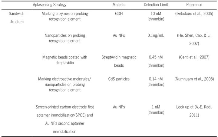
1.5.1. Sandwich Assay (SA)
The sandwich structure strategy presents several advantages as compared to the strategy that uses only one recognition element to capture and label the target biomolecules. These advantages include its high sensitivity and simple operation in what regards the biosensor fabrication for biosensor fabrication. In a sandwich structured biosensor, the sequence is firstly immobilized on the electrode to hybridize with a certain part of the target DNA, and then the probing sequence is available to react with another part of the target sequence. The electroactive label at the probing sequence thus electrochemically transduces the presence/binding of the target sequence.
Therefore, as a strategy employed for aptasensor in sandwich manner, the target should have two or more recognition elements, namely the one that binds to the aptamer, used as capturing element to be immobilized on the electrode surface and catch target molecules; and the other one that serves as a probing element to mark the target with electroactive molecules or nanoparticles (Y. Xu et al., 2009).
To construct highly sensitive detection systems, some researchers have attempted the conjugation of nanoparticles (NPs), enzymes, quantum-dots (QDs) and carbon nanotubes (CNT) with aptamers (Abe et al., 2013).
1.5.2. Label-Free Aptamer Sensor -Using Electrochemical Impedance Spectroscopy (EIS) Electrochemical impedance spectroscopy (EIS) has been mainly applied to study corrosion systems, specifically to monitor the resistance components of the impedance response (Grieshaber et al., 2008; Y. Xu et al., 2009). Therefore, it reflects changes in a diffusion-limited electrochemical process, presumably due to steric hindrance created by the bound molecules to the immobilized aptamer and repulsion between electron mediators and immobilized molecules (Abe et al., 2013).
1.5.3. Aptamer Conformational Changes
In this detection strategy, the DNA ligands recognize their targets primarily by their special shape and not by their sequence. They are capable to fold into unique complex intramolecular secondary and tertiary structures, binding and crosslinking the target molecule in a large surface area. Therefore, they are able to distinguish a small structural change in the target molecules. Besides, aptamers change its conformation through Watson-Crick base pairing and drastically, in some cases, they reduce the electronic transfer distance of the electroactive probing group from the