University of Bologna
OPTIMIZING PHYSICOCHEMICAL PROPERTIES OF NATURAL
ANTIOXIDANTS AND GEROPROTECTORS: L-CARNOSINE AND MELATONIN
Oleksii Shemchuk
Dissertazione / Dissertation
Erasmus Mundus Laurea Magistrale in Innovazione Chimica e il Regolamento (Erasmus Mundus Master in Chemical Innovation and Regulation)
Il lavoro svolto sotto la guida di: Work supervised by Prof. Fabrizia Grepioni
Prof. Teresa Duarte
OPTIMIZING PHYSICOCHEMICAL PROPERTIES OF NATURAL
ANTIOXIDANTS AND GEROPROTECTORS: L-CARNOSINE AND MELATONIN
Declaration of Authorship
I declare that I am the author of this work, which is original. The work cites other authors and works, which are adequately referred in the text and are listed in the bibliography.
____________________________________ Oleksii Shemchuk
Copyright: Oleksii Shemchuk, the University of Bologna has the right to keep and publicise
this work through printed copies in paper of digital form, or any other means of reproduction, to disseminate it in scientific repositories and to allow its copy and distribution with educational and/or research objectives, as long as they are non-commercial and give credit to the author and editor.
ACKNOWLEDGEMENTS
I am grateful to the European Commission for the scholarship funded within the Erasmus+ KA1 Programme, ref. 2013-0241 - Erasmus Mundus Joint Master Degree in Chemical Innovation and Regulation.
I would like to express my sincere gratitude to my supervisors – Prof. Teresa Duarte and Prof. Fabrizia Grepioni for their full support, expert guidance, understanding and encouragement throughout this project. Actually, this thesis has become a reality with the kind assistance and help of many individuals – Dr. Vânia André, Dr. Alexandra Antunes, Dr. Laura Chelazzi, Prof. Lucia Maini, Dr. Katia Rubini. My heartfelt gratitude to Prof. Dario Braga for the great privilege of working in his lab and his undying support.In addition, I express my appreciation to all the people I had pleasure to work with.
I would also like to express my deepest appreciation to our coordinators – Prof. Isabel Cavaco and Prof. Emilio Tagliavini.
Finally, I would like to thank my parents, my brother, my wife and my grandmother for their unconditional love and support. I would not have been able to complete this thesis without their continuous love and encouragement.
ABSTRACT
The issue of “Healthy Ageing” has become a significant challenge due to the continuous grow of World Population Ageing. Advancing health and well-being into old age and ensuring supportive environments is a main objective of NATO’s Department of Economic and Social Affairs Population Division [1].
Natural antioxidants such as L-carnosine and melatonin have been successfully used as geroprotectors. They reduce the risk of developing ageing-related diseases which is one the main aspects of healthy ageing. New formulation of the existing APIs is a widely used approach to optimize their physicochemical properties. Carnosine underwent reactions with a number of organic acids resulting in the discovery of seven new salts. The obtained salts were characterized by X-ray powder diffraction and thermal analyses. The structures were determined using X-ray Powder Diffraction (XRPD). The carnosine salts with glycolic and succinic acids might be of higher interest since both components possess antioxidant activity and carnosine, when used as a nutraceutical is usually combined with other antioxidants. Thus, the new formulation of carnosine as a salt with an acid possessing antioxidant activity can enhance its biological activity. Melatonin was crystallized with a number of co-formers from various chemical groups. Two new co-crystals of melatonin with DABCO and piperazine were obtained by using mechanochemical technique (kneading). The structure of melatonin-DABCO co-crystal was established from a single crystal obtained by recrystallization from dichloromethane. The structure of melatonin-piperazine was solved using XRPD. Though only one of the obtained co-crystals can find a direct application in Pharmaceutical industry (DABCO does not belong to the GRAS list), our results confirm the reactivity of melatonin in co-crystallization processes, and emphasizes the need to continue the search for an improved formulation.
Abstract
La questione di un "sano invecchiamento" è diventato una sfida significativa a causa del continuo aumento dell’età della popolazione mondiale. L’obiettivo principale del Dipartimento per gli Affari Economici e Sociali della NATO Divisione per la Popolazione è quello di garantire il benessere e ambienti favorevoli con il progredire dell’età [0]. Antiossidanti naturali come la L-carnosina e la melatonina sono stati utilizzati con successo come geroprotettori. Riducono il rischio di sviluppo di malattie legate invecchiamento, che è uno degli aspetti principali di un invecchiamento sano. La nuova formulazione di API già esistenti è un approccio ampiamente utilizzato per l’ottimizzazione delle loro proprietà chimicho-fisiche. La reazione tra la Carnosina ed un certo numero di acidi organici, ha prodotto la formazione di sette nuovi sali organici. I sali ottenuti sono stati caratterizzati mediante da diffrazione da raggi X su polveri ed analisi termiche (TGA e DSC). Le strutture cristalline sono state determinate utilizzando diffrazione da raggi X su polveri (XRPD). I sali ottenuti con gli acidi glicolico e succinico potrebbero essere di interesse maggiore poiché entrambi i componenti possiedono attività antiossidante e la carnosina, quando utilizzata come nutraceutico è solitamente combinata con altri antiossidanti. Così, la nuova formulazione di carnosina come un sale con un acido che possiede attività antiossidante può migliorare la sua attività biologica e, di conseguenza, consentire il suo utilizzo come farmaco indipendente / nutraceutico. La melatonina, invece, è stata co-cristallizzata con un numero di
co-former. Due nuovi co-cristalli di melatonina con diazabiblicloottano (DABCO) e
piperazina (PIP) sono stati ottenuti per via meccanochimica (kneading). La struttura cristallina del co-cristallo melatonina-DABCO è stato determinata da diffrazione da raggi X su cristallo singolo, cresciuti da soluzioni di diclorometano; mentre la struttura del co-cristallo melatonina-PIP è stata risolta con XRPD. Anche se solo uno dei co-cristalli ottenuti può trovare applicazione nell'industria farmaceutica (DABCO non appartiene alla lista GRAS), i nostri risultati confermano la reattività della melatonina in processi di co-cristallizzazione, e sottolinea la necessità di proseguire nella ricerca di formulazioni migliori.
PAROLE CHIAVE: carnosina, melatonina, geroprotectors, ingegneria di cristallo, co-cristalli, sali
CONTENT
DECLARATION OF AUTHORSHIP AND COPYRIGHT……….2
ACKNOWLEDGEMENTS………..3
ABSTRACT………...4
TABLE INDEX..……....………...8
FIGURE INDEX……....………...9
LIST OF ABBREVIATIONS AND ACRONYMS………..11
1. INTRODUCTION………...12
1.1 HEALTHY AGEING AND GEROPROTECTORS……….12
1.2 L-CARNOSINE AND MELATONIN………..13
1.3 CRYSTAL ENGINEERING……….15
1.4 PHARMACEUTICAL SALTS AND CO-CRYSTALS………..17
1.5 SYNTHESIS OF CO-CRYSTALS………...19
2. EXPERIMENTAL PART………..22
2.1 POLYMORPHIC SCREENING……..………..22
2.2 SYNTHESIS USING SOLUTION METHODS………22
2.3 SYNTHESIS USING GRINDING METHODS……….23
2.4 X-RAY POWDER DIFFRACTION……….23
2.5 SINGLE CRYSTAL X-RAY DIFFRACTION………23
2.6 DIFFERENTIAL SCANNING CALORIMETRY (DSC)………..24
2.7 THERMAL GRAVIMETRIC ANALYSIS (TGA)………..24
2.8 STRUCTURE DETERMINATION FROM PXRD ……….……….24
3.1 CARNOSINE………..28
3.2 MELATONIN……….43
4. CONCLUSIONS………..49
BIBLIOGRAPHY………...51
TABLE INDEX
Table 2.2.1 Preparation of carnosine salts………22
Table 2.8.1 The obtained salts and a co-crystal solved using X-ray powder diffraction…….24
Table 2.8.2 Structure solution using powder X-ray diffraction data………25
Table 3.1.1 Carnosine polymorphic screening……….28
Table 3.1.2 Co-crystallisation of carnosine………..29
Table 3.1.3 Carnosine - organic acids interaction………32
Table 3.1.4 Carnosine with linear saturated dicarboxylic acids………..35
Table 3.2.1 Melatonin polymorphic screening……….44
Table 3.2.2 Melatonin; solution method; solvent – methanol………..44
FIGURE INDEX
Fig.1.2.1 L-Carnosine………...14
Fig.1.2.2 Melatonin………...14
Fig. 1.4.1 A gabapentin salt with 2-hydroxybenzoic acid………18
Fig. 1.4.2 A gabapentin co-crystal with 3-hydroxybenzoic acid………...19
Fig. 1.5.1 Representative supramolecular synthons……….20
Fig. 2.8.1 Experimental (blue curve), calculated (red curve), and difference (violet curve) powder pattern for carnosine-fumaric acid salt. Peak positions are marked in blue...26
Fig. 2.8.2 Packing diagram of carnosine:fumarate salt detailing hydrogen bond interactions...27
Fig. 3.1.1 Powder diffraction data of the salt obtained with carnosine - fumaric acid….……29
Fig. 3.1.2 Powder diffraction data of the salt obtained with carnosine - trimesic acid………30
Fig. 3.1.3 L-carnosine: zwitter-ionic form………30
Fig. 3.1.4 Powder diffraction data of the salt obtained with carnosine - salicylic acid..……..32
Fig. 3.1.5 Powder diffraction data of the salt obtained with carnosine – maleic acid.…...…..33
Fig. 3.1.6 Powder diffraction data of the salt obtained with carnosine - pimelic acid....…….33
Fig. 3.1.7 Powder diffraction data of the salt obtained with carnosine - azelaic acid….…….34
Fig. 3.1.8 Powder diffraction data of the salt obtained with carnosine - benzoic acid.………35
Fig. 3.1.9 Powder diffraction data of the salt obtained with carnosine - succinic acid.……...36
Fig. 3.1.10 Powder diffraction data of the salt obtained with carnosine - glutaric acid……...36
Fig. 3.1.11 Powder diffraction data of the salt obtained with carnosine - adipic acid………..37
Fig. 3.1.12 Powder diffraction data of the salt obtained with carnosine - glycolic acid….….37 Fig. 3.1.13 TGA of carnosine - succinic acid salt……….39
Fig. 3.1.15 Packing diagram of carnosine:glutarate salt detailing hydrogen bond interactions………40 Fig. 3.1.16 Packing diagram of carnosine:maleate salt detailing hydrogen bond interactions………....41 Fig. 3.1.17 Packing diagram of carnosine:salicylate salt detailing hydrogen bond interactions………...…….41 Fig. 3.1.18 Packing diagram of carnosine:glycolate salt detailing hydrogen bond interactions………...……….42 Fig. 3.1.19 Packing diagram of carnosine:succinate salt detailing hydrogen bond interactions……….43 Fig. 3.2.1 Powder diffraction data of Melatonin – DABCO interaction in methanol………45 Fig. 3.2.2 Powder diffraction data of Melatonin – DABCO interaction in ethanol…………45 Fig. 3.2.3 Powder diffraction data of Melatonin – DABCO co-crystallisation in ball mill…46 Fig. 3.2.4 Powder diffraction data of Melatonin – piperazine co-crystallisation in ball mill………..47 Fig. 3.2.5 Packing diagram of Melatonin-DABCO co-crystal………...…..47 Fig. 3.2.6 Packing diagram of Melatonin-piperazine co-crystal………...48
LIST OF ABBREVIATIONS AND ACRONYMS API(s) – active pharmaceutical ingredient(s)
CSD – Cambridge Structural Database
CQE, IST – Centro de Química Estrutural, Instituto Superior Técnico DABCO - 1,4-Diazabicyclo[2.2.2]octane
DMSO – dimethyl sulfoxide
DSC – Differential scanning calorimetry GRAS – Generally regarded as safe EM – Erasmus Mundus
EMMC-ChIR or ChIR – Erasmus Mundus Master Course in Chemical Innovation and Regulation
IR – Infrared spectroscopy
NMR – Nuclear magnetic resonance
PXRD or XRPD – Powder X-ray Diffraction TGA – Thermal gravimetric analysis
1. INTRODUCTION
1.1 HEALTHY AGEING AND GEROPROTECTORS
Population ageing is such a phenomenon when the average age of people living in the given region increases because of the extension of life duration and/or birth rates decrease. The United Nations report [1] indicates that the currently observed population ageing reaches the highest rates ever and according to the provided prediction, the number of older people worldwide will exceed that of younger ones by 2050. The increase of life expectancy has become possible due to a number of important factors, for instance, improvements in nutrition, sanitary conditions, and medical care to mention a few. Therefore, the most highly developed countries are the ones that already face this phenomenon: in 2000 approximately 20 percent of the population were people aged 60 or older and by 2050 it is expected that every third person will be at this age. Though in the less developed countries the amount of people in the same age group is significantly lower, a tendency of population ageing can also be observed [1].
Taking into account the fact that recent scientific and clinical advances have led to the increase of life longevity the issue how to live these years with an appropriate quality of life has become of paramount importance. Increasing life expectancy is accompanied by a higher danger of development of age-associated diseases (e.g. coronary heart disease, cerebrovascular disease, cancer, arthritis, dementia, cataract, osteoporosis, type II diabetes, hypertension, Alzheimer's disease, Parkinson’s disease, etc.) [2]. Obviously, all the illnesses mentioned above notably decrease quality of life. Thus, a new notion – “Healthy Ageing” emerged as a complex of strategies which have to be applied in order to avoid or, at least, to postpone the development of these diseases and, consequently, to improve the wellbeing of older people.
Healthy ageing can be determined as a permanent process which is aimed at maintaining and protecting physical, social, and mental health that, as a result, will allow sustaining independence and a higher quality of life. The governments of Western countries apply a number of various policies and information campaigns to emphasize the relevance of healthy ageing. The increase of awareness on health self-management, the benefits of physical activity, the “right” lifestyle and preventative health strategies is one of the main goals pursued by the governments. To summarize, the fundamental aim of those information
campaigns is to make ageing individuals pay more attention on personal contribution to the maintenance of health and to initiate the “anti-ageing” technologies [3].
Anti-ageing medicine has recently emerged as a new branch of medicine first in the US and afterwards spread around the world. Nowadays it has become a continuously expanding multimillion-dollar industry. According to anti-ageing medicine, senescence is referred to as a disease since it affects the economic, social and personal wellbeing of a person. Moreover, it is assumed that ageing might be even “cured” by a combination of diverse approaches: from diet and food supplements to antioxidants, hormone replacement therapies, and stem cell therapies [3].
In order to “treat” or, at least, influence on ageing it is necessary to understand what stays behind this process. Despite the fact that a large number of theories explaining the nature of senescence exists none of them has been accepted yet [4]. However, the free radical theory which was proposed by Harman in 1956 [5] and modified by other authors has become the most accepted one [6]. The general idea of this theory can be described in the following way: as oxygen-derived free radicals possess high reactivity they can damage biological molecules such as lipids, proteins and DNA that results into their functional impairment. And since the oxidative lesion of the macromolecules mentioned above accumulates with age, the caused damage becomes irreversible [7]. Of course, no one will deny the fact that oxygen radicals play an important role in a large number of metabolic and physiological processes. Therefore, the equilibrium between free radicals production and their antioxidant-linked inactivation is essential to protect a person’s health [6]. Actually, there are a number of mechanisms within the organism to maintain the reactive oxidants at the desired level or to reduce/treat the possible damage they might cause (e.g. enzymes capable of inactivating reactive oxidants or enzymes that repair the oxidized DNA, proteins, lipids) [7]. Besides the endogenous enzymatic defensive mechanisms, the consumption of nutrient antioxidants plays a crucial role in the maintenance of oxidants-antioxidants equilibrium. Fruits and vegetables are usually considered to be the main source of dietary antioxidants. However, it is hardly possible to consume them in the recommended quantities every day to maintain the required levels of antioxidants in the blood. Probably, that is the reason why antioxidants have become widely used in dietary supplements [7].
1.2 L-CARNOSINE AND MELATONIN
NH N N H O OH O H 2N Fig 1.2.1 L-carnosine
Carnosine was first isolated and purified in 1900 [8]. It is found exclusively in animal tissues. Usually it is accumulated in vertebrate brain and muscles in amounts proportional to their functional activity [9]. There are particular enzymes - carnosine synthase and carnosinase that regulate the level of carnosine in the blood: the former is responsible for its synthesis, the latter – for hydrolysis. The numerous studies of carnosine biological activity showed that it is a potent natural hydrophilic antioxidant which preserves human tissues from oxidative stress and is extremely useful in brain function protection [8]. Carnosine works as a scavenger of hydroxyl and superoxide radicals and, even more efficiently, of the singlet oxygen molecule. It shows the ability to inhibit some of the biochemical changes that might be responsible for ageing and ageing-associated diseases (protein oxidation, glycation). Due to its biological activity, carnosine has been successfully employed to treat neurodegenerative diseases and to prevent accumulation of senescence-related oxidative lesion of the macromolecules [9]. Melatonin (N-acetyl-5-methoxy-tryptaminem, fig. 1.2.2) was first isolated and described by Lerner in 1958 [10]. It is the pineal hormone produced from the essential amino acid tryptophan mainly during the night [11].
N H O N H O Fig 1.2.2 Melatonin
Melatonin plays a vital role in the regulation of circadian and seasonal changes in various aspects of physiology and neuroendocrine function. In other words, melatonin is responsible for the body internal timekeeping system and for the regulation of the sleep-wake cycle and seasonal adaptation. In addition to its main functions, melatonin is also a powerful free-radical scavenger and wide-spectrum antioxidant. Like carnosine, melatonin in vitro scavenges hydroxyl and superoxide radicals and the singlet oxygen molecule, but, in contrast
to carnosine, it can also detoxify reactive nitrogen species. Besides its role as a direct scavenger, melatonin induces antioxidative enzymes (glutathione peroxidase, glutathione reductase, and catalase) and improves the effectiveness of the electron transport chain decreasing electron leakage and, as a result, free radicals generation [11]. All these properties allow melatonin to protect macromolecules (DNA, protein, lipids) from free radical damage. The investigations of melatonin influence on life span and longevity in rodents [12] and fruit flights [13] have been recently accomplished. In both cases the geroprotective effect of melatonin was observed. However, with ageing, the nocturnal synthesis of melatonin declines significantly in humans, which results into a deficit of this hormone [11].
To sum up, both carnosine and melatonin play an essential role in human healthy life and the shortage of any of these antioxidants may result into oxidative damage of their DNA, lipids and proteins. Consequently, it leads to the accelerated senescence and may cause a higher risk of the development of ageing-related disease, particularly neurodegenerative ones (Parkinson’s disease and Alzheimer’s disease). Therefore, the increasing interest in consumption of antioxidants is not surprising. Currently both carnosine and melatonin are used as nutraceuticals usually in combination with each other or some other antioxidants. There can be several reasons to explain this approach. First, the producers may intend to introduce to a patient a number of various antioxidants at once. Second, there is a chance that neither carnosine nor melatonin possesses enough antioxidant activity to cause significant geroprotective effect. Finally, there is some evidence that carnosine and/or melatonin may be acting synergistically with other antioxidants [14].
Another possible explanation why neither melatonin nor carnosine are commercialized as separate drugs may be the fact that no suitable formulation of these APIs have been found yet. Thus, we decided to investigate the solid-state properties of these antioxidants and, based on the obtained information, to try and develop new pharmaceutical formulations for them.
1.3 CRYSTAL ENGINEERING
The choice of an appropriate pharmaceutical formulation and/or a particular solid form of API is one of the most crucial aspects in the development of new drugs [15]. The application of crystal engineering approaches can be of use to solve this problem. Von Hippel explained in details the basics of crystal engineering using the term ‘molecular engineering’ in 1962 [16]. Subramanian et al. defined crystal engineering as “exploitation of non-covalent interactions between molecular or ionic components for the rational design of solid-state structures that
might exhibit interesting electrical, magnetic, and optical properties” [17]. In our work crystal engineering approaches were used from the point of view of design of molecular solids in order to have a chance to influence on specific physicochemical properties of the investigated APIs.
The increasing interest in a thorough investigation of solid-state properties of drugs can be explained by serious setbacks caused by insufficient understanding of them. A clear understanding of solid-state properties may be beneficial for both manufacturers (higher yields, lower expenses) and the customers (improved drug properties) [18]. A number of investigations should be done in the drug development process since the used API can exist in several crystal forms (polymorphs) [15]. But why the choice of the most suitable polymorphic form can be of paramount importance? It is critical that an optimal solid form must be chosen for production since it can drastically affect physicochemical characteristics (e.g. solubility, dissolution rate, melting point to mention a few) which, in turn, influence on their pharmaceutical activity, and pharmacodynamic properties. Stability of the formulated drug is another important issue since the chosen solid form may transform into another polymorph during formulation of the pharmaceutical or its storage.
Nowadays design, formulation and characterization of multiple crystal forms, especially co-crystals and molecular salts, have become one of the most widely studied areas in the broad field of crystal engineering [19]. Pharmaceutical co-crystals attracts scientists since they significantly diversify the amount of solid forms of APIs, can improve their physicochemical properties [15], and, finally, they satisfy the three criteria of patentability (novelty, non-obviousness and utility) [20]. The latter is of high interest for the manufacturers since it allows covering their scientific and technological inventions using patent system. Patenting is a mechanism to promote research for society’s benefit: “it gives the right to exclude others from practicing a patented invention affords an economic incentive to the inventor, while the limited term of that exclusionary right ultimately delivers the invention into the public domain” [21]. Here is an example of possible benefits the producers of APIs may receive due to thorough investigations of solid-state properties of a new API and patenting of its particular crystal form. When a research team realizes that the newly obtained API possesses the desirable biological activity and can find an application in Pharmaceutical industry, it patents the chemical structure of this API (primary patent protection for a marketed pharmaceutical product). The following search for polymorphs, co-crystals, salts, hydrates and solvates of this API can be of interest. If any of the obtained crystal forms shows preferable physicochemical
properties, the developer will receive additional options in patenting. On the one side, the novel solid forms of the API can be patented simultaneously with the chemical structure of API. This approach protects against other companies filing applications on the same solid forms and can serve as an additional obstacle for the development of technology that will allow circumventing the product intellectual property protection. On the other side, the patenting of the new crystal form can be postponed. This approach is more risky, but it can prolong the patent protection (the one for the solid form will still work after the core chemical structure patent expires) and, as a result, the patent holder may receive higher profits [21].
1.4 PHARMACEUTICAL SALTS AND CO-CRYSTALS
Since both pharmaceutical salts and co-crystals find a wide application in Pharmaceutical industry it is important to understand the difference between them. The important difference between a salt and a co-crystal formation is the role they play in pre-formulation activities and chemical development of a drug. Salts and co-crystals can be considered to be at the opposite extremes of multi-component structures. A salt, as a formulation, is preferable for those APIs which are free acids or bases, since it can improve their crystallinity, solubility and stability; whereas a co-crystal serves as an alternative in case when a salt cannot be formed due to the absence of ionisable moieties in the API or when the obtained salts do not possess the proper solid state or required physicochemical properties [22].
The Compendium of Chemical Technology defines a salt as a “chemical compound comprising an assembly of cations and anions” [23]. Consequently, a pharmaceutical salt consists of an API which can be either cationic or anionic and of a counterion. To date, there is no generally accepted definition for a co-crystal. The definition “co-crystals are structurally homogeneous crystalline materials containing two or more components present in definite stoichiometric amounts” [24] seems to make sense in opinion. However, it is worth mentioning one important thing: components of co-crystal are discrete neutral molecular reactants solid at ambient temperature. This qualification allows distinguishing co-crystals from solvates and hydrates. One must be cautious since this clarification can be misleading: an API hydrate is not a co-crystal whereas a solid-state API hydrate can form a co-crystal with a solid co-former [25]. Consequently, a pharmaceutical co-crystal consists of an API and a co-former. Dario Braga et al. [19] defined a pharmaceutical co-crystal as “a multi-component crystalline solid formed when the molecule of interest can be stoichiometrically crystallized with a co-former molecule so that the API retains its chemical nature, but the
resulting solid acts as a different functional material due to changes in solubility, morphology, thermal stability, hygroscopicity, etc.” An important issue is the fact that in order to be applicable as a part of a drug a co-former must be non-toxic. For this purpose, a list of Generally Regarded as Safe (GRAS) co-formers was formed [26].
The general approach to distinguish salts from co-crystals is based on charged or neutral molecular components. However, sometimes this task becomes quite complicate since there is a chance that during the co-crystallisation process a proton involved in the hydrogen-bonding interaction can be transferred from the donor (acid) to acceptor (base) resulting into a formation of a salt [20]. In order to have a better chance to predict the final product of interaction, an “empirical rule of two” may be useful. According to this rule, one should expect to obtain a salt when the difference between pKa values of the acid and conjugate acid of the base is equal to two or higher. Of course, there are some exceptions and it is important to realize the influence of environmental factors on the possible protonation [20]. Nonetheless, we applied this rule in our work and it appeared to be very helpful.
In order to confirm the fact that the distinction between a co-crystal and a salt is a difficult task I decided to give two examples: a gabapentin salt with 2-hydroxybenzoic acid (fig. 1.4.1) and a gabapentin co-crystal with 3-hydroxybenzoic acid (fig. 1.4.2) described by Reddy et al [27].
Fig. 1.4.1 A gabapentin salt with 2-hydroxybenzoic acid H atoms not involved in hydrogen bonding have been omitted for clarity
From fig.1.4.1 it becomes clear that the transfer of proton (marked in green) from carboxylic group of salicylic acid to amino group of gabapentin took place and, correspondingly, the salt was formed.
Fig. 1.4.2 A gabapentin co-crystal with 3-hydroxybenzoic acid H atoms not involved in hydrogen bonding have been omitted for clarity
However, fig. 1.4.2 shows that the carboxylic group proton of 3-hydroxybenzoic acid (marked in green) was not transferred to the amino group of gabapentin. The protonation of amino group of gabapentin became possible due to intramolecular protonation of gabapentin: we can clearly see that in this solid form it exists as a zwitter-ion.
1.5 SYNTHESIS OF CO-CRYSTALS
Unlike salt formation wherein an acid is obviously required to make a salt upon a reaction with a base and vice versa, the process of co-crystals construction is more complex. Intermolecular interactions such as van der Waals interactions, π-π stacking interactions, and hydrogen bonding play crucial role in co-crystal formation. Thus, the task of crystal engineering is to alter these intermolecular interactions and, in this way, to influence the formation of non-covalent bonds, i.e. the crystal packing of a solid material [25]. The possibility to obtain a co-crystal is to a great extent based on reliable patterns of directional non-covalent interactions, which keep molecules together generating zero-, one-, two- or
three-dimensional aggregates in the crystal [28,29]. These patterns are called supramolecular synthons – the term introduced by Desiraju [30] as “structural units within supermolecules which can be formed and/or assembled by known conceivable synthetic operations involving intermolecular interaction”. In other words, supramolecular synthons are spatial arrangements of intermolecular interactions and the task of crystal engineering is to identify and to apply them in the design of co-crystals [31].
The fig. 1.5.1 represents typical synthons employed in pharmaceutical co-crystals formation.
O O H O O H N O H N O H H H N O H O O H H O O H H N O O H N N H H 1 2 4 5 3
Fig. 1.5.1 Representative supramolecular synthons
Synthons 1 and 2 are homosynthons exhibited by carboxylic acid and amide dimers, respectively; synthon 4 is an example of heterosynthons formed by acid-amide dimers. Synthon 3 has strong N–H⋅⋅⋅O and O–H⋅⋅⋅N interactions whereas 5 is a less favoured synthon with one weak C–H⋅⋅⋅O and one strong O–H⋅⋅⋅N hydrogen bond [31].
Despite the fact that many different approaches to obtain co-crystals have been reported to date, the most common of them can be to a certain extent united into two main groups: formation method based on solution and grinding [25,32]. Solution method is of particular interest since it is the only way to grow a single crystal and to fully characterize it using single crystal X-ray diffraction testing. Solution methods comprise the following techniques: evaporation of a heterometric solution, reaction crystallisation, and cooling crystallisation. Evaporation of stoichiometric solutions is one of the most applicable approaches for co-crystal screening. The technique is based on reactant solubility levels in a particular solvent: if both reagents possess similar solubilities in the used solvent they are mixed at equimolar amounts and a 1:1 pure co-crystal can be obtained after slow evaporation of the solvent from
the solution. A number of co-crystals have been obtained using this approach [33]. Reaction crystallisation method is applied when reagents have different solubilities in the used solvent. For such cases co-crystallisation using evaporation of an equimolar solution cannot be employed since the supersaturation of a less soluble reagent will occur earlier which can lead to the formation of its crystals while the second component will be still in solution. As a result, reactants can be crystallised separately or as a mixture with a co-crystal. Therefore, the reaction co-crystallisation method becomes helpful for such cases. It is performed by adding one of the reactants to a saturated solution of the other one. In this way the solution becomes supersaturated with respect to co-crystal. Cooling crystallisation technique is based on varying the temperature of the crystallisation system. This approach has a great potential for a large-scale of co-crystal production. In this method large amounts of starting materials and solvent are mixed in a reactor. Afterwards the mixture is heated to ensure both reagents are completely dissolved in the solvent. The next step is a controllable cooling of the system. When the temperature decreases, the solution becomes supersaturated with respect to a co-crystal, its crystals start to precipitate [25].
Grinding methods consist of grinding and kneading. Grinding method represents a manual (using a mortar and a pestle) or mechanical (using a ball mill or a vibratory mill) mixing of the stoichiometric amounts of reagents which are expected to form a co-crystal. Kneading (liquid-assisted grinding, solvent-drop grinding, wet co-grinding) is similar to grinding with the only difference that minor amounts of solvent is added to the grinded mixture. However, considerable improvements in kinetics of co-crystal formation by grinding can in many cases be attained by the addition of a drop of the suitable solvent [34,35]. The main drawback of grinding method, in my point of view, is the fact that in order to characterize the obtained co-crystal it is necessary either to reco-crystallise it to have a single co-crystal or to solve the structure from X-ray powder diffraction which is pretty difficult. Recrystallization of the co-crystal sometimes leads to its separation to the starting materials.
Besides these methods, a number of new co-crystallisation approaches have recently emerged (crystallisation using supercritical fluid, hot-stage microscopy, and ultrasound-assisted co-crystallisation) [25].
2. EXPERIMENTAL PART
All reagents and solvents used in this work were purchased from Sigma-Aldrich and used without further purification.
2.1 POLYMORPHIC SCREENING
50 mg of L-carnosine were dissolved stirred in 4 mL of several aqueous mixtures of different organic solvents miscible with water (ethanol, methanol, acetone, propanol-2, and acetonitrile). Solutions were left to evaporate at room temperature, yielding microcrystalline powders. The obtained powders were analysed using X-ray powder diffraction.
A similar procedure was used for Melatonin: 50 mg were dissolved in 4 mL of ethanol, methanol, dichloromethane, acetone, chloroform and water solutions. The solutions were left to evaporate at room temperature, yielding single crystals in all the vials but the one with water, where microcrystalline powder was formed. The obtained crystals were analysed using single crystal X-ray diffraction.
2.2 SYNTHESIS USING SOLUTION METHODS
50 mg of L-carnosine and 17 mg of Fumaric acid (1:1 stoichiometry) were dissolved in 3 mL of water; the solution was left to evaporate at room temperature, yielding microcrystalline powder of a monohydrated salt of fumaric acid and carnosine (Table 2.2.1). Using the same approach (slow evaporation from aqueous solutions using 1:1 stoichiometry) carnosine salts with adipic, glutaric, succinic, glycolic, salicylic and maleic acids were obtained. The only difference was the fact that evaporation process resulted into oil formations. These oils precipitated as microcrystalline powders of salts in 1-2 weeks. Thus, we obtained monohydrated salts of carnosine with succinic, salicylic, and maleic acids and anhydrous salts of carnosine with glutaric, adipic and glycolic acids.
Table 2.2.1 Preparation of carnosine salts
# Acid Quantity for 50
mg of Carnosine Solvent Product 1 Fumaric 25.5 mg H2O [Hcar]*[Hfum]*H2O 2 Salicylic 30.5 mg H2O [Hcar]*[sal]*H2O 3 Maleic 25.5 mg H2O [Hcar]*[Hmal]*H2O 4 Glutaric 29 mg H2O [Hcar]*[Hglu] 5 Succinic 26 mg H2O [Hcar]*[Hsuc]*H2O 6 Glycolic 17 mg H2O [Hcar]*[gly]
The folic acid:carnosine salt was obtained using 1:2 ratio. This salt and the one with adipic acid are not listed in the table given above since there crystal structures have not been determined yet.
2.3 SYNTHESIS USING GRINDING METHODS
The co-crystals of melatonin with DABCO and piperazine were obtained by kneading experiments using some drops of ethanol. 50 mg of melatonin and 24 mg of DABCO or 18.5 mg of piperazine correspondingly (1:1 stoichiometry in both cases) were ground using a Retsch MM400 grinder mill operated at a frequency of 20 Hz for 30 minutes adding a few drops of solvent.
2.4 X-RAY POWDER DIFFRACTION
Room temperature powder X-ray diffraction (PXRD) patterns were collected on two different diffractometers: on a Bruker D2 phaser X-ray diffractometer in the 2θ range from 3° to 37° using a Cu-Kα (λ=1.54 Å) source equipped with a LinxEye detector, nickel filter and operated at 30 kV and 10 mA and on a PANalytical X’Pert PRO automated diffractometer equipped with a X’celerator detector in the 2θ range 2.5–60° (step size 0.011, time/step 50 s, VxA 40x40). Data analysis was carried out using the Panalytical X’pert Highscore Plus program. The identity between the bulk material obtained via the solution and solid-state processes was verified by comparing calculated and observed powder diffraction patterns.
2.5 SINGLE CRYSTAL X-RAY DIFFRACTION
Melatonin-DABCO single crystals were (fig. 3.2.5) obtained by slow evaporation after dissolving in dichloromethane a mechanochemically prepared co-crystal. X-ray single diffraction was conducted on Bruker D8 and X8 Apex II diffractometers equipped with MoKα X-ray sources and graphite monochromators. Multi-scan absorption correction (SADABS) was applied. Structure was solved by direct methods and refined using SHELX-97.
2.6 DIFFERENTIAL SCANNING CALORIMETRY (DSC)
DSC measurements were performed for carnosine salts with a Perkin–Elmer Diamond. Samples (3–5 mg) were placed in hermetic aluminium pans. Heating was carried out at 5 °C min-1 for all samples.
2.7 THERMAL GRAVIMETRIC ANALYSIS (TGA)
TGA measurements of carnosine salts were performed using a Perkin-Elmer TGA7 in the temperature range 30-400 °C under an N2 gas flow, at a heating rate of 5 °C min-1.
2.8 STRUCTURE DETERMINATION FROM PXRD
Powder diffraction data were analysed with the software “X’Pert HighScore Plus”. 15-25 peaks were chosen in the 2θ range 2.5-42.5°, and unit cell parameters were found using DICVOL4 or DICVOL algorithms (table 2.8.1 and table 2.8.2).
Table 2.8.1 The obtained salts and a co-crystal solved using X-ray powder diffraction
Co-former Stoichiometric ratio Product
COOH HOOC
Fumaric acid, [H2fum]
1:1 [Hcar] [Hfum] *H2O
COOH HOOC
Succinic acid, [H2suc]
1:1 [Hcar] [Hsuc] *H2O
COOH HOOC
Glutaric acid, [H2glu]
1:1 [Hcar] [Hglu]
OH HOOC
Glycolic acid, [Hgly]
1:1 [Hcar] [gly]
OH COOH
Salicylic acid,[Hsal]
1:1 [Hcar] [sal] *H2O
COOH HOOC
Maleic acid, [H2mal]
1:1 [Hcar] [Hmal] *H2O
N
H NH
Piperazine, pip
Carnosine-fumaric acid salt is described as an example. Identical procedures were used for all the systems whose structure was solved from powder diffraction data. This salt has a triclinic unit cell with a volume of 418.742 Å3. It was established that in the asymmetric unit one molecule of carnosine, fumaric acid and water were present. Space group determination with HighScore Plus was not necessary since carnosine has a chiral centre and P1 space group is the only possible option in this case. Z=1, Z’=1. The structure was solved by simulated annealing using all independent ions and molecules. Simulated annealing, that runs with structure fragments, was performed with “EXPO2014” using one carnosine, fumaric acid and water molecules. Ten runs for simulated annealing trial were set, and a cooling rate (defined as the ratio Tn/Tn-1) of 0.95 was used. Best solutions were chosen for Rietveld refinements, which was performed with the software “TOPAS4.1”. A shifted Chebyshev function with 16 parameters and a Pseudo-Voigt function were used to fit background and peak shape, respectively. Soft constraints were applied for all bond distances and angles of the fumaric acid molecule, and a planar group restraint was applied to the aromatic ring. An overall thermal parameter was adopted for all atoms of the carnosine and fumaric acid molecules. All the hydrogen atoms were fixed in calculated positions. Refinement converged with χ2 = 3.718 and R_wp = 4.760. Figure 2.8.1 shows the experimental, calculated and difference diffraction patterns of this salt and figure 2.8.2 shows its structure.
Fig. 2.8.1 Experimental (blue curve), calculated (red curve), and difference (violet curve) powder pattern for carnosine-fumaric acid salt. Peak positions are marked in blue.
3. RESULTS AND DISCUSSIONS
3.1 CARNOSINE
The issue of “Healthy Ageing” has become a significant challenge because of the continuous population ageing. Natural antioxidants L-carnosine and melatonin have been successfully applied as geroprotectors. They reduce the risk of the development of ageing-related diseases that is one the main aspects of healthy ageing. New formulation of the existing APIs is a widely used approach to optimize their physico-chemical properties.
The first step of our research was a polymorph screening (Table 3.1.1). According to the Cambridge Structural Database (CSD) only one form of L-carnosine is currently described in the literature. In order to accomplish the screening we used a number of organic solvents (ethanol, methanol, acetone, acetonitrile, propanol-2, dichloromethane and chloroform) and water to dissolve carnosine. The idea was to obtain crystals/powder after the slow evaporation from the solvents and to analyse them using X-ray Single Crystal or X-ray Powder diffraction. Unfortunately, carnosine appeared to be insoluble in any of the applied organic solvents or their mixtures. However, carnosine is highly soluble in water. Thus, we chose the organic solvents miscible with water and used their mixture to dissolve carnosine. After the evaporation process was complete and the obtained powders were analysed (in none of the cases crystals suitable for single crystal analysis were obtained) it was found that in all the occasions we had the solid form already described in the literature.
Table 3.1.1 Carnosine polymorphic screening
# Solvent Result
1 H2O The known solid form
2 Methanol/ H2O The known solid form
3 Ethanol/ H2O The known solid form
4 Acetonitrile/ H2O The known solid form
5 Propanol-2/ H2O The known solid form
6 Dichloromethane Insoluble
7 Chloroform Insoluble
The next step of our investigations was to co-crystallize carnosine with ten co-formers from different chemical groups (morpholine, piperazine, fumaric acid, quinoxaline, citric acid, salicylic acid, 4,4’-bipyridine, trimesic acid, L-asparagine and DABCO) using 1:1
stoichiometry. We decided to start with the solution method using water or water/ethanol mixture as a solvent (Table 3.1.2).
Table 3.1.2 Co-crystallisation of carnosine
# Co-former Solvent Results
1 Morpholine H2O Starting materials
2 Piperazine H2O Starting materials
3 Fumaric acid H2O New salt
4 Quinoxaline H2O Starting materials
5 Citric acid H2O Oil
6 Salicylic acid H2O/EtOH Oil
7 4,4’-bipyridine H2O/EtOH Starting materials
8 Trimesic acid H2O/EtOH New salt
9 L-asparagine H2O Starting materials
10 DABCO H2O Starting materials
Once again, in none of the cases a single crystal was obtained. Moreover, a reaction with citric and salicylic acids resulted into the formation of transparent oils. The other eight samples were analysed using X-ray powder diffraction. The diffraction patterns of carnosine with fumaric (fig. 3.1.1) and trimesic acids (fig. 3.1.2) showed that the reaction occurred and it was complete since there were no peaks belonging to the starting materials.
Fig. 3.1.2 Powder diffraction data of the salt obtained with carnosine - trimesic acid
Despite the numerous attempts to recrystallise the newly obtained co-crystals/salts we failed to do that since they appeared to possess similar solubility characteristics to the ones of carnosine. Therefore, we had to use water or water/organic solvents mixtures to dissolve them, but no crystals suitable for single crystal analysis were obtained. Furthermore, while carnosine-fumarate salt remained stable, the one with trimesic acid separated to the starting materials in some of the experiments. In all the other samples we just observed a physical mixture of the starting materials. Thus, we conclude that carnosine might react with organic acids.
Obviously, we decided to use a range of various organic acids to check whether our conclusion was correct. In order to better understand how carnosine might interact with acids it was necessary to predict the possible mechanism of this reaction. Carnosine in water exists in a zwitter-ionic form (fig. 3.1.3): the amino-group is protonated by the carboxylic group of carnosine.
NH N N H O O O N H 3 +
Fig. 3.1.3 L-carnosine: zwitter-ionic form
The pyridine-type nitrogen of imidazole ring of carnosine molecule (marked in red) is the target atom for organic acids in this reaction. Thus, an important issue was to establish whether the protonation of this atom occurred and a salt or a co-crystal was formed via interaction of this nitrogen with the carboxylate/carboxylic group of the chosen acid. To answer this question we decided to apply an “empirical rule” [20]: we calculated the difference pKa (base) – pKa (acid), where pKa (base) is the dissociation constant of the pyridine-type nitrogen and pKa (acid) – of carboxylic group. If the obtained value was ≥ 2 then the probability of having a salt as the result of the reaction was higher, and the bigger this difference, the greater the chance. In all the cases where we succeeded to obtain new substances this value varied from 2.52 to 5.05. We therefore concluded that the NH+
⋅⋅⋅COO -hydrogen bonding interaction had to be present in the newly obtained salts.
Using the same evaporation technique with water or water/ethanol mixture as a solvent we found out that carnosine in these conditions does not interact with any of the selected amino acids (L-glutamic acid, L-asparagine, L-tryptophan) – just a physical mixture of starting materials was observed. The majority of the substances obtained by reaction with the acids as formers appeared to be oils. The oil formation is a relatively common event in co-crystallisation with organic acids. However, these oils behaved differently upon standing at ambient conditions (Table 3.1.3).
Table 3.1.3 Carnosine - organic acids interaction
Stable oils Unstable oils – precipitated as starting materials
Unstable oils – precipitated as new salts
Citric acid L-Glutamic acid Pimelic acid
Gallic acid Biphenyl-4,4’dicarboxylic
acid Azelaic acid
Homophthalic acid L-Tryptophan Maleic acid
Malic acid Nicotinic acid Salicylic acid
Tricarboxylic acid Isonicotinic acid Benzoic acid (a mixture with starting materials) Vanillic acid
Tartaric acid Oxalic acid Aconitic acid
Based on their behaviour we divided the oils into three main groups. To the first groups belong those oils which remained stable at ambient conditions and did not change their appearance with time (with oxalic, citric, malic, aconitic, tricarboxylic, vanillic, gallic and tartaric acids). The second group consists of the oils that precipitated as a physical mixture of starting materials after 10-30 days (with isophthalic, trans-cinnamic, 5-hydroxyisophthalic, nicotinic and isonicotinic acids). The last group includes those oils which precipitated as new salts – with salicylic, maleic, azelaic and pimelic acids (fig. 3.1.4-3.1.7).
Fig. 3.1.5 Powder diffraction data of the salt obtained with carnosine - maleic acid
Fig. 3.1.7 Powder diffraction data of the salt obtained with carnosine - azelaic acid
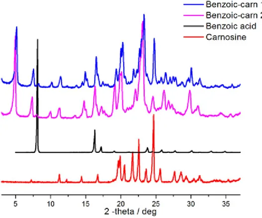
Unfortunately, once again no crystals suitable for X-ray single crystal diffraction were obtained. All our attempts to recrystallize the new samples were unsuccessful: a combination of various solvents with water had to be used to enable the dissolution of the salts that led to oil formation. Most likely, this happened due to the lower volatility rate of water in comparison to the organic solvents employed. Consequently, organic solvents left the solution first and the conditions became the same as the initial ones. It is worth mentioning that benzoic acid as a co-former at 1:1 stoichiometry gave a mixture of a new salt and carnosine. Increasing the amount of benzoic acid to a 2:1 ratio did not solve this problem: we could still observe the peaks of the salt and carnosine; moreover, the peaks of benzoic acid were also present in the diffraction pattern (fig. 3.1.8).
Fig. 3.1.8 Powder diffraction data of the salt obtained with carnosine - benzoic acid
Based on the information we got from the experiments mentioned above it is obvious that carnosine reacts with dicarboxylic acids (salicylic acid was the only exception). Thus, we decided to accomplish the reaction of carnosine with a homologous series of dicarboxylic acids HOOC-(CH2)n-COOH (n = 0-8, 10) using the same technique. According to the obtained results, we established that there is a noticeable relationship between the length of the aliphatic chain of dicarboxylic acids and the products of the reactions (Table 3.1.4).
Table 3.1.4 Carnosine with linear saturated dicarboxylic acids
# Acid pKa [36] Solvent Result
1 Oxalic acid 1.23; 4.19 H2O/EtOH Stable oil
2 Malonic acid 2.85; 5.05 H2O Stable oil
3 Succinic acid 4.21;5.41 H2O New salt
4 Glutaric acid 4.34; 5.41 H2O New salt
5 Adipic acid 4.41; 5.41 H2O New salt
6 Pimelic acid 4.51; 5.58 H2O New salt with starting materials
7 Suberic acid 4.53; 5.50 H2O/EtOH New salt with starting materials
8 Azelaic acid 4.55; 5.5 H2O New salt with starting materials
9 Sebacic acid 4.59; 5.59 H2O/EtOH New salt with starting materials 10 1,12-dodecanedioic acid 5.70; 6.60 H2O/EtOH New salt with starting materials
The interaction of carnosine with oxalic and malonic acids (n=0 and n=1, respectively) resulted in the formation of quite stable oils. Succinic, glutaric and adipic acids (n=2-4) reacted with carnosine producing oils which precipitated in 1-2 weeks into new substances (fig. 3.1.9-3.1.11).
Fig. 3.1.9 Powder diffraction data of the salt obtained with carnosine - succinic acid
Fig. 3.1.11 Powder diffraction data of the salt obtained with carnosine - adipic acid
All the other acids (n=5-8, 10) with carnosine formed oils which precipitated in 1-4 weeks as a mixture of new salts with the starting materials. Even pimelic (n=5) and azelaic (n=7) acids, which seemed to be pure upon a preliminary analysis using X-ray powder diffraction, appeared to be contaminated with the starting materials when they were analysed in a capillary (in transmission mode) for a much longer time.
Taking into account the fact that carnosine possesses antioxidant activity, the following step of our research was a trial to test the possibility of its interaction with an acid that will also have an analogous biological activity. Actually, we had already obtained such a salt – the one with succinic acid. Thus, we decided to try and obtain more salts with other acids with antioxidant properties. Ascorbic, glycolic and folic acids were chosen to react with carnosine using 1:1 stoichiometry and slow evaporation from solution technique. The interaction of carnosine with ascorbic acid resulted into an oil which seemed to be unstable, since bubbles of gas trapped in the oil appeared within a few days after oil formation and the amount of those bubbles kept increasing. The reaction with folic acid in 1:1 stoichiometry was found to be incomplete since the X-ray powder diffraction pattern showed the presence of the peaks of folic acid. Consequently, we decided to repeat the experiment using folic acid:carnosine in 1:2 ratio. In this way we managed to solve the problem. The substance was amorphous (the peaks of folic acids and carnosine were not visible in the diffraction pattern), but this
behaviour – amorphization upon salt formation - had been observed previously [37], and we were not surprised. The most successful result was achieved by the reaction of carnosine with glycolic acid: an oil that precipitated into a new salt was obtained (fig. 3.1.12).
Fig. 3.1.12 Powder diffraction data of the salt obtained with carnosine - glycolic acid
After numerous unsuccessful trials to recrystallize the obtained salts had been made, we decided to focus all our efforts on solving structures from X-ray powder diffraction patterns. This is still, and usually, a more complicate and time consuming approach, with respect to single crystal X-ray diffraction, and represents the frontier of structural solution in molecular solid-state and materials chemistry. To achieve this goal it was necessary first to perform thermal analyses - thermal gravimetric analysis (TGA) and differential scanning calorimetry (DSC). In this way we managed to understand which of the salts might contain solvent in their structures, and based on the percentage of solvent lost upon heating (TGA) to find out its stoichiometric amount. The figures 3.1.13 and 3.1.14 represent the TGA of succinic and glycolic acids correspondingly. They are given as an example of hydrated and anhydrous salts. From the TGA data collected for succinic acid-carnosine salt we can notice the loss of approximately 2.5% of samples weight at approximately 100 °C. Taking into account the fact that melting point of both carnosine and succinic acid are higher than 100 °C and that the salt was obtained from water we can conclude that this weight loss occurs due to water evaporation. At the same time, for carnosine-glycolic acid salt there is no significant weight loss at this temperature range, which indicates that water is absent in this sample.
Fig. 3.1.13 TGA of carnosine - succinic acid salt
Using TGA and DSC data we were able to show that four salts (with fumaric, maleic, succinic and salicylic acids) out of seven contained one water molecule per formula unit.
The following software was used to fully characterize all seven solids: with “X’Pert High Score Plus” and “Dash 3.3.3” the unit cells were determined, whereas “Topas 4.1” and “EXPO 2014” were applied for the structure solution.
The detailed information of the solved structures can be found in the experimental part of the thesis (table 2.8.1 and table 2.8.2). Using this characterization approach we managed to establish the structures of all the salts but the one with adipic acid. Despite all the numerous trials to solve it, we failed to do that so far. It confirms the fact that this approach is a complicate one and requires enough patience and skills. The figures 3.1.15-17 shows the structures of carnosine salts with glutaric, maleic and salicylic acids.
Space group P21 Unit cell parameters: a 8.16 b 38.82 c 5.41 α 90.0 β 90.37 γ 90.0 Cell volume 1713.84
Space group P 1; Unit cell parameters: a 17.31 b 8.90 c 5.76 α 75.63 β 102.49 γ 98.59 Cell volume 830.432
Fig. 3.1.16 Packing diagram of carnosine:maleate salt detailing hydrogen bond interactions
Space group P21; Unit cell parameters: a 12.08 b 10.52 c 7.19 α 90.0 β 90.18 γ 90.0 Cell volume 913.715
The use of mechanochemistry in our work with carnosine was limited because of the several obstacles we faced while using it. First, grinding never worked properly – only physical mixtures of the starting materials were detected by X-ray powder diffraction. Second, the use of drops of water in kneading experiments usually led to formation of sticky substances which did not dry out. The use of some drops of ethanol as a solvent looked more promising. By kneading with ethanol we managed to obtain the same product with fumaric acid as we did from solution. In the case of salicylic acid, however, the X-ray powder diffraction pattern recorded after ball milling for 30 minutes with carnosine showed that the reaction was not complete. Similar results were obtained for pimelic and azelaic acids: the intensity of the peaks of the starting materials was higher than that measured using solution method. Nevertheless, we cannot conclude that mechanochemistry is inapplicable for carnosine – organic acid interaction, since more thorough investigations in the field are required.
The carnosine salts with glycolic (fig. 3.1.18) and succinic acids (fig. 3.1.19) might be of higher interest since both of them also possess antioxidant activity and carnosine is used as a nutraceutical usually in combination with other antioxidants. Thus, the new formulation of carnosine as a salt with an acid possessing antioxidant activity can enhance its biological activity and, as a result, enable its usage as a separate drug/nutraceutical.
Space group P 21 Unit cell parameters: a: 10.372 b: 14.187 c: 4.689
α: 90.0 β: 100.75 γ 90.0 Cell volume 677.848
Space group P 1 Unit cell parameters:
a: 10.964 b: 10.454 c: 4.844 α: 62.20 β: 106.81 γ 120.89 Cell volume 420.558
Fig. 3.1.19 Packing diagram of carnosine:succinate salt detailing hydrogen bond interactions
Furthermore, there is some evidence that glycolic acid [38] and some derivatives of succinic acid [39] can act synergistically with other antioxidants. Therefore, it can be interesting to investigate biological activity of these salts. Another important issue is the stability of all the obtained salts. It was not investigated thoroughly, but their structures remained stable after storage for a couple of month at ambient conditions (the obtained X-ray powder diffraction patterns were exactly the same as the ones recorded just after the salts were formed). It can be beneficial since according to Sigma Aldrich requirements [40] pure carnosine must be stored in the freezer at -20 °C. The carnosine salt with folic acid can be even more interesting since it is soluble in water whereas folic acid is almost insoluble. Unfortunately, it is impossible to solve its structure using X-ray diffraction. Probably, it might be investigated in the future using solid-state NMR, IR or Raman techniques.
3.2 MELATONIN
The next step of our research dealt with the second antioxidant – melatonin. Based on the information from CSD there is only one known polymorph of melatonin so far. Thus, we decided to perform a polymorph screening. Unlike carnosine, melatonin is soluble in the majority of organic solvents whereas it is hardly soluble in water. Melatonin was dissolved in ethanol, methanol, dichloromethane, acetone, chloroform, DMSO and water (table 3.2.1). After the solvents evaporated crystals of melatonin were formed in all the vials, but the ones
with water and DMSO. The obtained crystals were analysed using X-ray Single Crystal diffraction. Unfortunately, in all the cases we dealt with the same already known crystal. Afterwards the crystals were ground and analysed using X-ray Powder diffraction. However, in all the occasions we had the known solid form.
Table 3.2.1 Melatonin polymorphic screening
# Solvents Single crystal Powder X-ray
1 EtOH the known polymorph ---
2 MeOH the known polymorph ---
3 Acetone the known polymorph ---
4 Dichloromethane the known polymorph
5 Chloroform the known polymorph ---
6 H2O --- the known polymorph
7 DMSO --- the known polymorph
Then we decided to try and co-crystallize melatonin in 1:1 ratio with eight co-formers from different chemical groups (morpholine, piperazine, DABCO, fumaric acid, nicotinic acid, isophthalic acid and 4,4’-bipyridine) using the solution method with methanol as a solvent (Table 3.2.2).
Table 3.2.2 Melatonin; solution method; solvent - methanol
# Co-former Single crystal Powder X-ray
1 Quinoxaline Melatonin crystals Starting materials
2 Morpholine Melatonin crystals Starting materials
3 Piperazine Melatonin crystals Starting materials
4 Fumaric acid --- Starting materials
5 DABCO Melatonin crystals Starting materials (a new peak
available)
6 4,4’-bipyridine --- Starting materials
7 Isophthalic acid Isophthalic acid crystals
Starting materials 8 Nicotinic acid Nicotinic acid crystals Starting materials
Unfortunately, with none of these co-formers we managed to obtain pure co-crystals. Furthermore, only with DABCO and piperazine there were some signs of interaction – a few peaks different from those of the starting materials were observed in the X-ray powder diffraction pattern (fig. 3.2.1).
Fig. 3.2.1 Powder diffraction data of Melatonin - DABCO interaction in methanol
All the other diffraction patterns indicated the presence of physical mixtures of the starting materials. Thus, we decided to change the solvent. However, with ethanol the results were similar to the ones described above, with the only difference that the intensity of the new peaks we observed in DABCO and piperazine diffraction patterns became much higher (Fig. 3.2.2). The next attempt was to use acetone, but this time there was no interaction in all cases.
On the basis of these results we decided to change not the solvent, but the method applied. The idea was to use ball milling with ethanol as a solvent, since it appeared to be the most suitable solvent based on the research described above. This time we succeeded to obtain melatonin co-crystals with DABCO and piperazine after milling for 20 minutes. The fact that we have new substances was confirmed by X-ray powder diffraction (fig. 3.2.3, 3.2.4).
Fig. 3.2.4 Powder diffraction data of Melatonin – piperazine co-crystallisation in ball mill
Our task was to solve the structure of the obtained co-crystals. Although numerous attempts to recrystallize the obtained co-crystals have been done, it appeared to be really tough to obtain a single crystal since, once the co-crystals were dissolved, mainly precipitation of the starting materials was observed. Finally, we managed to recrystallize melatonin - DABCO co-crystal from dichloromethane and to solve its structure using X-ray Single Crystal diffraction (fig. 3.2.5).
Space group P 21/c Unit cell parameters: a: 17.685 b: 9.925 c: 9.296 α: 90.0 β: 103.39 γ 90.0 Cell volume 1587.31
Fig. 3.2.5 Packing diagram of Melatonin-DABCO co-crystal DABCO hydrogen atoms have been omitted for clarity
Attempts at recrystallization of melatonin - piperazine co-crystals following the same procedure were unsuccessful. So it became obvious that the only way to solve its structure was from powder pattern. This time we were helped by the fact that DABCO and piperazine molecules are isomorphous. They are similar both in shape and hydrogen bonding propensity, and we found out that the obtained co-crystals had very similar unit cells and, finally, it was established that the intermolecular interactions in melatonin – DABCO and melatonin – piperazine co-crystals was identical (fig. 3.2.6).
Space group P 21/c Unit cell parameters: a: 17.052 b: 9.833 c: 9.335 α: 90.0 β: 105.10 γ: 90.0 Cell volume 1511.18 Fig. 3.2.6 Packing diagram of Melatonin-piperazine co-crystal
Some of piperazine hydrogen atoms have been omitted for clarity
Afterwards we tried to co-crystallize melatonin with a number of other co-formers using ball milling as the preferred approach. However, we failed to obtain new co-crystals so far (Table 3.2.3). Though only one of the obtained co-crystals can find a direct application in Pharmaceutical industry (DABCO does not belong to the GRAS list), our results confirm the reactivity of melatonin in co-crystallization processes, and emphasizes the need to continue the search for an improved formulation.
Table 3.2.3 Melatonin; ball milling; solvent - ethanol
# Co-former X-ray powder diffraction
1 L-Glutamine Starting materials
2 L-aspartic acid Starting materials
3 4,4'-trimethylenedipiperidine Starting materials
4 L-Glutamic acid Starting materials
5 4-aminosalicylic acid Starting materials
6 e- Caprolactam Sticky, impossible to analyse
7 L-Histidine Starting materials
8 L-tryptophan Starting materials