ii
DIREITOS DE AUTOR E CONDIÇÕES DE UTILIZAÇÃO DO TRABALHO POR TERCEIROS
Este é um trabalho académico que pode ser utilizado por terceiros desde que respeitadas as regras e boas práticas internacionalmente aceites, no que concerne aos direitos de autor e direitos conexos. Assim, o presente trabalho pode ser utilizado nos termos previstos na licença abaixo indicada.
Caso o utilizador necessite de permissão para poder fazer um uso do trabalho em condições não previstas no licenciamento indicado, deverá contactar o autor, através do RepositóriUM da Universidade do Minho.
Licença concedida aos utilizadores deste trabalho
Atribuição CC BY
iii
ACKNOWLEDGMENTS
I would like to give a special thanks to everyone that has, somehow, helped me facing all the academic and non-academic challenges, throughout the last five years. Every single one was of a huge relevance in my life, giving me strength and pushing me forward to complete my journey.
Also, I would like to acknowledge my supervisors Doutora Adriana Sampaio e Doutora Angela Bartolo for all the support, and for showing me that it is possible to achieve our goals if we work hard and if we stay positive.
To my friends, for all the moments we spent together, for all the things I have learned from you “Obrigada!”.
To my family, thank you all for giving me the opportunity of going further in my life, seeking a better version of myself, I promise I will always do my best so I will never let you down.
“No man is an island.” John Donne, 1624
v
Na busca das bases neuronais de Pantomimes: Evidência de fMRI Resumo
Como qualquer processo que ocorre no cérebro humano, o reconhecimento de gestos não é um mecanismo de fácil entendimento. Têm sido descritos vários resultados contraditórios no que diz respeito ao conhecimento das bases neuronais responsáveis pelo processamento de gestos que não envolvem manipulação de objetos. Mais ainda, continuam desconhecidos os correlatos neuronais relativos ao processamento cerebral de gestos que mimetizam a dita manipulação. No presente estudo estudamos os padrões de ativação cerebral aquando da observação de gestos de mímica e gestos intransitivos, produzidos com diferentes orientações – em direção ao corpo da pessoa que faz o gesto (p.e. usar batom) ou na direção oposta (p.e. conduzir um carro).
Globalmente, foi encontrado um padrão distinto de ativação cerebral para a observação dos dois tipos de gestos. Gestos de mímica orientados ao próprio corpo mostram uma ativação no córtex parietal (i.e., precuneus e circunvolução parietal inferior), enquanto que para a condição oposta observamos maior atividade no córtex occipital (i.e., circunvolução fusiforme e lingual). Finalmente, não se observam diferenças nos mapas de ativação cerebral para os gestos intransitivos.
vi
In Search of Neural Bases of Pantomimes: fMRI Evidence Abstract
As every neural process that happens in the human brain, gesture recognition is not of easy understanding. Controversial results have been found in the literature regarding the neural bases recruited for the recognition of non-object related gestures. With that in mind, we have now tried a distinct approach on the study of gestures hoping to unfold the mechanisms behind pantomimes’ recognition. The present paradigm consisted in the observation of pantomime and intransitive gestures performed either oriented towards the body (e.g. “using lipstick”) or away from it (e.g. “driving a car”).
Globally, we found increased activity in the anterior regions for the two gestures presented towards the body, and a more posterior activation for those away from the body. Additionally, we found that pantomimes and intransitive gestures are recruiting distinct neural. Results unveiled a more left-lateralized activation for pantomime observation, while intransitive gestures produced a more widespread pattern of activation along the brain. More specifically, pantomimes toward the body require the involvement of parietal regions (e.g. precuneus and inferior parietal gyrus) whilst pantomimes away activate occipital areas (e.g. fusiform and lingual gyrus).
vii TABLE OF CONTENTS Introduction ... 1 Method... 6 Participants ... 6 Procedure ... 6 Stimuli ... 6 Experimental task ... 7 Data Acquisition ... 8 Cognitive Measures ... 9 Data Analysis ... 10 Results ... 11
Behavioral Data Analysis ... 11
Neuroimaging Data Analysis ... 12
Discussion ... 17 Future Directions ... 21 Limitations ... 22 References ... 23 List of Figures Figure 1- Timeline
Figure 2- Sagittal and axial view of brain activity Figure 3- Sagittal and axial view of brain activity Figure 4- Sagittal and axial view of brain activity Figure 5- Sagittal and coronal view of brain activity Figure 6- Right and left brain activation
viii
Figure 7- Sagittal view of brain activity
Figure 8- Sagittal, coronal and axial view of brain activity Figure 9- Sagittal, coronal and axial view of brain activity LIST OF TABLES
Table 1-Type of gestures entailed in the experimental procedure Table 2- Recognition of Intransitives (away)
Table 3- Recognition of Pantomimes (toward) Table 4- Recognition of Pantomimes (away) Table 5- Recognition of Pantomimes away Table 6- Recognition of Intransitives away Table 7-Recognition of Pantomimes toward Table 8- Recognition of Pantomimes Table 9- Recognition of Intransitives LIST OF ACRONYMS
DRM - Dual-Route Model IFG – Inferior frontal gyrus POJ – Parieto-occipital gyrus PMC – Premotor cortex PPC – Posterior parietal cortex
VLPFC – Ventrolateral prefrontal cortex DLPFC – Dorsolateral prefrontal cortex STS – Superior Temporal Sulcus STG – Superior Temporal Gyrus PCS – Postcentral sulcus SMG – Supramarginal gyrus IFC – Inferior frontal cortex SMA – Supplementary motor area
NEURAL BASES OF PANTOMIMES
1
Introduction
Gestures play a central role in human’s daily life communication. We need them, in order to interact with our equivalents, as members of a sociable species. Being so, it is not surprising that our brain structures are intrinsically devoted to detect all types of informational gestures that one can either perform or observe, in a social interaction context.
It is undeniable that gestures are used, mainly, to convey social information. This is the reason why gestures are categorized according to their meaning, i.e. the information they hold. If they hold no meaning, they are called meaningless or unfamiliar gestures, distinguished from meaningful or familiar ones.
Meaningful gestures can be divided into two main categories: transitive (i.e. object-related gestures) and intransitive (i.e. non-object related gestures).
Intransitive gestures convey a social meaning (e.g. waving goodbye), and it is an expressive and symbolic action. Albeit related to objects, pantomimes are a mimicry of a tool use, performed in the absence of an object, which might give these gestures a classification in between of object/non-object related. This thesis project will focus on pantomime and intransitive gestures.
To account for the complexity and conceptual characterization of praxis processing, some complex cognitive models emerged. The Dual Route Model (DRM) (Gonzalez Rothi et al., 1991) proposed the existence of a lexical and a sub-lexical route for the processing of meaningful gestures and for gestures imitation, respectively, as they rely upon different brain structures. According to Rumiati et al. (2005) the lexical route is sustained by the ventral stream, which includes the left inferior frontal gyrus (IFG), while the sublexical route is supported by the dorsal stream, including the right parieto-occipital junction (POJ). Thus, suggesting that a left-lateralized network must be responsible for the processing of meaningful actions. In accordance, deficits in gesture processing are studied in the field of limb apraxia, a neurologic condition resultant from left brain lesion that leads to deficits in gesture production in the absence of other sensory-motor defects (Bartolo, Cubelli, & Sala, 2008).
Regarding meaningful actions, transitive and intransitive gestures are not only classified as different in purpose, but they are also taking distinct pathways when processed by the human brain. Focusing on the production of transitive gestures, superior motor areas including the premotor cortex (PMC) and the posterior parietal cortex (PPC) are remarkably activated (Balconi, Crivelli, & Cortesi, 2017). On the other hand, for intransitive gestures, areas such as the ventrolateral prefrontal cortex (VLPFC)
NEURAL BASES OF PANTOMIMES
2
along with the amygdala (i.e. emotional processing) seem to be responsible for the brain processing of this type of gesture (Lotze et al., 2006), highlighting the role of socio-affective brain areas for intransitive gestures processing.
On the search for the neural markers of gesture processing, Villareal et al. (2008) pointed out a remarked activity in the left inferior frontal gyrus (IFG) for intransitive gestures, along with activation in the left dorsolateral prefrontal cortex (DLPFC). Additionally, the authors found greater activity in the PMC and also some left temporal activation for pantomime gestures. In addition, an increased brain activity in the left superior temporal sulcus (STS) was equally found during the observation of both gesture types. Hence, suggesting a left-hemispheric lateralization for the processing of meaningful gestures.
Moreover, Bohlhalter et al. (2009), described a left frontoparietal network similarly activated for gesture planning of pantomimes and intransitive gestures. However, an increased left activation in the PMC and PPC was found for intransitive gestures, whereas pantomimes planning recruited more areas located bilaterally on the brain. Intransitive gestures seem to recruit more left-lateralized brain areas than pantomimes. Authors came forward with the hypothesis that this activation pattern could be due to the communicative nature of intransitive gestures.
Furthermore, when we look closer to studies on brain injuries, the dissociation between pantomimes and intransitive gestures becomes demarked. Generally, healthy subjects have more difficulty performing pantomimes than intransitive gestures (Mozaz, Rothi, Anderson, Crucian, & Heilman, 2002). Additionally, patients with limb apraxia showed more difficulties in pantomime production whereas the production of intransitive gestures is generally more preserved.
Mozaz et al. (2002) claims that the neural processing of intransitive gestures accounts for a more widely distributed network across the brain (i.e. perhaps bilaterally), and so, more resilient to local damage, as it happens in limb apraxia disease.
In the same line, Stamenova et al. (2010) evaluated the gestural production of pantomimes and intransitive gestures in stroke patients. Interestingly, authors found that patients suffering from left hemisphere damage were impaired in the production of pantomime but not of intransitive gestures, whilst at least 4 patients with right hemisphere damage showed deficits in the production of intransitive gestures while pantomime production was preserved, thus reinforcing the existence of distinct brain networks supporting pantomime and intransitive gestures in the brain.
NEURAL BASES OF PANTOMIMES
3
Similarly, Helon and Króliczak (2014) found that when intransitive congruent visual cues were presented in the left hemifield (i.e. processed by the right hemisphere), gesture categorization was significantly faster in comparison to when the cues were presented in the right hemifield. The authors pointed out that intransitive gesture processing would rely on right-sided brain areas, apart from the previously established ones in the left hemisphere, and specially engaging brain areas related with social cognition.
Bartolo, Cubelli, Salla and Drei (2003) reported a case-study of a patient that displayed impairment on the production of pantomimes coupled with working memory deficits, in the presence of an intact cognitive profile. Since pantomimes are artificial gestures whose motor programme has to be created de novo, the authors suggested that to produce pantomimes one needs to integrate the conceptual information about what the object is for with the procedural information about its use to generate the new gesture.
Given the current findings, the authors proposed a modified version of the classical model of gestural processing. The authors included a “workspace” component along the lexical route to account for this integration process. The current hypothesis can be valuable to explain the specific deficit found for pantomimes, proving that they might be calling for specific neural mechanisms (Bartolo et al., 2003).
Here, one can say that new evidence for the dissociation of intransitives and pantomimes was certainly found.
Even so, as stated beforehand, there is still no clear distinction between the neural correlates ascribed either to pantomimes or intransitive gestures processing. As noticed by Bartolo and Stieglitz Ham (2016) pantomimes and intransitive gestures are double dissociated in the neuropsychological studies. Nonetheless, no evidence has been found about the neural basis underlying this dissociation.
Gallagher and Frith (2004) found distinct neural pathways supporting brain processing of what the authors called expressive and instrumental gestures, that are basically intransitive gestures. Expressive gestures are used to express inner states (e.g. “I am bored”; “I don’t know”), while instrumental gestures production aims to induce behavioural changes on others’ current state of affairs (e.g. “Be quiet”; “Look up”). In this study whereas most of the expressive gestures were performed toward the body, instrumental were performed away from the body.
Basically, the authors proposed that expressive and instrumental gestures rely on dissociable brain circuits. The so-called “social brain” network responsible for mentalizing and theory of mind (ToM)
NEURAL BASES OF PANTOMIMES
4
aspects (e.g. involving the right superior temporal sulcus), is enrolled in the recognition of expressive gestures, whereas a more left-sided and language-specific network (e.g. left inferior frontal cortex), is recruited for the brain processing of instrumental gestures. In other words, the social nature of a gesture determines what areas will be recruited.
Bartolo and Stiegliz Ham (2016) on their revision of the literature on limb apraxia, more specifically on meaningful gestures, proposed that if intransitive gestures are associated with mentalizing and social brain areas (Gallagher & Frith, 2004) and if pantomimes are rooted on a “workspace” area (Bartolo, Cubelli, & Salla, 2003), then distinct brain mechanisms must be ascertained to each one of them.
Moreover, we now suggest that the current gap between neuroimaging measures - overlap of left-sided neural regions for pantomimes and intransitive gestures - and the behavioural studies on limb apraxia - major impairment in pantomimes with left-brain damage – might be due to the very nature of the gestures used to test this dissociation. As mentioned in Bartolo and Stieglitz Ham (2016), the majority of the gestures tested on limb apraxia, and related studies, share the same instrumental feature (i.e. are performed in order to change the behaviour of others).
Following this statement, we have so decided to give a closer look to the fMRI neuroimaging studies that have attempted to demonstrate the dissociation presented above. Besides the instrumental nature, usually, ascribed to the gestures used in the previously reported research studies, gestures share another characteristic in a very consistent way, across all literature. Either tasks on gesture production or gesture observation, made use of gestures resembling actions which take place away from the body of the person that is executing the gesture, and in a much lesser extent, gestures are performed toward the body, which hinders the possibility of drawing stringent conclusions.
In this train of thoughts, in the current study we propose a different approach on the categorization of gestures. That might help us, not only, to answer the inquiry raised by Bartolo and Stieglitz Ham (2016), but also, enable us to tell apart pantomime and intransitive gestures’ brain networks. We categorized gestures based on body-gesture relation, the differentiation is as follows:
o Toward the body: gestures performed within the body, either touching the body, directly, or having hand direction oriented towards oneself;
o Away from the body: oriented toward others, hand directed to an external point comparatively to the individual’s body.
NEURAL BASES OF PANTOMIMES
5
Therefore, and for the purpose of our study, pantomime and intransitive gestures were divided into toward and away categories. Intransitive gestures performed toward the body would be related to the expression of inner states, and consequently, would engage the ToM brain network, related to mentalizing and empathy. Furthermore, pantomimes performed toward the body are associated with the personal intimate sphere of individuals, mimicking gestures, since they are in relation to personal activities such as self-nutrition, self-protection or hygiene.
For the latest, an increased activation is expected in posterior parietal areas. Although, at this point, we might not have a clear hypothesis on the neural basis of pantomimes carried out toward the body, due to scarcity of literature on this domain, our assumption is based on the literature of a particular condition called spatial hemineglect.
Patients having such condition, usually, are victims of right brain damage and are unable to detect stimuli in the environment, when presented on the contralateral side of the lesion (Pouget & Driver, 2000).
In hemineglect the posterior-inferior parietal regions seem to be involved in the human capacity to process space or body-space information (Vallar, 1998). The supramarginal gyrus (SMG), the post-central sulcus (PCS), and the posterior superior temporal gyrus (STG) are linked to the regulation of space and integration of proprioceptive information, which can modulate actions performed toward the body (Committeri et al., 2007). Thus, we expect that these cortical areas might be enrolled in the brain processing of pantomimes toward the body.
Based on previous findings, an increased activation on the right hemisphere is expected, for intransitives gestures performed toward the body (e.g. “I am cold”; “I am tired”), namely, in ToM related areas (i.e. medial prefrontal cortex, anterior cingulate, temporoparietal junction). On the other hand, for pantomimes directed toward the body (e.g. “using lipstick”; “answering the phone”) an increased activation is expected on the SMG, PCS and STG. And lastly, for both pantomimes performed away from the body (e.g. “driving a car”; “throwing a ball”) and intransitives away from the body (e.g. “waving goodbye”; “come here”), a similar activation is expected in left frontal and parietal areas, related with semantic processing.
In this study, we aim to achieve clear-cut neuroanatomical information, capable to diminish the current incongruency observed in the literature. Hopefully, with this innovative gesture classification, we can now achieve supporting fMRI data on the dissociation of pantomimes and intransitive gestures.
NEURAL BASES OF PANTOMIMES
6
Looking to the brain processing of gestures with a new perspective, maybe the underpinnings of gestural processing can be further clarified.
Method
➢ Participants
Fifteen volunteers (8 men; age range 16–40; M= 23,47; SD= 5,81) with no history of neurological or psychiatric disease, agreed to participate in the present study. These participants were recruited through the accreditation system of the Psychology School and through solicitations posted on social media. An informed written consent was obtained from all the subjects prior to experimental procedure. Except for one subject, all remaining ones were right-handed as assessed by the Edinburgh Handedness Inventory (Oldfield, 1971), as well as Portuguese native-speakers. The study was approved by the Ethics Committees of the University of Minho (SECVS; CEUM) and is in agreement with the ethical standards defined in 1964 Declaration of Helsinki.
➢ Procedure
The experimental procedure described below, was based in a previously conducted study (Soares Pereira, 2018, unpublished).
The first session was conducted at a Clinical Center (SMIC-Boavista) in the city of Porto. Besides the informed consent signature, participants were also invited to fill a sociodemographic questionnaire and the Edinburgh Handedness Inventory (Oldfield, 1971). Afterwards, participants underwent trough an fMRI task, in which the gestures recognition task was presented. All the security measures were first granted, prior to the participants’ placement into the fMRI scanner. Overall, the first session lasted about 90 minutes.
➢ Stimuli
All the presented stimuli were selected from a previously validated database. A pilot study was conducted, in which 16 Portuguese volunteers (50% males; M=29.38, SD=4.83) visualized a sample of 166 video-clips and were then asked to classify the depicted gestures as meaningless or meaningful. The videos mentioned above, displayed a human character performing gestures either with the left or right hand. Every stimulus was a video-clip with 4s time-duration. On total, 96 video-clips were assigned to the database. The inclusion criteria were based in a 75% rate of agreement among judges. No statistical differences were found whether the gestures were produced with the left or right hand, this fact agrees with previous literature on hand-related gestures (Króliczak & Frey, 2009).
NEURAL BASES OF PANTOMIMES
7
The created sample includes 48 meaningless gestures (24 performed toward the body; 24 performed away from the body) and 48 meaningful gestures – 24 pantomimes (12 toward the body; 12 away from the body) and 24 intransitive gestures (identically sub-divided). Each gesture was presented two times, either performed with the right or the left hand. Taken together, the stimuli yielded the formation of the 6 experimental conditions entailed in the study.
The totality of the gestures was used in the experimental setting and, within each condition, they were presented in a randomized order. Experimental conditions were counterbalanced across participants.
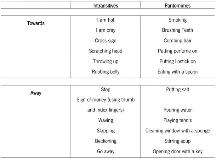
Table 1. Type of gestures entailed in the experimental procedure ➢ Experimental task
Stimuli were shown on a monitor inside the fMRI scanner. The experimental design consisted on an event-related fMRI procedure. Thus, the total stimuli of the experimental conditions were randomly assigned to every trial. Experimental conditions are defined as follows: (1) meaningless gestures toward the body; (2) meaningless gestures away from the body; and for the meaningful category - (3) pantomimes
Intransitives Pantomimes
Towards I am hot Smoking
I am cray Brushing Teeth
Cross sign Combing hair
Scratching head Putting perfume on
Throwing up Putting lipstick on
Rubbing belly Eating with a spoon
Away Stop Putting salt
Sign of money (using thumb
and index fingers) Pouring water
Waving Playing tennis
Slapping Cleaning window with a sponge
Beckoning Stirring soup
NEURAL BASES OF PANTOMIMES
8
toward the body; (4) pantomimes away from the body; (5) intransitive gestures toward the body; (6) intransitive gestures away from the body.
As mentioned beforehand, the stimuli presented depicted a human female person performing a gesture, placed in the center of a green background. Immediately before and after the stimulus presentation the screen turned green, displaying only a white cross (+) in the middle (Figure 1.). Participants were instructed to fixate the white cross that could remain on the screen from 4 to 16 seconds – jittered design.
Figure1. Timeline
Globally, the task consisted on the participants’ observation of gestures presented in the video-clips, while remaining inside the scanner. Meanwhile, they must reply to the following question: “Is this gesture meaningful?”. For that, they must press a button choosing “Yes” or “No” as their answer. Once again, the buttons’ order will be counterbalanced across subjects.
➢ Data Acquisition
Magnetic Resonance (MR) images were obtained through a Siemens 3 T scanner. Structural and functional sequence parameters of MRI acquisition were established in such terms: the T1-weighted 3D volumetric acquisition was obtained with a 3D MPRAGE (Magnetization Prepared Rapid Gradient Echo) sequence performed with the following protocol - time of repetition (TR)/ time of inversion (TI)/ time of echo (TE) = 2700 ms/1000 ms/2,33 ms/, flip angle (FA)= 7º, field of view (FoV)=240x256 mm2, 240 sagittal slices and isotropic voxel size = 0.8x0.8x0.8mm3. MPRAGE images were required as an auxiliary for the spatial normalization of the functional imaging data. Relatively to the functional acquisition, a 2D echo planar imaging (EPI) blood-oxygen-level dependent (BOLD) sensitive sequence with the following parameters was used: TR/TE = 2000ms/29ms, FA=90º, FOV=256 mm2, voxel size=3x3x3 mm,3 41 ascending interleaved axial slices with no gap, 535 slices.
4-16s
4-16s
4s
NEURAL BASES OF PANTOMIMES
9
➢ Cognitive Measures
The second session was devoted to the cognitive measures’ assessment. To begin with, participants were enrolled in a Theory of Mind (ToM) task, described by Sebastian and collaborators (2012). As stated by the authors, this task purpose is to access social cognition and participants’ empathic abilities. It consists of 30 cartoon vignettes displayed on the computer screen, 10 of those related to cognitive ToM, 10 others to affective ToM and the remaining ones concerned with physical causality (PC). Each cartoon tells a different story and is split in 3 distinct frames, presented sequentially and lasting 2 seconds each. Once it gets to the last frame, participants must judge on the portrayed characters’ feelings or intentions, i.e. infer their mental states, and choose the ending scenario they think more appropriate. The last frame remains 5 seconds on the screen and participants must give an answer within this time window. Task completion took participants about 8 minutes.
It seemed also relevant to test Phonemic Fluency, to assess language related abilities. We used the task proposed by Cavaco and collaborators (2012). Participants were asked to generate as many words as possible within the time-period of 1 minute. They were instructed to generate words beginning with specific letters, but were not allowed to say words that, generally, begin with capital letters, such as names of cities/places or personal names. Thus, the task has 3 trials and individuals must, sequentially, produce words starting with the letters M, R and P. Participants took one point for every correct word. Total score was obtained with the sum of each trial score. The task lasted 3 minutes (5 if we consider task instructions).
Albeit our study is not focused on the action production, action production and observation cannot be segregated (see Viana, 2015). Being so, it was equally relevant to have access to participants’ production abilities. For that we started by introducing a simple task called Motor Imagery. The task is an adaption of the original version proposed by Decety and Michael (1989). Motor imagery is the capacity to mentally simulate the production of a given act or movement. This cognitive ability is known to be, specially, recruited for pantomimes (Bartolo & Stieglitz Ham, 2016). Moreover action imagination and execution share similar brain regions activation.
Furthermore, imagining writing a sentence should take the same time as to writing it down on a paper (Decety & Michael, 1989). In the “execution phase”, participants were asked to hold a pen and point it to the fixation point (i.e. a black dot) in a white sheet. Their task was to write three sentences (actual movement) pronounced by the examiner. The sentences were the following: 1) “I am Portuguese”; 2) “I am Portuguese, and I live in Portugal”; 3) “I am Portuguese, and I live in Portugal with my family”.
NEURAL BASES OF PANTOMIMES
10
These sentences were chosen as they have a similar and intuitive meaning to every participant in the study. The time taken to complete the task was recorded.
On a second stage of this task participants had to imagine writing the same sentences on the paper (mental movement). Individuals were instructed not to move their hands (lay them on the table) nor say the sentences out loud. Once participants had complete the imagined writing, they had to say “STOP”. Response times were measured. Globally, 10 minutes were required to complete the task.
Furthermore, we wanted to test the individuals’ capacity to reproduce gestures. In Viana (2015) a new battery of tests for gestures assessment was created. The protocol was specially designed to test pantomimes and intransitive gestures production by means of a visual context. Meaningless gestures were assessed through an imitation task. Twelve drawings were shown to the participants, each picture depicting a scenario in which one or more characters are about to perform a gesture, either using an object (i.e. eliciting a pantomime, N=6) or doing some social gesture (i.e. eliciting an intransitive gesture, N=6). Drawings illustrating social interactions, and therefore inducing the use of an intransitive gesture came along with the following instruction: “What gesture do you think the character on the picture would do?”. Conversely, for the pantomime drawings condition, instructions were as follows: “Think of the object you would need if you were to use it in the portrayed situation. What gesture would you perform holding it?”.
Pictures were presented in a counterbalanced order across participants. Two additional drawings were presented as trial-ins, one for each gesture type. After that, every correct answer scores one point. The participants’ performance was video recorded.
All these measures provided additional information for the fMRI data interpretation. ➢ Data Analysis
All the fMRI data obtained were analyzed with the toolbox for neuroimaging analysis SPM12 extension (Statistical Parametric Mapping, Wellcome Trust Center for Neuroimaging, London, UK). In all the data obtained was performed a slice timing and a motion correction effect, was also applied the procedure of normalize, estimate and reslice followed by a spatial smoothing with an 8 mm filter. Afterwards, random-effect second-level analysis, one-sample t-test, was conducted with a cluster-extent based threshold of p < .001 (uncorrected). Established contrasts were brain mapped with the Automated Anatomical Labeling (AAL) atlas, based on MNI (Montreal Neurological Institute) coordinates. The contrasts created for the present analysis were as follows: pantomime towards > pantomime away;
NEURAL BASES OF PANTOMIMES
11
pantomime away > pantomime towards; intransitive towards > intransitive away; intransitive away > intransitive towards; pantomime towards > intransitive towards; pantomime away > intransitive away; intransitive towards > pantomime towards; intransitive away > pantomime away; pantomime > intransitive; intransitive > pantomime.
All the statistical analysis of behavioral data was conducted with SPSS Statistics 22 software (Statistical Package for the Social Science, IBM Corp.). The behavioral assessment of the study included a ToM task, followed by the Phonemic Fluency task, the Motor Imagery and Gesture’s assessment tests.
Results
➢ Behavioral Data Analysis
Considering performance in ToM task, participants’ scores revealed a range comprised between 22 and 30 points (M = 27.3, SD = 2.14) and in terms of RT’s they were from a minimum of 3205.5 ms to a maximum of 1217.4 ms (M = 1984.6; SD = 482.3). For the Phonemic Fluency task scores ranged between 13 and 51 (M = 34.7, SD = 9.24), and for the Motor Imagery task time values went from 0.55 s to 9.64 s (M = 3.90, SD = 2.45). In gesture assessment minimum score obtained was 3 reaching to a maximum of 6 (M = 5.1, SD = 0.71).
Correlation analyses between ToM scores (i.e. for either accuracy (AC) and reaction times (RT)) and the production of intransitive gestures were run. No significant correlation was found for either AC (r = .26, p = .35) and RT’s (r = .17, p = .65). There was also a non-significant correlation between ToM Ac and ToM RT’s (r = -.30, p = .28).
We have then proceeded to the analysis in which pantomimes production was related with scores in Phonemic Fluency and Motor Imagery. At first, pantomime production was tested for a possible association with phonemic fluency, analysis revealed no significant correlation (r = .37, p = .18). Similarly, results on pantomime production and the ones obtained in motor imagery task appeared non-related (r = -.17, p = .55).
Additionally, we conducted a paired samples t-test to verify whether there were significant differences between the production of pantomimes and intransitive gestures. No significant difference emerged (t (14) = .22, p = .82, d = .09).
NEURAL BASES OF PANTOMIMES
12
➢ Neuroimaging Data Analysis
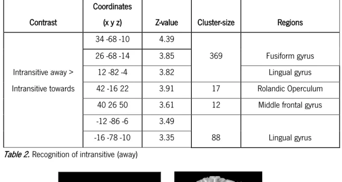
Initially, we contrasted activations related to the observation of intransitive gestures oriented towards > away from the body. In this contrast we did not observe no suprathreshold voxels. The opposite contrast, intransitive gestures away > toward the body revealed multiple regions activation. A pattern of increased activation was observed in the medial occipitotemporal gyrus, including the right fusiform gyrus (cluster = 369; Z = 4.39; x = 34, y =-68, z = -10) and lingual gyrus bilaterally (cluster = 369; Z = 3.82; x = 12, y = -82, z = -4). Additionally, we found activation in the right middle frontal gyrus (cluster = 12; Z = 3.61; x = 40, y = 26, z = 50) and the right rolandic operculum (cluster = 17; Z = 3.91; x = 42, y = -16, z = 22) in the insular lobe – see table 2; figure 2.
Table 2. Recognition of intransitive (away)
Figure 2. Sagittal and axial view of brain activity
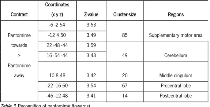
Regarding pantomimes, when we contrasted pantomime gestures toward with away from the body, we observed a more frontally localized activation. Brain areas that include the left supplementary motor area (SMA) (cluster = 85; Z = 3.63; x = -6, y = -2, z = 54), left precentral (cluster = 67; Z = 3.54; x = -22, y = -16, z = 60) and postcentral lobes (cluster = 14; Z = 3.41; x = -46, y = -12, z = 48), the right cingulum
Contrast
Coordinates
(x y z) Z-value Cluster-size Regions
34 -68 -10 4.39
369 Fusiform gyrus
26 -68 -14 3.85
Intransitive away > 12 -82 -4 3.82 Lingual gyrus
Intransitive towards 42 -16 22 3.91 17 Rolandic Operculum
40 26 50 3.61 12 Middle frontal gyrus
-12 -86 -6 3.49
Lingual gyrus
NEURAL BASES OF PANTOMIMES
13
(cluster = 20; Z = 3.42; x = 10, y = 8, z = 48) and cerebellum (cluster = 49; Z = 3.43; x = 16, y = -54, z = -44) showed increased activation – see table 3; figure 3.
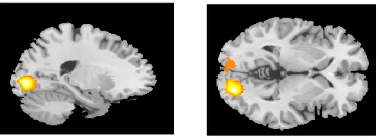
Table 3. Recognition of pantomime (towards)
Figure 3. Sagittal and axial view of brain activity
When considered pantomimes away from the body > toward the body, a distinctive pattern of activation was found, in particular in the occipital lobe, including the right and left calcarine sulcus (cluster = 838; Z = 5.02; x = 20, y = -86, z = 0), and the left fusiform gyrus (cluster = 269; Z = 3.42; x = -18, y = -88, z = -8) – see table 4; figure 4.
Table 4. Recognition of pantomime (away) Contrast
Coordinates
(x y z) Z-value Cluster-size Regions
-6 -2 54 3.63
85 Supplementary motor area Pantomime towards -12 4 50 3.49 22 -48 -44 3.59 49 Cerebellum > 16 -54 -44 3.43 Pantomime
away 10 8 48 3.42 20 Middle cingulum
-22 -16 60 3.54 67 Precentral lobe
-46 -12 48 3.41 14 Postcentral lobe
Contrast
Coordinates
(x y z) Z-value Cluster-size Regions
Pantomime away 20 -86 0 5.02 838
Calcarine sulcus
> -6 -94 -6 3.59
269
NEURAL BASES OF PANTOMIMES
14
Figure 4. Sagittal and axial view of brain activity
The remaining analysis concerns the direct comparison between pantomimes and intransitive gestures. Distinctive brain’s hemodynamic responses were found whenever the observed gestures where performed either toward or away from the body.
When pantomimes were compared with intransitives gestures (both gestures type performed away from the body), the left middle temporal gyrus (cluster = 16; Z = 4.06; x = -52, y = -74, z = 6) emerged as a key brain area – see table 5; figure 5.
Table 5. Recognition of pantomimes away
Figure 5. Sagittal and coronal view of brain activity
Contrast Coordinates (x y z) Z-value Cluster-size Regions Pantomime away
>
NEURAL BASES OF PANTOMIMES
15
Regarding the contrast of intransitive gestures > pantomimes (i.e. away condition),we observed an increased activation of the precentral gyrus (cluster = 79; Z = 3.50; x = 24, y = -28, z = 64), bilaterally.
Table 6. Recognition of intransitives away
Figure 6. Right and left brain activation
Several brain areas were activated for the contrast of pantomimes > intransitive gestures, for the towards the body condition. Overall, an increased left-lateralized brain network in the left precuneus (cluster = 15; Z = 3.40; x = -10, y = -70, z = 60), inferior parietal gyrus (cluster = 44; Z = 3.58; x = -44, y = -32, z = 36) and precentral lobe (cluster = 75; Z = 3.88; x = -28, y = -4, z = 56) was observed. Also, activity was seen in the right superior frontal gyrus (cluster = 10; Z = 3.17; x = 24, y = 4, z = 58).
Table 7. Recognition of pantomimes toward Contrast
Coordinates
(x y z) Z-value Cluster-size Regions
-22 -24 70 3.33
40
Precentral gyrus Intransitive away -20 -28 58 3.16
> 36 -18 56 3.43
Pantomime away 24 -28 64 3.50 79 Precentral gyrus
Contrast Coordinates (x y z) Z-value Cluster-size Regions Pantomime
towards 24 4 58 3.17 10 Superior frontal gyrus
> -10 -70 60 3.40 15 Precuneus
Intransitive towards -44 -32 36 3.58 44 Inferior parietal gyrus
NEURAL BASES OF PANTOMIMES
16
Figure 7. Sagittal view of brain activity
For the observation of intransitive gestures > pantomimes toward the body, no suprathreshold voxels emerged as significant.
As illustrated in the Table 8 and 9, pantomimes, as well as intransitive gestures were then directly contrasted without considering the gesture direction (toward or away from the body).
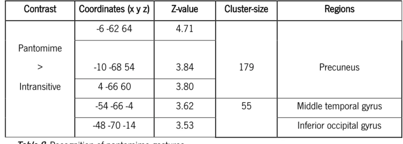
For the contrast pantomimes > intransitive gestures an increased activation was observed in the left precuneus (cluster = 179, Z = 4.71; x = -6, y = -62, z = 64), left middle temporal gyrus (cluster = 55; Z = 3.62; x = -54, y = -66, z = 54) and also left inferior occipital gyrus (cluster = 55; Z = 3.53; x = -48, y = -70, z = -14) – see table 8; figure 8.
Table 8. Recognition of pantomime gestures
Figure 8. Sagittal, coronal and axial view of brain activity
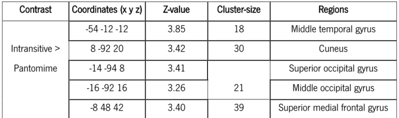
Concerning the inverse contrast, i.e. intransitive > pantomime we observed an increased activation in the left middle temporal gyrus (cluster = 18; Z = 3.85; x = -54, y = -12, z = -12), right cuneus
Contrast Coordinates (x y z) Z-value Cluster-size Regions
-6 -62 64 4.71
Pantomime
> -10 -68 54 3.84 179 Precuneus
Intransitive 4 -66 60 3.80
-54 -66 -4 3.62 55 Middle temporal gyrus
NEURAL BASES OF PANTOMIMES
17
(cluster = 30; Z = 3.42; x = 8, y = -92, z = 20), the left superior occipital gyrus (cluster = 21; Z = 3.41; x = -14, y = -94, z = 8) and middle occipital gyrus (cluster = 21; Z = 3.26; x = -16, y = -92, z = 16), and superior medial frontal gyrus (cluster = 39; Z = 3.40; x = -8, y = 48, z = 42) – see table 9; figure 9.
Contrast Coordinates (x y z) Z-value Cluster-size Regions
-54 -12 -12 3.85 18 Middle temporal gyrus
Intransitive > 8 -92 20 3.42 30 Cuneus
Pantomime -14 -94 8 3.41
21
Superior occipital gyrus
-16 -92 16 3.26 Middle occipital gyrus
-8 48 42 3.40 39 Superior medial frontal gyrus
Table 9. Recognition of intransitive gestures
Figure 9. Sagittal, coronal and axial view of brain activity Discussion
The human brain’s complexity is undeniable. This premise is, usually, confirmed by studies conducted with fMRI and other brain imaging techniques, in which multiple areas appeared to be recruited for one single brain function. Moreover, if we take the case of gesture recognition a lot of researches have been conducted that can state the complexity inherent to this process. As said above, while neuroimaging studies found that, at least, partially common networks are responsible for the brain processing of gestures whether they are object or non-object related (Bohlhalter et al., 2009), neuropsychological evidence reveals the distinctive characteristics of pantomimes (Bartolo et al., 2003), or even the social and communicative nature that is exclusive of intransitive gestures (Mozaz et al., 2002; Stamenova et al., 2010). Therefore, additional clarifications are now demanded to obtain a full comprehension of the neural networks requested by both intransitive and pantomime gestures processing.
We contributed to this realm as we have studied not only the neural patterns for the brain processing of intransitive and pantomime gestures, but also we compared gestures within the same category, to verify whether gesture direction (i.e. towards the body or away from the body) would be
NEURAL BASES OF PANTOMIMES
18
associated with distinct brain networks, and thus provide additional information for the possible intransitive/pantomime neural dissociation.
Considering the previously described results, we can take some reflexions on how the present study constitutes valuable information and to what extent it helps to clarify the controversy presented by the discoveries made until now.
Interestingly, results did not yield any map of brain activation for the contrast intransitive gestures towards > away from the body. In turn, the opposite contrast (intransitive away > intransitive towards) showed an increased activity in posterior areas of the brain, including the occipital poles (i.e. fusiform and lingual gyrus), but also the right middle frontal gyrus.
Taken together these results do not support the ones found by Gallagher and Frith (2004). In this study, authors compared intransitive gestures by dividing them in expressive or instrumental. In fact, they found differences in brain activity between the two conditions. Whenever the gestures had a more expressive purpose, areas related with social cognition and ToM appeared highly activated, and in the case of the gestures with an instrumental function, areas related with semantic processing showed an increased activity.
Despite the brain activity observed in the right middle frontal gyrus, respecting the recognition of intransitives away, the remaining areas belonged to posterior areas of the brain, mostly in the occipital lobe. The dissociation created, in the present study, did not provide similar results to those found in 2004, probably meaning that Gallagher and Frith’s dissociation is not related with gesture direction, as we have did here. Instead, possible differences in hemodynamic activation must be due to the distinct meanings that intransitive gestures convey.
The neural bases of pantomimes processing have been less explored in the literature, to our knowledge there is no study that have ever tried to explore the neural correlates of pantomime observation with the approach of the current study. This group of contrasts, includes the pantomimes performed toward the body in relation to pantomimes away from the body (pantomime towards > pantomime away) and the opposite (pantomime away > pantomime towards). Regarding the first one, we observed an increased frontal activation namely, in the left SMA and the left precentral lobe. Additionally, right postcentral lobe and middle Cingulum were also activated.
Increased activation in the motor and somatosensory areas (precentral and postcentral) in pantomime towards the body are likely to be related with actions performed onto the body (i.e.
self-NEURAL BASES OF PANTOMIMES
19
nutrition and personal hygiene) known to be processed in the superior parietal lobe with the integration of proprioceptive information (Committeri et al., 2007). A different pattern was observed when activations from the second contrast were obtained. For pantomimes away from the body, an increased activity was seen in the occipital lobe, particularly in the left calcarine sulcus and the left fusiform gyrus.
Our results, derived from the comparison of intransitives (away and towards) and pantomimes (away and towards), suggest that gesture direction plays a relevant role in gestures brain processing, in particular for pantomimes, with posterior areas mostly activated for the recognition of gestures away from the body, whilst the towards condition produces the recruitment of frontal activation.
Activations in occipital areas (i.e. bilaterally) for gesture observation were already described in the literature. As Villareal and co-workers (2008) found visual-related areas appeared activated for the observation of both type of gestures. Regions from the anterior part of the brain, namely activity in the medial frontal cortex has been identified as relevant for social cognitive processing (Amodio & Frith, 2006).
Considering these results, we analysed the away from the body condition, regardless of being pantomimes or intransitive gestures. Pantomimes’ observation originated occipital activity, when compared with intransitive gestures (pantomime away > intransitive away). Here, activations regarding pantomime observation were consistent with the ones found in the first contrasts revealing a tendency to occipital activation when gestures are performed away from the body. In fact, the left middle temporal gyrus showed a demarked activation as well, reinforcing the perspective of a more left-lateralized activation for these mimicked gestures when compared with intransitives (Villarreal et al., 2008).
On the other hand, the contrast intransitive away > pantomime away was associated with an increased bilateral activation of the precentral gyrus, which is consistent with previous evidence on bilateral activation for intransitive gestures observation (Helon & Króliczak, 2014).
Turning our focus to the towards the body condition, for pantomimes oriented toward the body (pantomime towards > intransitive towards) the most significant activity was found in left parietal regions. Namely, left precuneus and inferior parietal gyrus. Even though our hypothesis concerning pantomime observation, in this condition, accounted for an activation in the supramarginal gyrus (Vallar, 1998), the left parietal areas mentioned above are in clear agreement with what expected.
NEURAL BASES OF PANTOMIMES
20
Moreover, indirect evidence from patients with left-parietal brain lesion reveals a particular deficit for gestures imitation, if they are performed in their own bodies (Halsband, Schmitt, Weyers, Binkofski, & Gru, 2001), reinforcing the role of parietal lobe for gesture imitation (pantomime).
Respecting the contrast intransitive towards > pantomime towards, we did not find any suprathreshold brain activation. Interesting enough, in our study, the main effect of intransitive towards the body never revealed significant brain activations, either compared with pantomime gestures toward or intransitives away. This is in contrast with our predictions, as we expected a brain pattern of activations in the “social brain”, that is, ToM related areas (Gallagher & Frith, 2004).
Finally, our behavioral data showed that participants performed fairly well in the ToM task, with a ceiling effect, and no correlation with intransitive gestures performance was found. Despite their good performance on this social cognition task, we did not observe the recruitment of the ToM network, for the processing of intransitive gestures. Furthermore, brain processing of intransitive gestures does not seem sensitive to information related with gesture direction, so the presented dichotomy was not able to produce any changes on brain activity patterns.
At this point, we can consider that observation of pantomime and intransitive gestures recruit distinct neural pathways whether they are performed towards or away from the body.
More than a dissociation between gestures with distinct directions, the question we aimed to address was whether, by controlling for gesture direction, a neural differentiation between intransitive and pantomime gestures could appear.
For the intransitive gestures (in contrast with pantomimes), we found a significant brain activity in the left middle temporal gyrus, superior and middle occipital gyrus, and superior medial frontal gyrus along with the right cuneus. These results are in accordance with the hypothesis of a widely distributed brain network for intransitive gestures recognition (Stamenova, Roy, & Black, 2010).
Regarding the pantomime gestures (in comparison with intransitive), we observed an increased activity in left-posterior areas of the brain, including the precuneus, middle temporal gyrus and also some activity registered in the inferior occipital gyrus. This posterior network involving parietal and occipital regions have, already, been identified as responsible for recognition of gestures related with objects (transitive), in fact, studies showed that these areas (i.e. precuneus, including SMG) entail information of object-function and motor-action representations (Balconi, Vanutelli, Bartolo, & Cortesi, 2015). Although pantomimes do not involve object interaction, they are certainly related to it.
NEURAL BASES OF PANTOMIMES
21
Once we get here, we are able to state that pantomimes and intransitive gestures are requesting the involvement of distinct brain regions for its observation.
Albeit in our behavioral assessment Motor Imagery task did not reveal any relation with pantomime production, the previous results illustrate the huge relevance of object-related and action-information factors when brain recognition of pantomimes is at stage.
In summary, our results allowed us to verify the existence of distinctive patterns of activation when gestures are performed in the towards vs away conditions, particularly in case of pantomime recognition. The same was not verified for intransitive gestures recognition, in that case gesture direction did not produce any changes in neural activity, it was merely verified a widespread network for the observation of such gestures.
Even though, we were able to verify our main hypothesis. The recruitment of left-posterior areas for the recognition of pantomimes with activations particularly focused in the parietal and occipital lobes, for pantomime gestures when compared to intransitives, was noticed in our data. As we have hypothesized recognition of pantomime (towards) is, indeed, ascribed to brain regions in the superior parietal cortex, related with bodily-space perception and integration of object-related information (Vallar, 1998).
Further data analyses are maybe needed to explore the relation between intransitive gestures and pantomime. In particular, it is possible to run some regression analyses by including the social behavioural tasks we considered and the activation pattern for intransitive gestures. The more individuals are better in the behavioural task the more the social brain regions are expected to be activated.
Given our current results, it becomes clear that we are still far from knowing all about the cerebral processing of gestures, either object or non-object related or even towards and away from the body. Nonetheless, we are now one step closer from uncovering, not only, the neural bases of pantomimes, but also the general underpinnings of gestural brain processing.
Future Directions
Certainly, several questions remain unanswered (after all we are talking about brains), but that is what makes it a very promising field of investigation. If we take into account the dissociation created in this article, investigation can proceed to address questions regarding gesture production. Since we have tested the towards-away dichotomy for gesture observation, it might be possible to attain further knowledge if the same paradigm is conducted for gestures production (e.g. imitation, visual or verbal
NEURAL BASES OF PANTOMIMES
22
command). In what concerns pantomimes (i.e. mimicked gestures) the present dissociation can be of greater help in determining neural correlates of this kind of gestures, on its different modalities.
Also, in our study no facial expression accompanied the observed gestures, that was a deliberate decision so the gestures could be totally neutral per se and all the activations obtained could be related, exclusively, to gesture orientation and not to possible emotional content held by the facial expressions. In the future, it would be of great interest to pursue an investigation related to gestures produced near the face, i.e. explore a bit more the towards condition as presented in this work. Some gestures, for example “Be silent!” can be rather ambiguous, despite being addressed to someone else (i.e. oriented to others, intent to modify others’ behaviour) is performed in one’s own body surface, precisely on the face.
Neural bases of pantomimes are still in need of deeper investigation. Even though, in our research pantomimes did not reveal activations of social areas, a possible social component intrinsic to this category of gestures must be explored. It is true that some studies have reported compelling evidence of the relation between social skills and the ability to produce pantomimes. In 2015, Gizzonio and collaborators investigated pantomime production in a group of children with ASD (autism spectrum disorder) and they found that children with severe social deficits displayed lower performances when they had to pantomime gestures (Gizzonio et al., 2015). Being so, a possible social purpose must be considered for pantomime gestures and further investigation should attend to their reliance in social cognitive mechanisms.
Limitations
Albeit we have established a clear definition on what is the towards and away orientation of the gestures, it can still be opened to multiple interpretations. In fact, in our stimuli, the towards condition represents all the gestures that were oriented to the own body of the actress in the videoclip, and still one might say that this constitutes a gesture performed away from the observer. In the same train of thoughts, gestures performed in the away condition are oriented towards the observer’s point of view. Despite our efforts to create clear and distinctive conditions, this might be a topic of controversy. Future research should establish strong parameters that can reduce the noticed ambiguity.
NEURAL BASES OF PANTOMIMES
23
References
Amodio, D., & Frith, C. (2006). Meeting of minds: The medial frontal cortex and social cognition. Nature Reviews Neuroscience, 7, 268 – 277.
Balconi, M., Crivelli, D., & Cortesi, L. (2017). Transitive Versus Intransitive Complex Gesture
Representation: A Comparison Between Execution, Observation and Imagination by fNIRS. Applied Psychophysiology Biofeedback, 42(3), 179–191. https://doi.org/10.1007/s10484-017-9365-1 Balconi, M., Vanutelli, M. E., Bartolo, A., & Cortesi, L. (2015). Transitive and intransitive gesture
execution and observation compared to resting state: the hemodynamic measures (fNIRS). Cognitive Processing, 16(1), 125–129. https://doi.org/10.1007/s10339-015-0729-2
Bartolo, A., Cubelli, R., & Della Sala, S. (2008). Cognitive approach to the assessment of limb apraxia. Clinical Neuropsychologist, 22(1), 27–45. https://doi.org/10.1080/13854040601139310 Bartolo, A., Cubelli, R., Sala, S. Della, & Drei, S. (2003). Pantomimes are special gestures which rely on
working memory. Brain and Cognition, 53(3), 483–494. https://doi.org/10.1016/S0278-2626(03)00209-4
Bartolo, A., & Ham, H. S. (2016). A Cognitive Overview of Limb Apraxia. Current Neurology and Neuroscience Reports, 16(8). https://doi.org/10.1007/s11910-016-0675-0
Bohlhalter, S., Hattori, N., Wheaton, L., Fridman, E., Shamim, E. A., Garraux, G., & Hallett, M. (2009). Gesture Subtype--Dependent Left Lateralization of Praxis Planning: An Event-Related fMRI Study, (June). https://doi.org/10.1093/cercor/bhn168
Committeri, G., Pitzalis, S., Galati, G., Patria, F., Pelle, G., Sabatini, U., … Pizzamiglio, L. (2007). Neural bases of personal and extrapersonal neglect in humans, 431–441.
https://doi.org/10.1093/brain/awl265
Gallagher, H. L., & Frith, C. D. (2004). Dissociable neural pathways for the perception and recognition of expressive and instrumental gestures. Neuropsychologia, 42(13), 1725–1736.
https://doi.org/10.1016/j.neuropsychologia.2004.05.006
Halsband, U., Schmitt, J., Weyers, M., Binkofski, F., & Gru, G. (2001). Recognition and imitation of pantomimed motor acts after unilateral parietal and premotor lesions: a perspective on apraxia, 39, 200–216.
Helon, H., & Króliczak, G. (2014). The effects of visual half-field priming on the categorization of familiar intransitive gestures, tool use pantomimes, and meaningless hand movements. Frontiers in Psychology, 5(MAY), 1–11. https://doi.org/10.3389/fpsyg.2014.00454
Króliczak, G. (2009). A Common Network in the Left Cerebral Hemisphere Represents Planning of Tool Use Pantomimes and Familiar Intransitive Gestures at the Hand-Independent Level, (October). https://doi.org/10.1093/cercor/bhn261
NEURAL BASES OF PANTOMIMES
24
Lotze, M., Heymans, U., Birbaumer, N., Veit, R., Erb, M., Flor, H., & Halsband, U. (2006). Differential cerebral activation during observation of expressive gestures and motor acts. Neuropsychologia, 44(10), 1787–1795. https://doi.org/10.1016/j.neuropsychologia.2006.03.016
Mozaz, M., Gonzalez Rothi, L. J., Anderson, J. M., Crucian, G. P., & Heilman, K. M. (2002). Postural knowledge of transitive pantomimes and intransitive gestures. Journal of the International Neuropsychological Society, 8(7), 958–962. https://doi.org/10.1017/S1355617702870114 Pouget, A., & Driver, J. (n.d.). Relating unilateral neglect to the neural coding of space, 242–249. Rumiati, R. I., Weiss, P. H., Tessari, A., Assmus, A., Zilles, K., Herzog, H., & Fink, G. R. (2005).
Common and Differential Neural Mechanisms Supporting Imitation of Meaningful and Meaningless Actions. Journal of Cognitive Neuroscience, 17(9), 1420–1431.
https://doi.org/10.1162/0898929054985374
Stamenova, V., Roy, E. A., & Black, S. E. (2010). Associations and dissociations of transitive and intransitive gestures in left and right hemisphere stroke patients. Brain and Cognition, 72(3), 483– 490. https://doi.org/10.1016/j.bandc.2010.01.004
Vallar, G. (1998). Spatial hemineglect in humans, 2(3), 87–97.
Villarreal, M., Fridman, E. A., Amengual, A., Falasco, G., Gerscovich, E. R., Ulloa, E. R., & Leiguarda, R. C. (2008). The neural substrate of gesture recognition. Neuropsychologia, 46(9), 2371–2382. https://doi.org/10.1016/j.neuropsychologia.2008.03.004
NEURAL BASES OF PANTOMIMES
25
NEURAL BASES OF PANTOMIMES