Pedro Nuno Galvão Ferreira
Dissertation presented to obtain the
Ph.D degree in Biochemistry – Neuroscience
Instituto de Tecnologia Química e Biológica António Xavier
Universidade Nova de Lisboa
Oeiras,
February, 2016
Behavioural and transcriptional
plasticity in striatal circuits
Pedro Nuno Galvão Ferreira
Dissertation presented to obtain the
Ph.D degree in Biochemistry – Neuroscience
Instituto de Tecnologia Química e Biológica António Xavier Universidade Nova de LisboaOeiras, February, 2016
Behavioural and transcriptional
plasticity in striatal circuits
From optogenetics to epigenetics
To my parents, who taught me how to be (Para os meus pais, que me ensinaram a ser)
The work presented in this dissertation was carried out under the International Neuroscience Doctoral Programme (INDP, at the Champalimaud Neuroscience Programme, Champalimaud Centre for the Unknown) under the supervision of Dr Rui M. Costa, and the thesis committee supervision of Dr. Susana Lima and Dr. Carlos Ribeiro. Financial support was given by a doctoral fellowship from Fundação para a Ciência e Tecnologia (SFRH/BD/33279/2007, attributed to Pedro Nuno Galvão Ferreira), ERA-NET (F4T), European Research Council (COG 617142).
Did it matter then, she asked herself, walking towards Bond Street, did it matter that she must inevitably cease completely; all this must go on without her; did she resent it; or did it not become consoling to believe that death ended absolutely? but that somehow in the streets of London, on the ebb and flow of things, here, there, she survived, Peter survived, lived in each other, she being part, she was positive, of the trees at home; of the house there, ugly, rambling all to bits and pieces as it was; part of people she had never met; being laid out like a mist between the people she knew best, who lifted her on their branches as she had seen the trees lift the mist, but it spread ever so far, her life, herself.
Virginia Woolf Mrs. Dalloway
Table of Contents
Page
Acknowledgments ……….. 7
Resumo ……… 11
Abstract ……….... 13
Chapter I – General Introduction: From behavioural to transcriptional plasticity ………... 17
Part 1 – Basal ganglia neurobiology ……… 23
1.1 The basal ganglia: tuning in on the striatum ………. 23
1.2 A tale of twos: the roles in reinforcement and motor behaviour of the striatonigral and striatopallidal pathways and the dorsomedial and dorsolateral striata ………. 25
1.3 New tools, new tales: striatonigral and striatopallidal molecular physiology and transcriptomics ……… 30
Part 2 – Epigenetics in brain function ……… 33
2.1 A small introduction to epigenetics ………... 33
2.2 Neuronal activity-dependent gene expression ……….... 36
2.3 RNA Polymerase phosphorylation dynamics: poised memories ……….. 40
2.4 RNA Polymerase II pausing in the brain ………... 44
References ……….. 46
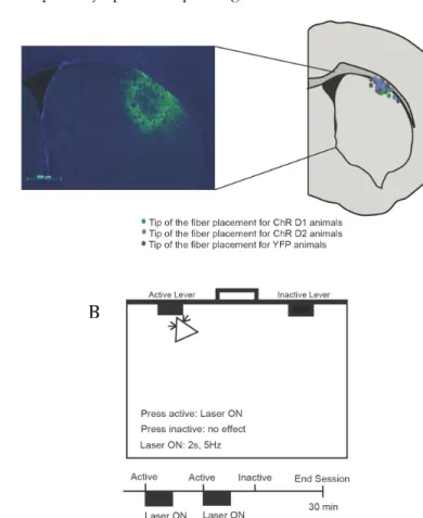
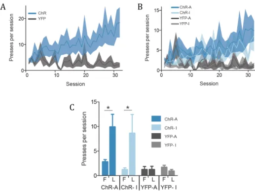
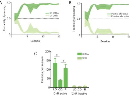
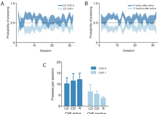
Chapter II – Differential role of striatonigral and striatopallidal dorsolateral striatum neurons in positive reinforcement ……….. 61
Introduction ………... 65
Results ……….... 67
Discussion ……….. 83
Experimental procedures ……….... 87
References ……….. 91
Chapter III – RNA Pol II phosphorylation dynamics in the striatum during motor skill learning ………. 97 Abstract ……….. 99 Introduction ………. 101 Results ……….. 103 Discussion ……… 117 Experimental procedures ……….. 121 References ……… 129
Chapter IV – General discussion: From striatal circuit function to RNA Pol II pausing ……... 135
Acknowledgements
It’s been a long, and at times arduous, road. I’ve heard of someone who lived two thousand years ago who seems to have experienced a similar ordeal, the differences being that 1) He was left out to dry, 2) that in His case it ended with a heavenward flight three days after His final examination and 3) that He finished without a PhD. Too bad for Him. However, such as He taught, as well as the Buddha, “when life throws lemons at you, you turn them into papers as best you can” (I might be paraphrasing here), and the only way I was able to turn these sour citruses into anything resembling a decent scientific output was through the help of a bunch of very dedicated friends and colleagues. This group of scientists and muggles pushed me forward, provided coffee (and alcohol) when I needed it, technical support when it was required, and lent an ear whenever one was needed. They almost made me love people as much as I do books. To them I owe this thesis.
First and foremost of all, my supervisor. If this PhD has taught me anything, then it is the absolute and inescapable value of good mentoring. I owe Rui a debt of gratitude equivalent in mass to a black hole, the day a black hole ate a planet too many. He has been the Yoda to my Luke, the Gandalf to my Frodo, the Dumbledore to my Harry. This doctorate would not exist were it not for his enthusiasm and relentless belief in the value of one as clumsy as I. If anything, the existence of this very thesis is proof of his magical powers, both as a supervisor, a scientist and a wholehearted human being. He gave me freedom and support, always in the measure I needed, even if I didn’t know it. If I am a scientist today, it is because of him. So thank you.
To my thesis committee, Susana Lima and Carlos Ribeiro, for the invaluable input and for keeping me on track. They taught me how smarts can meet
To my parents, aos meus pais. Assim sendo, este vai em Português. Para além de
terem-me dado biologicamente vida, os meus pais ensinaram-me a ser uma pessoa inteira, com todos os valores e idiosincrasias que isso acarreta. Eu não seria certamente a pessoa que hoje sou não fosse pelo constante apoio e carinho com o qual cresci, filho de pais que sempre quiseram que os filhos crescessem a ser tudo aquilo que conseguissem ser. Acima de tudo, por serem as pessoas mais completas que conheço, e por ensinaram-me que amar é mais que dizer – é viver e fazer. A eles esta tese é dedicada. (Como já mencionado umas páginas atrás, mas só para que não haja confusão, é mesmo para eles)
To my sister, the Dee Dee to my Dexter, and a continuous source of inspiration. Besides having shared the insides of the same mammal – with a two-year interval – we share the same darkly humorous brain and an inner sense of how great books are. She is probably the only person I like more than books.
To Rui (“o Ribeiro,” the cutest T. Rex), for putting up with me with the affection and patience of a saint, for showing me how to be both more human and a bit more generous, and for caring enough to teach me how to clean surfaces with three different types of detergent.
To the Costa lab, from the golden oldies of IGC days, to the new zygotes who are just beginning to appreciate how much they have struck gold here. A very very special thanks to Mafalda, my partner in crime, the Sam to my Frodo, without whom a part of this thesis would have had a much harder time coming to life (if at all).
To those special CNP people, team mete-nojo and extended family: Andreia, Mafalda, Grant, Ana (aka Belinha), João, Nicco, Susana, Anna, Libbi and Mai. If the end of the world ever came, I wouldn’t want to spend it with anyone else.
To the initial PGCN (now INDP) 10: Rodrigo (especially for five years of amazing friendship and shared roof), Maria, Patrícia, Isabel, Margarida, Patrício, Mariana, Iris and Zé. It was a great ride.
To Lena, Sílvia and Daniela. Two of you are flying in from Stockholm and London for my PhD party (and the third would do so from Boston, were it not cheaper to fly in from Mars). I’ll be the last one graduating, but I’m the cutest.
To the CNP, for creating this most amazing of creative atmospheres. I’m sure I’ll never work in a place with a greater vibe, a superior summed IQ or better parties. You made me the scientist I hope I am today.
To the IGC, my first scientific home, and to its people. There I learned what doing great science meant, and to that I owe another eternal debt of gratitude. To Mike, whose mentoring helped me tread my first steps in science, and whose recommendation helped me get into this programme. To Rute, Ana Luísa and Sílvia, whose pragmatic spirit kept me on the right path.
To those geniuses, from Virginia Woolf to Fernando Pessoa and Sergei Prokofiev; from Giordano Bruno to Marie Curie, Albert Einstein and Barbara McClintock; from Marguerite Yourcenar to Eimear McBride and Martha Argerich. Of all of you I am in awe.
To Johannes Gutenberg, for inventing mechanical movable type printing. To you I owe my bankruptcy. My feelings for you resemble my intestines’ towards warm chocolate: I love you and hate you.
This work was supported by Fundação Champalimaud, Fundação Gulbenkian, Fundação para a Ciência e Tecnologia (SFRH/BD/33279/2007), ERA-NET (F4T), European Research Council (COG 617142).
Resumo
O sistema nervoso central medeia a relação entre um organismo e o seu meio ambiente, construíndo uma ponte fisiológica moldada por uma série de estímulos que poderá envolver processos celulares e moleculares de diferentes ordens e diferente natureza. Esta tese explora plasticidade ao nível dos circuitos, bem como a nível molecular.
Na primeira parte desta tese, utilizando uma abordagem optogenética, exploramos o papel das duas principais vias de projecção dos gânglios da base no reforço de acções, mostrando que, ao contrário daquilo que foi previamente descrito, a via directa e indirecta suportam o reforço positivo de acções, mas reforçam estratégias de acção diferentes. Estes resultados mudam o conhecimento da fisiologia e função dos gânglios da base, e mostram que as vias directa e indirecta possuem papeis diferentes mas complementares na modulação das nossas acções.
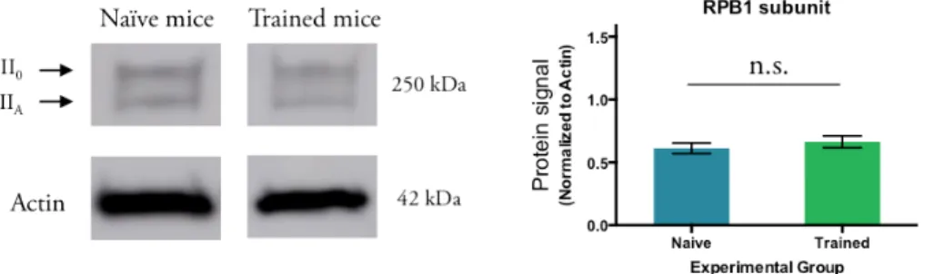
Na segunda parte desta tese, investigámos o impacto da aprendizagem no mecanismo conhecido por pausa da ARN Polimerase II (ARN Pol II), medindo como a aprendizagem de uma habilidade motora modula a dinâmica de fosforilação desta macromolécula. Mostrámos que a aprendizagem motora impacta a fosforilação in vivo da subunidade RPB1 da ARN Polimerase II, e que esta modulação ocorre em genes de activação imediata (immediate early genes). Estes resultados fornecem uma nova demonstração de plasticidade ao nível da transcrição, demonstrando, pela primeira vez, que aprendizagem modula a pausa da ARN Polimerase II em genes no cérebro adulto.
Abstract
The central nervous system mediates the relationship between an organism and the external and internal worlds, building a physiologic bridge that is shaped by a plethora of stimuli and may involve cellular and molecular processes of different orders and diverse nature. This thesis explores plasticity both at the circuit and molecular levels.
In the first part, using an optogenetic approach, we explore the role of the two main striatal output pathways in action reinforcement, showing that, unlike what has been previously described, the striatonigral and striatopallidal pathways both support reinforcement, but of different action strategies. These results introduce new insight into our knowledge of basal ganglia circuit function, and demonstrate the concomitant but complementary role of the direct and indirect pathways in shaping our actions.
In the second part of this thesis, we investigate the impact of learning on the mechanism known as RNA Polymerase II (RNA Pol II) pausing, by analyzing how learning a motor skill modulates the striatal phosphorylation dynamics of this macromolecule. We show that indeed learning a skill impacts on the in vivo phosphorylation of the RNA Pol II RPB1 subunit carboxy terminal domain, and that this modulation occurs at immediate early genes. These results provide a new demonstration of plasticity at the transcriptional level, demonstrating, for the first time, that learning modulates RNA Pol II pausing in the adult behaving brain.
Half way along the road we have to go, I found myself obscured in a great forest, Bewildered, and I knew I had lost the way. It is hard to say just what the forest was like, How wild and rough it was, how overpowering; Even to remember it makes me afraid. So bitter it is, death itself is hardly more so; Yet there was good there, and to make it clear I will speak of other things that I perceived.
Dante Alighieri The Divine Comedy – Inferno, Canto I
Chapter I
General introduction:
It is universally acknowledged that cells respond to different orders of stimuli, both internal and external, and modulate their gene expression programmes accordingly. As a cell type whose basic modus operandi consists in receiving information from its interconnected partners, integrating that information and mounting an appropriate response — be it producing an activity-induced action potential or shaping its very connectivity via dendritic spine remodeling — the neuron sits as the ideal laboratory for the study of activity-dependent gene expression. Ran and interpreted through a dynamically changing nervous system, experience molds anatomy and physiology.
The Oxford English Dictionary defines learning as “[an] acquisition of knowledge or skills through study, experience, or being taught.” If thought of as a translation of performance into neuronal coding, learning can be drawn as a dynamic and functional link between behavioural and neuronal plasticity (Faulk and Dolinoy, 2011; Fischer, 2014). In an anatomically complex brain, this junction may be studied in many different systems, one of them being the array of interconnected nuclei known as the basal ganglia. The striatum sits as the primary gateway into the basal ganglia, doubling its complexity into two main output pathways, distinguishable mainly by their gene expression profiles and projections to different structures (reviewed in Kreitzer and Malenka, 2008). The so-called “direct” and “indirect” pathways (also known as, respectively, “striatonigral” and “striatopallidal” pathways) have traditionally had differential roles assigned to them along the skill learning curve, with the direct pathway responsible for the initial phases of learning, and the indirect pathway involved in the later phases of skill learning, when the memory of how to do something is consolidated (Gerfen and Young, 1988; Albin et al., 1989; Alexander and Crutcher, 1990; DeLong, 1990; Gerfen et al., 1990; Le Moine et al., 1991; Bernard et al., 1992; Mink, 1996; Ince et al., 1997).
As anatomically complex as the basal ganglia may be, this complexity is perfectly mirrored in its function, which spans the regulation of motor behaviour to calibrating the motivation-related valency of action performance (reviewed in
Graybiel and Grafton, 2015). However, there has been considerable controversy with mapping the roles of the direct and indirect pathways to motor and reward behaviours. It is a partial aim of this thesis, then, to elucidate the involvement of the direct and indirect striatal output pathways in one of these behavioural features: positive reinforcement.
As mentioned above, striatonigral and striatopallidal neurons are morphologically similar and need, subsequently, to be genetically identified. Given that a study which tries to link neuronal to transcriptional plasticity would be extremely enriched by the examination of these mechanisms with cell type specificity — especially when the circuits under scrutiny have such opposing functions — in this thesis we took advantage of the genetical identifiability of these two groups of neurons to attempt both activity manipulation and extraction of pure populations for both pathways.
As is often said, “you never forget how to ride a bike,” for, once consolidated, motor skills can last a lifetime. As with many other types of learning, proper and long-lasting consolidation of a motor skill very likely requires adjustments to the way the genomes of the neuronal circuits supporting that learning are read. This being said, the need arises, not only to identify and capture the specific neurons underlying a learning process, but also the regions of the genome being dynamically read.
Behind the apparent simplicity of the cellular nucleus and its three billion base pairs (bp) or readily readable nucleotidic information, the mammalian genome presents an awesome challenge of molecular interpretation. Similar to opening the correct section on a two million-page book, reaching the specific nucleotide sequence embedded in a dynamic bundle of chromatin is a task of gargantuan precision. The multi-dimensional chromatin structures that contain and comprise the genome need not only to be decompacted and the underlying DNA sequence exposed, but the correct transcription machinery needs to reach and adequately bind to that specific site (reviewed in Hager et al., 2009; Levine et al., 2014). A multitude of molecular mechanisms — such as acetylation, ubiquitination or
methylation of the histone macromolecules of nucleosomes, or methylation of cytosines within DNA CpG dinucleotides — facilitate or hinder gene expression, in most cases by changing the biophysical relationships between DNA and the protein content of chromatin (reviewed in Wolf and Linden, 2012; Meaney and Ferguson-Smith, 2010). Once chromatin is open, scaffolding elements and transcription factors prime DNA for transcription, facilitating the binding of effector molecular complexes such as RNA Polymerase II (RNA Pol II) (reviewed in Hager et al., 2009; Levine et al., 2014). The entire process of transcription, including its prior and posterior events, can be regulated at different stages, one of them being the very progression of RNA Pol II throughout the transcription cycle. Initially discovered in Drosophila melanogaster during the 1970s and 1980s, RNA Pol II promoter-proximal “pausing” has emerged as a major player in transcriptional regulation at several levels of mammalian biology, from embryonic development to brain function (reviewed in Jonkers and Lis, 2015). Although a specific involvement of RNA Pol II pausing in brain physiology has been implied through the work of the Dudek lab (including a new taxonomical approach to “immediate early genes” [IEGs] based on their activity-dependent transcriptional dynamics) (Saha et al., 2011), no role has of yet been demonstrated for this mechanism in the adult, in vivo, brain in the context of learning.
As a biological mechanism that so intuitively links upstream cues and stimuli to transcriptional plasticity, RNA Pol II pausing sits as an ideal candidate to bridge dynamic neuronal activity — necessary for learning — to a flexibly read genome. With this in mind, we set out to explore the impact of learning a motor skill on RNA Pol II pausing in the mouse striatum. After training in a fast lever-pressing task, an initial examination of the global phosphorylation dynamics of RNA Pol II was followed by profiling of the binding kinetics of these phospho-variants to the promoters and gene bodies of IEGs. Our experiments provide, to the best of our knowledge, the first demonstration of RNA Pol II phosphorylation modulation in the adult brain in the context of learning.
Given the dual nature of this thesis, the general introduction will be divided into two parts. In the first part of the introduction, we will focus on the basic neurobiology of the basal ganglia and the underlying complexity — and polemics — behind the physiology and function of its two main output pathways. In the second part of the introduction, we will shift our attention to the intersection between epigenetic mechanisms and neurobiology, exploring in detail the interplay between nuclear architecture, RNA Pol II pausing and neuronal activity-dependent epigenetic transcriptional regulation.
Part 1 — Basal ganglia neurobiology
1.1 The basal ganglia: tuning in on the striatum
Santiago Ramon y Cajal once said “The brain is a world consisting of a number of
unexplored continents and great stretches of unknown territory.” Sitting underneath the
columnar complexity of the cortex, the group of interconnected nuclei known as the basal ganglia has lost many of its functional and anatomical mysteries throughout the years. Dating back to the 1660s work of Thomas Willis, which first identified and systematized different subcortical structures, the basal ganglia saw its intricate structure and nuclei dissected and acknowledged throughout the following centuries (Steiner and Tseng, 2010). In 1941, Cécile and Oskar Vogt proposed the distinction of striatum (named by Samuel Wilson in 1912 due to its striated appearance) into caudate nucleus, putamen and nucleus accumbens (NAc) (Steiner and Tseng, 2010): the caudate-putamen constituting the dorsal striatum (primarily involved in motor control and habit/skill learning), with the NAc corresponding to the ventral striatum (traditionally involved in motivation and reinforcement) (reviewed in Graybiel and Grafton, 2015). A master regulator of motor behaviour and of the reinforcement value of learned actions, the basal ganglia are part of a series of loops linking several cortical areas, via basal ganglia, to the thalamus and back to the cortex (Joel and Weiner, 1994; Parent, 1990).
The striatum sits as the main entry point to the basal ganglia, receiving glutamatergic excitatory inputs from cortex and thalamus which synapse onto two distinct classes of striatal medium spiny neurons (MSNs) (reviewed in Kreitzer and Malenka, 2008). These GABAergic MSNs compose approximately 95% of all striatal neurons, the remaining 5% comprising aspiny GABAergic neurons and cholinergic interneurons (Kawaguchi, 1995; Bolam et al., 2000). The striatum owes its uniqueness, in great part, to this complete lack of glutamatergic cells (Kawaguchi, 1995; Bolam et al., 2000).
According to the classical view of basal ganglia information flow, a balance of glutamatergic and dopaminergic neurotransmission at the level of its entry point, the striatum, splits into a twofold array of projections, as striatal MSNs connect to different downstream nuclei (reviewed in Kreitzer and Malenka, 2008). Activation of the so called “direct pathway,” composed of MSNs expressing the dopamine D1 (D1R) and muscarinic M4 receptors (Chrm4) that project directly to basal ganglia output nuclei, leads to GABAergic inhibition of these structures: the substantia nigra pars reticulata (SNr) and internal — or medial — globus pallidus (GPi or GPm); in turn inhibition of the SNr (the neurons of which are also GABAergic) results in disinhibition of its thalamic downstream glutamatergic targets and its excitatory transmission to the cortex (Gerfen and Young, 1988; Gerfen et al., 1990; Le Moine et al., 1991; Bernard et al., 1992; Ince et al., 1997). This direct connection of striatum to SNr gave the direct pathway its other name: striatonigral pathway (Albin et al., 1989; Alexander and Crutcher, 1990; DeLong, 1990). Inversely, activation of the “indirect pathway,” comprising MSNs expressing the dopamine D2 (D2R) and adenosine A2A receptors, indirectly projects to the SNr via the external — or lateral — globus pallidus (GPe or GPl) and subthalamic nucleus (STN); this inhibition of GABAergic GPe neurons leads to a disinhibition STN glutamatergic neurons, which in its turn activates the SNr-thalamus GABAergic neurons (Gerfen and Young, 1988; Gerfen et al., 1990; Schiffmann et al., 1991). The bypass, via GPe, of the striatum-SNr indirect pathway resulted in it being named striatopallidal pathway (Albin et al., 1989; Alexander and Crutcher, 1990; DeLong, 1990). The divergence in naming the striatonigral and striatopallidal as the direct and indirect pathways, respectively, hence stems from the direct pathway reaching SNr directly, while the indirect pathway bypasses it.
As mentioned above, these MSNs are divided into two separate populations — the direct and indirect pathways — based mainly on their genetic identities and subsequent protein expression for, morphologically, they are indistinguishable (Gerfen and Young, 1988; Gerfen et al., 1990; Le Moine et al.,
1991; Bernard et al., 1992; Ince et al., 1997). Each of the differentially expressed dopamine receptors (D1R and D2R) triggers distinct intracellular signaling pathways based on the G proteins it’s linked to (reviewed in Calabresi et al., 2014).
The above biochemical differences, as well as the nuclei to which each pathway projects, further fueled the direct/indirect pathway dichotomy. The impact this view of pathway divergence had on behavioural function will be the focus of the next section.
1.2 A tale of twos: the roles in reinforcement and motor behaviour of the striatonigral and striatopallidal pathways, and the dorsomedial and dorsolateral striata
A selection of optimal actions amongst alternatives is essential to the way an organism relates to, and interacts with, an ever-changing world. This ability to learn and act according to experience requires a system that can encode action-outcome associations, be plastic enough to adapt behaviour according to dynamic changes in value, and initiate specific actions while inhibiting non-selected ones. The striatum sits at this functional juncture as also evidenced by its anatomical connectivity, as it receives inputs from varied cortical and limbic brain regions (reviewed in Ena et al., 2011).
Historically, basal ganglia function has been based on the dichotomic nature of its two main striatal output pathways, onto which an information processing duality is mapped (Gerfen and Young, 1988; Gerfen et al., 1990; Le Moine et al., 1991; Bernard et al., 1992; Mink, 1996; Ince et al., 1997). This influential view sees the direct pathway as facilitating or selecting appropriate motor sequences, while the indirect pathway inhibits or hinders movement (Albin et al., 1989; DeLong, 1990; Mink, 1996; reviewed in Kreitzer and Malenka, 2008). This dichotomy also extends to the different regions of the dorsal striatum, the dorsomedial and dorsolateral striata (DMS and DLS, respectively), with the first involved in goal-directed behaviours and the latter in more habitual behavioural
strategies (a division supported by further anatomical data, which shows afferent projections from frontal and parietal cortical areas to DMS and sensorimotor areas projecting preferentially to DLS) (reviewed in Hilário and Costa, 2008). The orbitofrontal cortex (OFC) has recently been shown, together with the DMS, as being engaged in goal-directed actions, with the same DMS/OFC neurons that support this action decreasing in activity; this is concomitant with DLS neuron activity increase as the animal shifts from a goal-directed to a habitual action (Gremel and Costa, 2013). In 2009, Yin et al. demonstrated a differential involvement of the dorsal striatum during the different stages of skill learning, with DMS more engaged in the initial acquisition of a skill and DLS starting to be involved early and continuing to be involved in late consolidation. It has also been shown that extensive training correlated with long-lasting changes in plasticity in both striatonigral and striatopallidal neurons in DLS, supporting the hypothesis that skill consolidation is dependent also on potentiation of indirect pathway neurons (Yin et al., 2009). This separation of functions becomes more complicated when one tries to fit together the direct/indirect pathway and DMS/DLS dichotomies, as both the DMS and DLS possess D1R- and D2R-expressing MSNs (D1 and D2 MSNs), resulting in an intermingling of direct and indirect pathway neurons throughout the dorsal striatum (reviewed in Ena et al., 2011).
As mentioned above, the direct and indirect pathways are hypothesized to exert opposing effects on motor behavior (Albin et al., 1989; DeLong, 1990; Mink, 1996; reviewed in Kreitzer and Malenka, 2008). This differentiation in action coding is supported by the idiosyncrasies in connectivity within each pathway, as both converge on the SNr and subsequently influence thalamic activity, with, however, the indirect pathway activating SNr-thalamus GABAergic neurons via STN glutamatergic disinhibition (Calabresi et al., 2014). The recent coming of age of optogenetics facilitated a more efficient and, for the first time, cell type-specific examination of the role of striatal output pathways in motor control (as well as in other behavioural settings). Cell type-specificity (as is the case of D1 and D2 MSNs) is a major issue in functional dissection of many neural circuits, as cell types are
often anatomically intermingled and thus extremely hard to tell apart if not through their genetic identity (Ena et al., 2011). This being said, traditional pharmacological and electrical stimulation techniques are insufficient for more precise, circuit-specific approaches. With the recent introduction, into mammalian neural cells, of single-gene component light-gated protein ion channels (most popularly, channelrhodopsin-2 [ChR2]), and subsequent millisecond-scale activation or inactivation of genetically-defined neuronal cell-types, scientists obtained unprecedented resolution for both the “where” and “when” of in vivo neuronal activity manipulation and baptized this approach “Optogenetics” (Boyden et al., 2005; reviewed in Yizhar et al., 2011).
In 2010, Kravitz et al. elaborated on the dual role of the direct and indirect pathways in motor control by expressing a Cre recombinase-dependent version of ChR2 in the DMS of D1 and D2 Cre-expressing MSNs (D1-Cre and D2-Cre mice, respectively), observing an increase in motor activity with D1 stimulation and decrease when stimulating D2 MSNs. The picture is more complicated, though, for concurrent direct and indirect pathway activation is observed preceding action initiation and termination (Cui et al., 2013). Simultaneous operation of direct and indirect pathway neurons may be key to integrate all the necessary inputs for a proper and functional motor response.
As hinted above, a go/no-go functional understanding of basal ganglia circuits, as it relates to motor behaviour, is entirely too simplistic (Calabresi et al., 2014). Recent work enhances this view by awarding to corticostriatal circuits a wider role in action initiation, performance and termination. This results in a much more complex view of sequence-related activity processing in the direct and indirect pathways, in which movement units are chunked into action sequences, with D1 MSNs preferentially displaying continuous or sustained sequence-related activity and D2 MSNs decreased or inhibited activity during sequence performance (Jin et al., 2014).
As we’ve seen, the striatal output pathways possess quite an integrative role in action performance: the “when” and “how” an animal does something. However, these basal ganglia subcircuits play an additional, very important role in the selection of action strategies, the “why” we perform a certain action — that is, the relationship that exists between an action and its outcome (reviewed in Macpherson et al., 2014).
Reinforcement learning — learning by trial and error within a contingency between and action and an outcome (possessor of a certain expected value) — may be defined as learning by interacting with an environment, and is at the very basis or instrumental conditioning (the learning of a certain action — or set of actions — that results in obtaining a reward and/or avoid punishment) (Yin and Knowlton, 2006; Macpherson et al., 2014). The performance of actions may then be dependent on an action-outcome (A-O) association, with action execution sensitive to changes in outcome value. In these cases, the action is dubbed goal-directed (Yin and Knowlton, 2006; Balleine et al., 2009). However, if an action is repeated without substantial changes in outcome value, it may become a habit, with further performance insensitive to changes in action-outcome contingencies (interaction with a stimulus results in a set response [S-R]) (Yin and Knowlton, 2006; Balleine et al., 2009).
Circuits for striatal-dependent instrumental learning have been previously identified as mentioned above: DMS involvement in goal-directed behaviours and initial skill acquisition; and DLS involved in habit formation and late skill consolidation (Yin et al., 2009; reviewed in Graybiel and Grafton, 2015). These long-lasting changes in DLS task-related neural activity have been shown to be pathway-specific, as they occur mainly in D2 MSNs and are observed to a lesser extent in D1 MSNs with prolonged skill training (Yin et al., 2009). Recently, it has been shown that striatal-specific deletion of A2AR (which colocalizes with D2R) leads to deficits in habit learning (Yu et al., 2009). Animals may also shift between goal-directed and habitual action strategies, or generalize previously learned actions (instead or learning an action de novo) when faced with a novel challenge (Hilário et
al., 2012). Concomitantly, animals trained in a random interval schedule in an operant task (which biases behavior towards habits) show a similar rate of pressing for both a lever that has been associated with reward and a new, inactive lever. This generalization is abolished lesioning DLS (Hilário et al., 2012).
Actions may be performed in order to procure a stimulus — a resulting outcome — that may be rewarding, while avoiding other stimuli that are aversive. A positive and rewarding outcome for a performed action leads to a reinforcement of that specific action; with time and repetition, action performance may become less dependent on the previously learned reward value of that action and thus result in the formation of a habit. While D1 neurons have ubiquitously been associated with a reinforcement role in action performance, D2 neurons are usually implied in aversion learning. Optogenetic experiments have lent additional credibility to this claim, with D1 stimulation inducing consistent reinforcement and D2 stimulation resulting in aversion (Kravitz et al., 2012). Using a different approach for circuit-specific manipulation, Hikida et al. (2010) showed that inhibition of D1 neurons decreases conditioned place preference previously associated with a reinforcing valence. Additionally, Durieux et al. (2009) demonstrated that D2 neuron-specific ablation in the ventral striatum results in an increase in drug reinforcement (showing, similarly to Kravitz et al. [2010], an inhibitory role for striatopallidal neurons in motor activity with striatal-wide D2 neuron ablation).
As we’ve seen, there have been extensive efforts towards functionally and anatomically characterizing the striatonigral and striatopallidal basal ganglia output pathways. This work has taken the basal ganglia field to a new understanding of how and why animals perform actions, and sheds new light on the role of the striatum in regulating motor behaviour and the reward value of an action. This will be further explored in chapter 2. Immense progress has also been made on the biochemical dissection of the direct and indirect pathways, studies that extend the idiosyncrasies of the striatonigral and striatopallidal pathways from function to molecules and their activity-dependent regulation. The examination of the
transcriptional output of genetically-defined neural circuits is a natural result of these new experimental approaches, as we will see in the next section.
1.3 New tools, new tales: striatonigral and striatopallidal molecular physiology and transcriptomics
A fuller understanding of genome sequences and function has taken the biomedical sciences to a fuller understanding of what a cell type is and how it can be biologically defined (Arendt, 2008; Deneris and Hobert, 2014; Trapnell, 2015). The development of bacterial artificial chromosome (BAC)-carrying transgenic mice, in which fluorescent protein genes are selectively expressed in specific neuronal subtypes under cell type-specific promoters, has allowed neuroscientists to functionally tackle neuronal circuits that would otherwise be extremely hard to individualize within the complex mammalian brain (reviewed in Durieux et al., 2011; Ena et al., 2011). Throughout the past decade and a half, the basal ganglia field has benefited enormously from these efforts that bridge molecular biology to systems neuroscience, and it has done so for two different experimental purposes: activity manipulation and visualization of neural circuits.
Recent D1- and D2-driven expression of Cre recombinase brought the Cre-lox system to basal ganglia research (Durieux et al., 2011; Ena et al., 2011). Some of the experimental consequences of this were the optogenetics studies mentioned above, which took cell type specificity to neuronal activity manipulation.
As suggested in the previous section, the striatonigral and striatopallidal pathways form an intermingled set of projections that can only be differentiated based on the specific set of molecules each cell type exclusively expresses (Ena et al., 2011). Given the molecular dichotomy between D1R expression in striatonigral neurons and D2R and A2AR expression in striatopallidal cells, visualization of these two circuits was attained by targeting the expression of fluorescent reporter genes using the promoters for the above-mentioned receptors (Gong et al., 2003; Shuen et al., 2008). Besides confirming the in vivo functional divergence between the two
pathways, these studies opened new windows into the characterization of the striatonigral and striatopallidal circuits, for, in this context, visualization also means identification (Durieux et al., 2011). The creation of Drd1a-dtTomato and
Drd2-EGFP mice allowed for a finer in vivo probing of neurophysiological details for each
pathway, but it also made something entirely different possible: the cellular isolation of pure striatonigral and striatopallidal populations for biochemical analysis (reviewed in Lobo, 2009). Following this experimental line, Lobo et al. (2006) applied fluorescence-activated cell sorting (FACS) and microarray analysis to the striata of Chrm4-EGFP, Drd1a-EGFP and Drd2-EGFP mice. Besides identifying a new set of differentially expressed genes between striatonigral and striatopallidal neurons, some of which with previously described clinical implications, this study provided an experimental framework for profiling cell type-specific gene expression dynamics.
Given the extreme and intricate cellular heterogeneity found in the mammalian brain, cell type-specific analyses of gene expression provide an essential improvement in biological resolution (Ena et al., 2011; Trapnell, 2015). These tools open interesting avenues for further scientific scrutiny. One possible and quite interesting next step involves the application of these novel cell type-specific isolation techniques, and subsequent gene expression analysis, to behaviorally relevant neuronal circuits. This will allow for a full bridging of neuronal and behaviour plasticity, taking the previously observed complexity in neural circuit function to the neuronal nucleus and it’s dynamic expression.
During the preamble to this introduction, we mentioned the bike-riding metaphor of learning a long-lasting skill. We also examined what we define as “learning,” and considered how the acquisition of the knowledge of “how to do something” is conveyed through experience and repetition. It is precisely to that directed shaping of our brains by experience, that acquisition and consolidation of “how to,” mediated by functional changes within specific neuronal circuits, and the
indentation it leaves on their genomes (as we saw, the modulation of the way neuronal genomes are interpreted), that we turn to next.
Part 2 — Epigenetics in brain function 2.1 A small introduction to epigenetics
If one thinks of the history of biology — and that of genetics in particular — over the first half of the twentieth century, much of the struggle was concentrated on finding the chemical basis of heritability. The actual term “genetics” was only coined by William Bateson in 1905, quite after Gregor Mendel’s 1850s and 1860s seminal experiments on the rules and patterns behind the inheritance of traits were rediscovedred (Krebs et al., 2014). The jump from so-called “Mendelian” or classical genetics to molecular genetics — that is, the search for the nature and regulation of the cellular molecules behind inheritance — was made possible by the 1911 and 1913 work of Thomas Morgan and Alfred Sturtevant on chromosomes and genetic linkage (Krebs et al., 2014). Between 1928 and 1952, with Griffith’s discovery of bacterial transformation, the Avery-MacLeod-McCarty experiment (identifying deoxyribonucleic acid [DNA] as the chemical principle behind the phenomenon identified by Griffith) and the Hershey-Case experiment (demonstrating DNA, and not protein, as the genetic material mediating viral infection of bacteria), did molecular genetics come into full being (Krebs et al., 2014). On the 25th of April 1953, Watson and Crick published their seminal two-page paper proposing a chemical structure for DNA (using unpublished — and involuntarily supplied — DNA X-ray diffraction data obtained by Raymond Gosling and Rosalind Franklin) and its obvious impact on information transfer in living matter (Watson and Crick, 1953).
By 1942, though, C. H. Waddington was already coining the term “epigenetics” to describe the interaction between genes (regardless of their — at the time unknown — biochemical nature) and their environment to produce a phenotype (Waddington, 1942). This epigenetic environment, or “landscape,” served as a useful metaphor for biological development, intuitively illustrating the way genetic information interacts with environmental cues (Waddington, 1942).
Waddington’s epigenetic landscape illustrates this interaction during development and subsequent stepwise cell-fate decisions leading to differentiation into multiple cell types (Waddington, 1942). However, an epigenetic regulation of gene function does not — and indeed is not — only applicable to development, as much of current epigenetics research concerns itself with adult somatic cell function (reviewed in Faulk and Dolinoy, 2011; Wolf and Linden, 2012). This interaction between the genome of an adult, terminally differentiated cell and a set of intrinsic or extrinsic cues will now be discussed in more detail.
Ever since Alfred Sturtevant showed genes to be linearly distributed on chromosomes that scientists have wondered how this information is read (Sturtevant, 1913). In much the same way as we find and interpret a word in a book, in order for a gene to be “read” — i.e. transcribed — that section of the genome must be accessible to the necessary regulatory and transcriptional machinery (reviewed in Hager et al., 2009). If we understand transcription as a three-way process, then besides the “message” and the “reader” (that is, respectively, the gene and the transcription machinery, including the enzyme RNA Pol II), “markers” that keep the message available and readable — similarly to a finger keeping a book page open — must also be in place (Hager et al., 2009; Meaney and Ferguson-Smith, 2010). Consequently, the genome should be viewed as a three-dimensional structure, the accessibility of which is tightly regulated (reviewed in Chakalova and Fraser, 2012).
If we zoom in on a eukaryotic cell nucleus during interphase, we find a variably compacted mesh of chromatin, the macromolecular complex composed of DNA, protein and ribonucleic acid (RNA) that packages the genome and controls gene expression and replication (reviewed in Chakalova and Fraser, 2012). Either DNA or the protein complement of chromatin may be chemically modified to manipulate the accessibility of DNA within the chromatin fiber, often by modifying the electrostatic interactions between DNA and surrounding proteins (Wolf and Linden, 2012; Meaney and Ferguson-Smith, 2010). DNA methylation is the
best-described DNA modification that impacts gene expression, usually consisting in the addition of a methyl group to cytosines within CpG dinucleotides by DNA methyltransferases (DNMTs), resulting mainly in transcriptional repression (reviewed in Schübeler, 2015).
In eukaryotes, the basic unit of chromatin is formed by nucleosomes, macromolecular complexes composed of two sets of the four core histones (H2A, H2B, H3 and H4) and around 147 base pairs of DNA wrapped around the histone octamer (reviewed in Qureshi and Mehler, 2014; Hager et al., 2009). These histone proteins may be subject of several sets of post-translational modifications (PTMs), amongst which phosphorylation, glycosylation, ubiquitination, S-nitrosylation and methylation, but most famously N-terminal lysine acetylation (Meaney and Ferguson-Smith, 2010; Riccio, 2010). Histone acetylation by histone acetyltransferases (HATs) results in more relaxed and accessible chromatin, while removal of acetyl groups by histone deacetylases (HDACs) results in tighter and less accessible chromatin (Meaney and Ferguson-Smith, 2010; Riccio, 2010). Chromatin — and by extension DNA — accessibility, however, is only part of the transcriptional puzzle. The spatial organization and positioning of the eukaryotic genome within the nucleus is an extremely well regulated process, with some loci displaying dynamic relocation upon transcriptional activation (Chakalova and Fraser, 2012; Bickmore and van Steensel, 2013). A preferential interaction with euchromatic (transcriptionally active) or heterochromatic (transcriptionally inactive) nuclear regions may also be the partial result of histone PTMs and local chromatin structure, intimately connecting chromatin modifications and genome positioning (Chakalova and Fraser, 2012; Bickmore and van Steensel, 2013). As mentioned above, RNAs may also modulate gene expression at the level of chromatin (Hamby et al., 2008; Zaratiegui et al., 2007). Small interfering RNAs (siRNAs) have been shown to bind to DNA and enable siRNA-mediated gene silencing, also facilitating long-term gene silencing by recruiting HDACs and DNMTs and subsequently modulating chromatin structure (Zaratiegui et al., 2007).
In the next section we will introduce in more detail the fine interplay between chromatin dynamics and gene expression in the brain, and focus on the curious peculiarities of epigenetic regulation of neuronal activity-dependent gene expression.
2.2 Neuronal activity-dependent gene expression
Animals depend on the correct interpretation of external and internal cues into an appropriate behavioural output in order to survive, making the nervous system an anatomical and physiological relay station between an animal and the surrounding environment (reviewed in Wolf and Linden, 2012). This experience of — and interaction with — an environment is conveyed through changes in neuronal connectivity, structure and activity that mold neural circuits in an activity-dependent manner for short- or long-lasting changes (reviewed in West and Greenberg, 2011). This phenomenon of neuronal adaptability is known as “neuroplasticity” (reviewed in West and Greenberg, 2011; Lyons and West, 2011). With the increase in organismal and functional complexity, more and more intricate molecular mechanisms were selected by evolution as epigenetic adaptation systems, including the several layers of epigenetic regulation briefly mentioned in section 2.1 (Wolf and Linden, 2012). A good example of this environment-to-genes axis is the 2007 Fischer et al. study showing a direct connection between associative and spatial learning and chromatin remodeling (via environmental enrichment-induced hippocampal histone acetylation and methylation of histones H3 and H4).
Many of these mechanisms were built into the signaling cascades behind neuronal plasticity and learning/memory, hinting towards an intimate — and many times necessary — link between neuronal activity, epigenetic adaptation and the genome’s output (Wolf and Linden, 2012; West and Greenberg, 2011; Lyons and West, 2011). Epigenetic regulation mediates neuroplasticity via a multitude of molecular interactions between chromatin remodeling enzymes (such as HDACs), Ca2+-dependent signaling proteins and activity-dependent transcription factors
(Riccio, 2010; West and Greenberg, 2011; Lyons and West, 2011). The translation of an extracellular signal into an intracellular one (or second messenger) is dependent on the specific signal and receptor involved, with many of these receptor proteins coupled to intracellular second messenger systems that regulate, in their own term, the activity of effector enzymes and downstream target proteins (i.e. ion channels and transcription factors) (Lyons and West, 2011; Goldie and Cairns, 2012). The Ca2+ second messenger system is based on Ca2+-sensitive proteins (such as Ca2+/calmodulin-dependent protein kinases, CAMKs) phosphorylating downstream targets that modulate gene expression: CAMKII, for example, phosphorylates the cyclic adenosine monophosphate (cAMP) response element-binding protein (CREB), a transcription factor with a widely documented role in long-term memory formation and consolidation, resulting in the induction of specific gene expression programs; CREB may also be activated by the cAMP-dependent protein kinase (PKA) via a different second messenger system, involving a G-protein-coupled receptor (GPCR)-mediated intracellular increase in cAMP (Dash et al., 1990; Bourtchuladze et al., 1994; reviewed in Lyons and West, 2011). CREB then binds to cAMP response elements (CRE) DNA sequences, inducing the activity of many activity-regulated genes, such as the IEG c-Fos or the
brain-derived neurotrophic factor (BDNF) (Barco and Marie, 2011; Lyons and West, 2011).
One of the binding partners of CREB, called CREB-binding protein (CBP), is involved in the transcriptional co-activation of several transcription factors, has inherent acetyltransferase activity of histone and non-histone proteins and locally remodels chromatin structure, as well as recruiting and stabilizing RNA Pol II (Barco and Marie, 2011; Lyons and West, 2011).
As mentioned above, c-Fos presents a paradigmatic example of activity-dependent gene expression (reviewed in Flavell and Greenberg, 2008). An IEG,
c-Fos has been present in the neuroscientific toolbox for some time now as an
activity marker, for c-Fos messenger RNA (mRNA) — and downstream protein — upregulation implies recent neuronal activity (Flavell and Greenberg, 2008; Saha
and Dudek, 2013). c-Fos was the first gene for which Ca2+-dependent promoter-proximal cis-acting regulatory elements were identified, in this specific case located 100 base pairs upstream of the c-Fos transcriptional start site and named calcium response element (CaRE) (Montminy et al., 1986). The CaRE sequence is similar to CRE elements identified in other gene promoters (such as the somatostatin gene [Montminy et al., 1986]). A second regulatory element was identified within the
c-Fos promoter, also identified as Ca2+-dependent, termed serum response element (SRE) (Montminy et al., 1986; reviewed in Flavell and Greenberg, 2008). The activity-dependent transcriptional regulation of IEGs will be further explored in chapter 3.
As we’ve seen, a significant epigenetic layer is present and necessary for activity-dependent neuronal gene expression (reviewed in Puckett and Lubin, 2011). Many of these epigenetic events have been found — and were initially identified — outside the brain, but many do seem to be highly enriched in the nervous system (Lyons and West, 2011; Puckett and Lubin, 2011). The methyl CpG binding protein 2 (MeCP2), an additional subject of Ca2+-dependent activation by phosphorylation, is present in high levels in mature neurons where it both represses and — more controversially — activates gene expression (Chahrour et al., 2008; reviewed in Guy et al., 2011). MeCP2 recruits HDACs to preferentially methylated DNA (5-methylcytosine [5mC]) sites and is present at near histone octamer level in neuronal nuclei (Nan et al., 1993; Chahrour et al., 2008; Skene et al., 2010; Mellén et al., 2012). 5mC may be converted to 5-hydroxymethylcytosine (5hmC) by ten-eleven translocation (TET) enzymes, an epigenetic mark which is highly present in the brain, preferentially at active genes, where MeCP2 is the major 5hmC-binding protein (Mellén et al., 2012). Recently, Rudenko et al. (2013) showed that neuronal TET1 is necessary for the regulation of memory extinction and IEG expression, such as that of c-Fos, the activity-regulated cytoskeleton-associated protein (Arc) and the neuronal PAS domain protein 4 (Npas4) genes.
Many of these epigenetic mechanisms have also been found specifically in the basal ganglia. TET1 has been shown to be downregulated in mouse NAc as a result of cocaine administration (Feng et al., 2015). Still in motivation and reward learning, striatal CBP-mediated histone H4 acetylation at the fosB promoter (Levine et al., 2005), histone H3 acetylation at the BDNF promoter (Kumar et al., 2005) or CREB-dependent striatal microRNA metabolism (Hollander et al., 2010; Im et al., 2010) have all been linked to cocaine-induced plasticity, or altered spine plasticity in MeCP2-deficient mice due to amphetamine exposure (Deng et al., 2010). A specific chromatin PTM, phosphorylation of histone H3 on serine-10 (H3-Ser10P), has been widely studied in striatal neurons (reviewed in Matamales and Girault, 2011). H3-Ser10P is hypothesized as promoting chromatin decondensation and subsequent gene expression (Johansen and Johansen, 2006) and, when combined with H3 lysine-14 methylation, is associated with c-Fos hippocampal transcription in neurons (Crosio et al., 2003). H3-Ser10P has also been shown to occur specifically in striatonigral neurons after acute cocaine treatment (Bertran-Gonzalez et al., 2008).
Recently, a new set of cis-acting epigenetic regulatory mechanisms was identified in the brain. Kim et al. (2010) demonstrated activity-dependent CBP-binding to enhancers in mouse cortical neurons, resulting in RNA Pol II recruitment and transcription of a new class of enhancer RNAs (eRNAs), specifically at enhancers actively involved in mRNA synthesis (and marked by histone H3 monomethylation of lysine 4). Some of these activity-dependent enhancers have also been shown to require c-Fos binding (which was believe to bind mainly to promoters), suggesting that c-Fos (that, together with other IEGs, regulates the expression of late-response genes) also controls neuronal activity-dependent gene expression at the level of enhancers (Malik et al., 2014).
As we’ve seen, activity-regulated gene expression depends highly on the creation of a chromatin environment permissive for effective transcription (Lyons and West, 2011; Puckett and Lubin, 2011). In neurons, this massive coordination of molecular mechanisms links neuronal activity to the transcription of selected
groups of genes, and it may be regulated from the most upstream signaling cascades responding to ligand-mediated receptor activation or intracellular Ca2+ influx, to the facilitation or repression of chromatin binding by transcriptional effectors and regulators (Lyons and West, 2011; Puckett and Lubin, 2011). The behaviour of RNA Pol II and its phosphorylation dynamics along the transcription cycle is a story in its own right. That is the story of RNA Pol II pausing — one that will be told in the next section.
2.3 RNA Polymerase II phosphorylation dynamics: poised memories
In 2001, the first draft sequences of the entire human genome were published, sketching a highly intricate map of known and uncharted coding and regulatory elements (International Human Genome Sequencing Consortium et al., 2001; Venter et al., 2001). The identification and exhaustive description of the DNA sequence of a mammalian genome as published in 2001, though, is of limited use without a thorough knowledge of the function and regulation of the genetic elements in question (Orphanides and Reinberg, 2002). Genome biology is as much an exercise in reading (i.e. interpreting the genome and regulating its expression) as in writing (that is, changing the sequence and structure of the genome itself). The proof of the pudding, as is often suggested, is in the eating after all.
As tradition had it, gene expression obeyed a series of segregated and ordered steps (Orphanides and Reinberg, 2002). Cytoplasmic signaling to the nucleus, transcriptional regulation, RNA processing, mRNA trafficking and translation — all happened in a neat and linear sequence, one molecular process after another (Orphanides and Reinberg, 2002). The contemporary view of gene expression, however, is quite different, with several of these processes known to work together to correctly regulate the expression of target genes: the role of chromatin, not just in packing genetic information, but also in regulating its interpretation; the involvement of the transcriptional machinery in recruiting the
apparatus necessary for processing nascent RNAs; or the role played by pre-mRNA splicing in the promotion of transcriptional elongation (reviewed in Orphanides and Reinberg, 2002, Kornberg, 2007; Hager et al., 2009).
The three-dimensional organization of chromatin and its subsequent modulation in a transcriptional context is a major level of transcriptional regulation (Bickmore and van Steensel, 2013; Ferrai et al., 2010; de Wit and de Laat, 2012; Wendt and Grosveld, 2014). The formation and maintenance of DNA loops — formed over transcription initiation between enhancers and core promoters and stabilized by cohesin and the CCCTC-binding factor (CTCF) — seem to be key for regulating proper gene expression (Bickmore and van Steensel, 2013; Ferrai et al., 2010; de Wit and de Laat, 2012; Wendt and Grosveld, 2014; Lenhard et al, 2012).
From the moment a cell decides a certain gene is to be expressed, a complex cascade of molecular events must unfold. A series of recent studies has highlighted how astoundingly beautiful and intricate these processes are and to which degree the function and dynamic structure of the genome are integrated (Dowen et al., 2014; Chen et al., 2014; Liu et al., 2014; reviewed in Levine et al., 2014); this realization becomes easy to appreciate if one remembers the three-dimensional nature of the cellular nucleus and the necessary rules for unpacking, reading and expressing genetic information: in other words, the timings and rules that dictate how regulatory factors find their genomic binding partners. Once this molecular hide-and-seek has been played to fruition, the preinitiation complex (PIC) is formed by binding of sequence-specific regulatory proteins (i.e. transcription factors) and recruited coactivators, such as the Mediator complex, to specific DNA elements or transcription factor binding sites (TFBSs, such as enhancers), upstream of the transcription start site (TSS). This allows RNA Pol II to then bind to general transcription factors (GTFs) at the TSS (reviewed in Kornberg, 2007; Hager et al., 2009; Wendt and Grosveld, 2014; Levine et al., 2014). Recruited RNA Pol II will typically transcribe around 20-50 bp before pausing, a process controlled by the interacting factors DRB sensitivity inducing factor
(DSIF) and negative elongation factor (NELF) (reviwed in Adelman and Lis, 2012; Heidemann et al., 2013; Jonkers and Lis; 2015). The paused RNA Pol II molecules may then proceed towards productive elongation or terminate transcription (Adelman and Lis, 2012; Heidemann et al., 2013; Jonkers and Lis; 2015). Pause release and subsequent elongation is catalyzed by positive transcription elongation factor b (P-TEFb)-mediated phosphorylation of paused RNA Pol II (Adelman and Lis, 2012; Heidemann et al., 2013; Jonkers and Lis; 2015).
Constituting a 550 kDa complex of ten to twelve different subunits (depending on species), RNA Pol II catalyzes, in eukaryotes, the synthesis of mRNAs precursors, small nuclear RNAs (snRNAs) and microRNAs (reviwed in Hager et al., 2009; Kornberg, 2007; Wendt and Grosveld, 2014; Levine et al., 2014). The largest of these subunits, RPB1, contains a carboxy terminal domain (CTD) composed of approximately 52 repeats of a heptapeptidic consensus sequence Tyr-Ser-Pro-Thr-Ser-Pro-Ser (YSPTSPS) (Allison et al., 1985; Corden et al., 1985). If individual aminoacids are numbered according to their position along the heptapeptide — Y1S2P3T4S5P6S7 — we find three distinct serine residues: ser-2 (Ser2P), ser-5 (Ser5P) and ser-7 (Ser7P). The phosphorylation of each of these serines carries with it a regulatory role in transcriptional progression and is catalyzed by different kinases within different regulatory complexes (reviewed in Brookes and Pombo, 2009; Heidemann et al., 2013; Jonkers and Lis; 2015). Some histone-modifying enzymes use these dynamic CTD configurations to discriminate promoter-proximal from promoter-distal regions with, for example, a combination of lysine methylation marks signaling different gene regions (H3K4 trimethylation around the promoter region and H3K36 methylation across downstream transcribed regions) (Brookes and Pombo, 2009; Heidemann et al., 2013; Jonkers and Lis; 2015). Upon PIC formation, mediator promotes phosphorylation of RNA Pol II on Ser5P of RPB1 CTD by the transcription factor II D (TFIID) (reviewed in Brookes and Pombo, 2009; Heidemann et al., 2013; Jonkers and Lis; 2015; Levine et al., 2014). This phosphorylation mark is maintained for part of the transcription cycle, but decreases as transcription progresses; the 5’ mRNA capping
machinery uses CTD Ser5P residues to physically tether itself to a position close to the mRNA exit channel (Brookes and Pombo, 2009; Heidemann et al., 2013; Jonkers and Lis; 2015; Levine et al., 2014).
As RNA Pol II elongates, the balance between Ser5P and Ser2P changes (with a decrease of Ser5P and increase of Ser2P), as RNA Pol II is released from the paused state by the P-TEFb complex, which includes cyclin-dependent kinase 9 (CDK9), promoting the phosphorylation of RPB1 CTD Ser2 as well as that of NELF (which dissociates from RNA Pol II upon phosphorylation) (Heidemann et al., 2013; Levine et al., 2014; Jonkers and Lis; 2015). Similarly to Ser5P, Ser2P anchors RNA processing factors as well, interacting with polyadenylation machinery at mRNA 3’ ends (Brookes and Pombo, 2009; Heidemann et al., 2013; Jonkers and Lis; 2015). Ser7 phosphorylation seems to be necessary for expression of snRNAs, but a fuller functional understanding of this phosphorylation mark is still in its infancy (reviewed in Heidemann et al., 2013).
Our understanding of RNA Pol II promoter-proximal pausing and its wider role in the regulation of gene expression is still incomplete. With the recent and current boom of functional genomic tools — from those integrating genome architecture and organization, to the visualization of in vivo single-molecule dynamics and their real-time three-dimensional movements — a more comprehensive bigger picture of genome wide expression regulation is emerging (Kieffer-Kwon et al., 2013; Liu et al., 2014; Chen et al., 2014; Crosetto et al., 2015; Schwartzman and Tanay, 2015). Most of the research into the mechanisms behind RNA Pol II pausing has focused on the “when” and “how” the necessary regulatory and effector machinery does its poised magic. However, some of the most intriguing and interesting insights into the overarching impact of this molecular phenomenon have come down to — and out of — the “where” it occurs. Our Waddingtonian brain is where we shall turn our attention to next.
2.4 RNA Polymerase II pausing in the brain
As has been a recurring theme throughout the second half of this introduction, we come back full circle to gene-environment interactions. Metazoans have evolved a complex myriad of mechanisms built specifically to translate developmental and environmental cues into appropriate transcriptional outputs (Wolf and Linden, 2012; Lyons and West, 2011; Puckett and Lubin, 2011). RNA Pol II pausing is such a mechanism (Gilmour and Lis, 1986). Transcription elongation was first identified as a rate-limiting step in gene expression in the 1970s and 1980s, but it was only with the analysis of the Drosophila melanogaster heat shock protein (Hsp) genes that already transcribing RNA Pol II was shown to accumulate downstream of gene promoters while associated with 20-50 bp-long nascent RNAs, naming this phenomenon, for the first time, as RNA Pol II “pausing” (Gilmour and Lis, 1986; Rougvie and Lis, 1988; Rougvie and Lis, 1990; Rasmussen and Lis, 1993). Throughout the years, other promoters were shown to display RNA Pol II pausing, in other species (including mammals) and ontogenetic contexts, from the transcriptional regulation of developmental genes to gene regulation in the adult organism (reviwed in Adelman and Lis, 2012; Levine et al., 2014; Jonkers and Lis; 2015). As a molecular mechanism that so beautiful and intuitively links activity-dependent transcriptional regulation to its upstream cues, RNA Pol II promoter-proximal stalling was naturally hypothesized as being at the basis of one or another biological phenomena. In the case of neuronal IEG expression, they just happened to be right.
In 2011, the lab of Serena Dudek published a paper in which it was shown, for the first time, that RNA Pol II pausing exists in the brain and that it is required for IEG rapid induction (further dividing IEGs into those with a rapid or delayed induction profile, that is, respectively, with or without promoter-proximal poised RNA Pol II), suggesting that this molecular mechanism may be involved in other cellular processes dependent on tightly regulated and fast transcriptional induction,
including the vast molecular and functional world of learning and memory (Saha et al., 2011). Aside from demonstrating that poised RNA Pol II does indeed play a role in neuronal activity-induced transcription as examined in in vitro neuronal cortical cultures (subjected to a prolonged treatment with tetrodotoxin [TTX], a sodium channel blocker, which upon washout induces quasi-synchronous neuronal activity), Saha et al. also detected RNA Pol II pausing in IEGs in the rat cortex and hippocampus in vivo, showing that this mechanism is additionally involved in RNA Pol II pausing-dependent IEG fast induction as a result of exposure to novel environments.
In chapter 3, we will explore the work of Saha el al. a bit further and frame, within the wider field of activity-induced gene transcription, our findings on the impact of learning on RNA Pol II pausing in the mouse striatum. We demonstrate that learning a motor skill shifts the phosphorylation dynamics of RNA Pol II, as well as its binding kinetics to IEGs, in the adult in vivo brain, providing a first instance of learning-dependent modulation of RNA Pol II pausing.