Stevia rebaudiana
Bertoni cultivated in Portugal: A prospective study
of its antioxidant potential in different conservation conditions
Marisa Barroso,1,2 Lillian Barros,1,3 M. Ângelo Rodrigues,1 Maria João Sousa,1 Celestino Santos-Buelga,2 Isabel C.F.R. Ferreira1,*
1
Mountain Research Centre (CIMO), ESA, Polytechnic Institute of Bragança, Campus de Santa Apolónia, 1172, 5301-855 Bragança, Portugal.
2
GIP-USAL, Facultad de Farmacia, Universidad de Salamanca, Campus Miguel de Unamuno, 37007 Salamanca, Spain.
3
Laboratory of Separation and Reaction Engineering - Laboratory of Catalysis and Materials (LSRE-LCM), Polytechnic Institute of Bragança, Campus de Santa Apolónia, 1134, 5301-857 Bragança, Portugal.
Abstract
Studies with well-established cultivation conditions are of utmost importance in order to standardize the plants production and, therefore, its chemical composition. However, the conservation conditions may influence some of the bioactive compounds present in those plants. Stevia rebaudiana Bertoni samples cultivated in the north-eastern of Portugal, were exposed to different conservation conditions (oven-dried at 30 °C, for six days, or kept fresh by freezing (-20 °C) in the same period), and then studied for their antioxidant potential (including antioxidant compounds such as reducing sugars, tocopherols and phenolic compounds, free radical scavenging activity (DPPH) and reducing power (RP)). Oven-dried samples gave the highest antioxidant activity (DPPH EC50 = 22.87 µg/mL and RP EC50 = 28.79 µg/mL) and the highest total phenolic compounds concentration. The most abundant phenolic compounds in both samples were two caffeic acid derivatives, 5-O-caffeoylquinic acid (chlorogenic acid) and 3,5-O-dicaffeoylquinic acid. Otherwise, the highest values of total tocopherols and total sugars were registered in frozen fresh samples.
This study gives clues to choose the most appropriate methodology to preserve primary or secondary metabolites of S. rebaudiana, involved in its antioxidant activity.
1. Introduction
The demand for natural sweeteners has been gaining more and more importance due to the great controversy associated with the use of some synthetic sweeteners as cyclamate, aspartame or acesulfame-K. The steviol glycosides (E-960) are a group of natural sweeteners of generalized availability. These compounds are obtained from Stevia rebaudiana Bertoni, a sweet plant native to South America (Carocho et al., 2015), belonging to the Compositae (Asteraceae) family. It is one of the 154 members of the Stevia genus and one of the only two species that produce sweet glycosides (Madan et al., 2010). This plant is also cultivated in China and Southeast Asia (Koyama et al., 2003).
Previous studies have shown that stevia leaves contain different antioxidant compounds, such as ascorbic acid (Kim et al., 2011), phenolic compounds (Shukla et al., 2009) including flavonoids (Jahan et al., 2010; Abou-Arab and Abu-Salem, 2010) and tannins (Savita et al., 2004). However, it is important to evaluate if the chemical composition of the plant is affected by the conservation process.
In fact, the plants with commercial value are often stored for some time before use and an efficient conservation is very important to avoid the loose of bioactive molecules during this process. One of the main conservation methodologies is drying, which prevents the growth of microorganisms and avoids certain biochemical reactions that may alter the plant organoleptic characteristics (Pinela et al., 2011).
Accordingly, the present work evaluated the effects of dehydration process on the S. rebaudiana antioxidant potential, by comparing the free radical scavenging activity, reducing power, and antioxidant compounds of fresh frozen and dried plants. It should be highlighted that the studied samples were obtained in an experimental field with defined cultivation conditions in order to standardize S. rebaudiana production and, therefore, chemical composition.
2. Materials and methods
2.1 Cultivation procedure, collection and treatment of the samples
sand), slightly acidic [pH(H2O) 6.3], and low in organic carbon (Walkley-Black) 5.6 g kg -1
. The soil was covered with anti-weed screen punched with holes spaced at 50 x 40 cm after the installation of the drip irrigation system. Cuttings of 10-15 cm height, previously placed on rooting in a greenhouse, were planted in the holes opened in the screen. The cuttings were obtained from a farmer’s field grown with cv. Morita. Crop plantation occurred in June 13th, 2014. Plants were watered twice a week throughout the growing season. The harvest took place in August 5th, 2014 and subsamples consisting of three whole plants were separated in fresh in stems and leaves.
After the collection, the specimens (leaves) were separated in two approximately equal halves and placed in paper bags. Half of the samples were dried at 30 °C (Memmert oven, Edelstahl Rostfrei, Germany) for six days, being designated as oven-dried samples, while the other half was immediately frozen (-20 °C) for the same period, being only lyophilized (FreeZone 4.5, Labconco, Kansas City, MO, USA) before analysis, in order to express the results in dry weight basis to be comparable with the oven-dried samples. These last samples were designated as fresh frozen samples.All the samples were kept in a desiccator and protected from light for subsequent use.
2.2. Standards and reagents
chemical suppliers. Water was treated in a Milli-Q water purification system (TGI Pure Water Systems, Greenville, SC, USA).
2.3. Free sugars
Free sugars were determined by high-performance liquid chromatography coupled to a refraction index detector (HPLC-RI) after an extraction method described by Barros et al. (2013), using melezitose as internal standard (IS). The equipment consisted of a combined system with a pump (Knauer, Smartline system 1000; Berlin, Germany), degasser system (Smartline manager 5000), auto-sampler (AS-2057 Jasco, Easton, MD) and an RI detector (Knauer Smartline 2300). The chromatographic separation was achieved with a Eurospher 100-5 NH2 column (4.6 ´ 250 mm, 5 µm, Knauer),
operating at 35 ºC (7971 R Grace oven). The mobile phase used was a mixture of acetonitrile/deionized water, 70:30 (v/v), at a flow rate of 1 mL/min. Sugars were identified by chromatographic comparison with standards and quantitation was performed by the IS methodology using a Clarity 2.4 Software (DataApex, Prague, Czech Republic). Results were expressed in g per 100 g of dry weight (dw).
2.4. Tocopherols
Tocopherols were determined after an extraction procedure previously described by Fernandes et al. (2013), using tocol as IS. The analysis was carried out using the HPLC system described above connected to a fluorescence detector (FP-2020; Jasco, Tokyo, Japan) programmed for excitation at 290 nm and emission at 330 nm. The chromatographic separation was achieved with a Polyamide II normal-phase column
Tocopherols were identified by chromatographic comparison with standards and quantitation was performed by the IS methodology using a Clarity 2.4 Software. The tocopherols content were expressed in g per 100 g of dry weight (dw).
2.5. Extracts preparation
Hydroalcoholic extractions were performed by stirring the plant material (leaves) (1 g) with 30 mL of methanol/water (80:20, v/v) at 25 ºC, with a stirring agitation of 150 rpm for 1 h, and filtered through Whatman No. 4 paper. The residue was re-extracted with an additional 30 mL portion of the hydroalcoholic mixture. The combined extracts were evaporated at 35 ºC under reduced pressure (rotary evaporator Büchi R-210, Flawil, Switzerland) and then further lyophilized.
The lyophilized extracts of both samples were re-dissolved in methanol/water (80:20, v/v, final concentration 5 mg/mL), for phenolic compounds determination and antioxidant activity evaluation. The final solutions were further diluted to different concentrations to be submitted to distinct in vitro assays.
2.6. Phenolic compounds
Double online detection was carried out in the DAD using 280 nm and 370 nm as preferred wavelengths and in a mass spectrometer (MS) connected to HPLC system via the DAD cell outlet.
MS detection was performed in an API 3200 Qtrap (Applied Biosystems, Darmstadt, Germany) equipped with an ESI source and a triple quadrupole-ion trap mass analyser that was controlled by the Analyst 5.1 software. Zero grade air served as the nebulizer gas (30 psi) and turbo gas for solvent drying (400 ºC, 40 psi). Nitrogen served as the curtain (20 psi) and collision gas (medium). The quadrupols were set at unit resolution. The ion spray voltage was set at −4500 V in the negative mode and spectra were recorded between m/z 100 and 1500. The MS detector was programed for recording in two consecutive modes: enhanced MS (EMS) and enhanced product ion (EPI) analysis. EMS was employed to show full scan spectra, so as to obtain an overview of all of the ions in sample. Settings used were: declustering potential (DP) -450 V, entrance potential (EP) -6 V, collision energy (CE)-10 V. EPI mode was performed in order to obtain the fragmentation pattern of the parent ion(s) in the previous scan using the following parameters: DP -50 V, EP -6 V, CE -25 V, and collision energy spread (CES) 0 V.
performed through the calibration curve of a compound from the same phenolic group. The results were expressed in mg/g of extract.
2.7. Evaluation of in vitro antioxidant properties
DPPH radical-scavenging activity was evaluated by using an ELX800 microplate reader (Bio-Tek Instruments, Inc; Winooski, VT, USA). Various concentrations of the extracts (30 µL) were mixed with a methanol solution containing DPPH radicals (6x10⁻⁵ mM, 270 µL) in a 96-well plate and then were left to stand for 60 min in the dark. The reduction of the DPPH radical was determined by measuring the absorption at 515 nm and calculated as a percentage of DPPH discoloration using the formula: [(ADPPH−AS)/ADPPH] × 100, where AS is the absorbance of the solution containing the sample at 515 nm, and ADPPH is the absorbance of the DPPH solution.
Reducing power was evaluated by the capacity to convert Fe3+ to Fe2+, measuring the absorbance at 690 nm in the microplate reader mentioned above. Various concentrations of the extracts (500 µL) were mixed with sodium phosphate buffer (pH 6.6, 200 mM, 500 µL) and potassium ferricyanide (1% w/v, 500 µL) in an Eppendorf (2 mL) and incubated at 50 ºC for 20 min. Trichloroacetic acid (10%, 500 µL) was added and then the upper layer (800 µL) was mixed with deionised water (800 µL) and ferric chloride (0.1%, 160 µL) in a 48-well plate; the absorbance was further measured at 690 nm.The results were expressed in EC50 values (sample concentration providing 50% of antioxidant activity or 0.5 of absorbance in the reducing power assay). Trolox was used as standard.
Three samples were used for each preparation and all the assays were carried out in triplicate. The results are expressed as mean values and standard deviation (SD). The results were analyzed using a Student´s t-test to determine the significant difference among two different samples, with α = 0.05. This treatment was carried out using SPSS v. 22.0 program (IBM, Armonk, NY, USA).
3. Results and discussion
3.1. Tocopherols and sugars composition
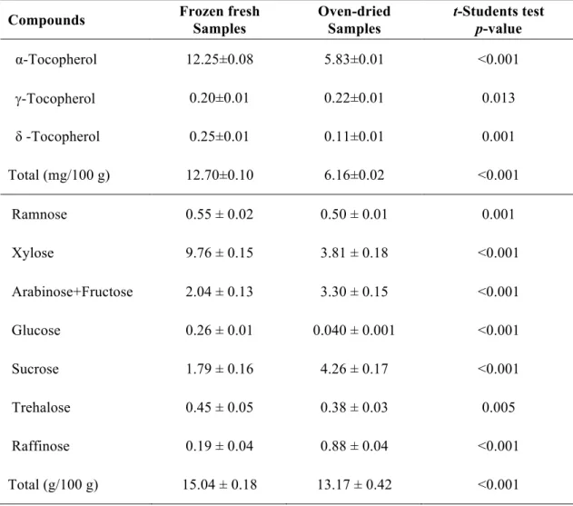
The tocopherol contents determined in the oven-dried and frozen fresh samples of S. rebaudiana are given in Table 1, with the presence of three isoforms (α-, γ- and δ -tocopherol), being α-tocopherol the majority compound in both samples. The highest value of total tocopherols was obtained in the frozen fresh samples, which is in concordance with Pinela et al. (2011), who reported a decrease in tocopherols content in Cytisus and Genista plant species after shade drying. The higher concentration of α -tocopherol in frozen fresh samples in comparison with the oven dried ones is also supported by the work of the previously mentioned authors.
Regarding free sugars composition (Table 1), oven-dried and frozen fresh samples revealed the presence of eight different sugars: rhamnose, xylose, arabinose+fructose,
glucose, sucrose, trehalose, and raffinose. In general, the highest value of total sugars
was registered in frozen fresh samples, mostly due to its higher content of xylose, the
Pinela et al. (2011) also reported a decrease in free sugars content of plant species from Cytisus genus and Genista genus, after shade drying. Not only free sugars and tocopherols are affected by the drying process but also other important compounds in S. rebaudiana such as steviol glycosides (Periche et al., 2015).
Sucrose was the major free sugar in the oven-dried samples (4.26 g/100 g), which is in agreement with the results reported by Pereira et al. (2015), who studied a dried and commercial sample of S. rebaudiana (3.93 g/100 g). However, the profile obtained was dissimilar between both samples, with the commercial one showing only sucrose, fructose, glucose and trehalose. Karimi et al. (2015) also studied soluble sugars in S. rebaudiana, describing an increasing content (mainly glucose) with soil moisture depletion. Fructose and sucrose were also identified by those authors, but in low levels. Indeed, differences in the chemical composition might be related with photoperiod (Ceumen and Geuns, 2013), planting times, plant stands and harvest regime (Serfaty et al., 2013; Karimi et al., 2015). It should be highlighted that the majority of studies in S. rebaudiana that are available in literature address to the evaluation of steviol (diterpene compound) glycosides (Brandle and Telmer 2007; Gardana et al. 2010; Geuns et al. 2003; Periche 2015), the responsible for the sweet taste of this plant, and not the herein studied free sugars.
3.2. Phenolic composition
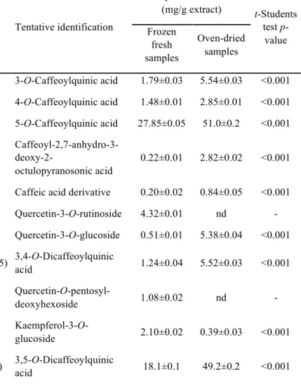
acid, quercetin-3-O-rutinoside, quercetin-3-O-glucoside and kaempferol-3-O-glucoside respectively, by comparison with authentic standards and analysis of their MS fragmentation pattern, retention time and UV-vis characteristics. The presence of these compounds has already been reported in S. rebaudiana by different authors (Cacciola et al., 2011; Muanda et al., 2011; Karaköse et al., 2011, 2015;Shivanna et al., 2013; Barba et al., 2015; Gaweł-Bęben et al., 2015).
Compounds 1 and 2 ([M-H]- at m/z 353) were identified as 3-O-caffeoylquinic acid and 4-O-caffeoylquinic acid based on the fragmentation pattern and characteristics reported by Clifford et al. (2003, 2005). Peaks 8, 11 and 15 ([M-H]- at m/z 515) could be assigned as 3,4-O-, 3,5-O- and 4,5-O-dicaffeoylquinic acids, respectively, based on their elution order, fragmentation pattern and relative abundances (Clifford et al., 2003, 2005). All those compounds have also been described in S. rebaudiana (Muanda et al., 2011; Karaköse et al., 2011, 2015; Shivanna et al., 2013), also confirming their identification in this sample. Peaks 4 ([M-H]- at m/z 381) and 5 ([M-H]- at m/z 391) presented a fragmentation pattern and UV-vis spectra similar to caffeic acid. Compounds showing a pseudomolecular ion at m/z 381 were detected in other Asteraceae like Erigeron breviscapus and identified as caffeoyl-2,7-anhydro-3-deoxy-2-octulopyranosonic acids (Zhang et al., 2007; Liao et al., 2011), identity that was tentatively assumed for the compound 4 detected herein. No structure could, however, be matched for peak 5 that remained unidentified as a caffeic acid derivative.
-at m/z 417), and 18 ([M-H]- at m/z 431) to kaempferol derivatives. They presented MS2 fragments corresponding to distinct losses of hexosyl (-162 u), deoxyhexosyl (-146 u) or pentosyl (-132 u) residues, although mass spectral characteristics did not allow characterising the position and nature of the sugar moieties. Similar compounds have already been reported in leaves of S. rebaudiana (Cacciola et al., 2011; Muanda et al., 2011; Karaköse et al., 2015; Shivanna et al., 2013). Among them, peaks 13 and 14 might be tentatively associated to xyloside and quercetin-3-O-rhamnoside, previously identified in leaves of S. rebaudiana by Shivanna et al. (2013) and Cacciola et al. (2011), respectively.
The most abundant phenolic compounds in the studied samples were 5-O-caffeoylquinic acid (chlorogenic acid) and 3,5-O-di5-O-caffeoylquinic acid. Oven-dried samples presented the highest total phenolic compounds concentration, due to the presence of higher concentration of phenolic acids, whereas the concentration of total flavonoids in both samples is similar. As for the qualitative composition, oven-dried samples did not reveal the presence of quercetin-3-O-rutinoside (rutin) and quercetin-O-pentosyl-deoxyhexoside, while the frozen fresh sample did not present peak 12 (kaempferol-O-hexoside).
3.3. Antioxidant activity
antioxidant activity in frozen fresh samples of four shrubby flowering plants widely consumed in folk medicine. However, our results are in accordance with the contents of phenolic compounds observed in both samples, once the extract of the oven-dried samples possessed higher concentration of total phenolic compounds (Table 2). Other authors have reported the high potential of S. rebaudiana leaves as a natural antioxidant, with associated health benefits (Xi et al 1998; Tadhani et al., 2007; Jahan et al 2010; Kim et al 2011).
S. rebaudiana is widely known for its natural sweeteners content, although this plant could also be considered as a good source of antioxidants, such as phenolic compounds. However, appropriate methodologies have to be applied to preserve the chemical constituents of S. rebaudiana. This study proved that oven-dried samples showed higher antioxidant activity and phenolic compounds contents than frozen fresh samples, whereas these latter possessed higher levels of tocopherols and free sugars. Overall, it is very important to establish suitable conditions to cultivate S. rebaudiana in order to standardize its chemical composition, but not least important is the preservation process selected for this plant.
Acknowledgments
The authors are grateful to the Foundation for Science and Technology (FCT, Portugal) and FEDER under Programe PT2020 for financial support to CIMO (UID/AGR/00690/2013), LSRE (Project UID/EQU/50020/2013) and L. Barros (SFRH/BPD/107855/2015) grant.
Abou-Arab, E.A., Abu-Salem, F.M., 2010. Evaluation of bioactive compounds of Stevia rebaudiana leaves and callus. Afr. J. Food Sci. 4, 627-634.
Barba, F.J., Grimi, N., Vorobiev E., 2015. Evaluating the potential of cell disruption technologies for green selective extraction of antioxidant compounds from Stevia rebaudiana Bertoni leaves. J. Food Eng. 149, 222-228.
Barros, L., Pereira, E., Calhelha, R.C., Dueñas, M., Carvalho, A.M., Santos-Buelga, C., Ferreira, I.C.F.R., 2013. Bioactivity and chemical characterization in hydrophilic and lipophilic compounds of Chenopodium ambrosioides L. J. Funct. Food. 5, 1732-1740.
Brandle, J.E., Telmer, P.G., 2007. Steviol glycoside biosynthesis. Phytochem. 68, 1855-1863.
Cacciola, F., Delmonte, P., Jaworska, K., Dugo, P., Mondello, L., Rader, J.I., 2011. Employing ultra high pressure liquid chromatography as the second dimension in a comprehensive two-dimensional system for analysis of Stevia rebaudiana extracts. J. Chromatogr. A 1218, 2012–2018
Carocho, M., Morales, P., Ferreira, I.C.F.R., 2015. Natural food additives: Quo vadis? Trends Food Sci. Technol. 45, 284-295.
Ceunen, S., Geuns, J.M.C., 2013. Influence of photoperiodism on the spatio-temporal accumulation of steviol glycosides in Stevia rebaudiana (Bertoni). Plant Sci. 198, 72-82.
Clifford, M.N., Johnston, K.L., Knight, S., Kuhnert, N.A., 2003. A hierarchical scheme for LC-MSn identification of chlorogenic acids. J Agric. Food Chem. 51, 2900-2911.
Dacome, A.S., Silva, C.C., Costa, C.E.M., Fontana, J.D., Adelmann, J., Costa, S.C., 2005. Sweet diterpenic glycosides balance of a new cultivar of Stevia rebaudiana (Bert) Bertoni: isolation and quantitative distribution by chromatographic, spectroscopic and electrophoretic methods. Process Biochem. 44, 3587-3594.
Fernandes, Â., Barreira, J.C.M., António, A.L., Santos, P.M.P., Martins, A., Oliveira, M.B.P.P., Ferreira, I.C.F.R., 2013. Study of chemical changes and antioxidant activity variation induced by gamma-irradiation on wild mushrooms: Comparative study through principal component analysis. Food Res. Int. 54, 18-25.
Gardana, C., Scaglianti, M., Simonetti, P., 2010. Evaluation of steviol and its glycosides in Stevia rebaudiana leaves and commercial sweetener by ultra-high-performance liquid chromatography-mass spectrometry. J Chromatogr. A 1217, 1463-1470. Ghanta, S., Banerjee, A., Poddar, A., Chattopadhyay, S., 2007. Oxidative DNA damage
preventive activity and antioxidant potential of Stevia rebaudiana (Bertoni) Bertoni, a natural sweetener. J. Agric. Food Chem. 55, 10962-10967.
Gaweł-Bęben, K., Bujak, T., Nizioł-Łukaszewska, Z., Antosiewicz, B., Jakubczyk, A., Karaś M., Rybczyńska, K., 2015. Stevia rebaudiana Bert. leaf extracts as a multifunctional source of natural antioxidants. Molecules 20, 5468-5486.
Geuns, J.M.C., Augustijns, P., Mols, R., 2003. Metabolism of stevioside in pigs and intestinal absorption characteristics of stevioside, rebaudioside A and steviol. Food Chem. Toxicol. 41, 1599-1607.
Karakose, H., Jaiswal, R., Kuhnert, N., 2011. Characterization and quantification of hydroxycinnamate derivatives in Stevia rebaudiana leaves by LC-MSn†. J. Agric. Food Chem. 59, 10143-10150.
Karakose, H., Muller, A., Kuhnert, N., 2015. Profiling and quantification of phenolics in Stevia rebaudiana leaves. J. Agric. Food Chem. 63, 9188-9198.
Karimi, M., Ahmadi, A., Hashemi, J., Abbasi, A., Tavarini, S., Guglielminetti, L., Angelini L.G., 2015. The effect of soil moisture depletion on Stevia (Stevia rebaudiana Bertoni) grown in greenhouse conditions: Growth, steviol glycosides content, soluble sugars and total antioxidant capacity. Sci. Hort. 183, 93-99.
Kim, I., Yang, M., Lee, O., Kang, S., 2011. The antioxidant activity and the bioactive compound content of Stevia rebaudiana water extracts. Food Sci. Technol. 44, 1328-1332.
Komissarenko, N.F., Derkach, A.I., Kovalyov, I.P., Bublik, N.P., 1994. Diterpene glycosides and phenylpropanoids of Stevia rebaudiana Bertoni. Rastitel'Nye Resursy 1, 53-64.
Koyama, E., Kitazawa K., Ohori, Y., Izawa, O., Kakegawa, K., Fujino, A., Ui, M., 2003. In vitro metabolism of the glycosidic sweeteners, Stevia mixture and enzymatically modified Stevia in human intestinal microflora. Food Chem. Toxicol. 41, 359-374.
Madan, S., Ahmad, S., Singh, G.N., Kohli, K., Kumar, Y., Singh, R., Garg, M., 2010. Stevia rebaudiana (Bert.) Bertoni - A Review. Indian J. Natural Prod. Res. 1, 267-286.
Muanda, F.N., Soulimani, R., Diop, B., Dicko, A., 2011. Study on chemical
composition and biological activities of essential oil and extracts from Stevia rebaudiana Bertoni leaves. Food Sci.Technol. 44, 1865-1872.
Pereira, C., Barros, L., Ferreira, I.C.F.R., 2015. A comparison of the nutritional contribution of thirty-nine aromatic plants used as condiments and/or herbal infusions. Plant Foods Hum. Nutr. 70, 176-183.
Periche, A., Castelló, M.L., Escriche, I., 2015. Influence of drying method on steviol glycosides and antioxidants in Stevia rebaudiana leaves. Food Chemistry, 173, 1-6.
Pinela, J., Barros, L., Carvalho, A.M., Ferreira, I.C.F.R., 2011. Influence of the drying method in the antioxidant potential of four shrubby flowering plants from the tribe Genisteae (Fabaceae). Food Chem. Toxicol. 49, 2983-2989.
Savita, S.M., Sheela, K., Sunanda, S., 2004. Stevia rebaudiana - A functional component for food Industry. J Hum. Ecol. 15, 261-264.
Serfaty, M., Ibdah, M., Fischer, R., Chaimovitsh D., Saranga, Y., Dudai N., 2013. Dynamics of yield components and stevioside production in Stevia rebaudiana grown under different planting times, plant stands and harvest regime. Ind. Crop Prod. 50, 731-736.
Shukla, S., Mehta, A., Bajpai, V.K., Shukla, S., 2009. In vitro antioxidant activity and total phenolic content of ethanolic leaf extract of Stevia rebaudiana Bert. Food Chem. Toxicol. 47, 2338-2343.
Tadhani, M., Patel, V., Subhash, R., 2007. In vitro antioxidant activities of Stevia rebaudiana leaves and callus. J. Food Com. Anal. 20, 323-329.
Xi, Y., Yamaguchi, T., Sato, M., Takeuchi, M., 1998. Antioxidant activity of Stevia rebaudiana. J. Japanese Soc. Food Sci. Technol. 45, 310-316.
Table 1. Composition of Stevia rebaudiana leaves in tocopherols and free sugars (mean ± SD).
Compounds Frozen fresh
Samples
Oven-dried Samples
t-Students test
p-value
α-Tocopherol 12.25±0.08 5.83±0.01 <0.001
g-Tocopherol 0.20±0.01 0.22±0.01 0.013
δ -Tocopherol 0.25±0.01 0.11±0.01 0.001
Total (mg/100 g) 12.70±0.10 6.16±0.02 <0.001
Ramnose 0.55 ± 0.02 0.50 ± 0.01 0.001
Xylose 9.76 ± 0.15 3.81 ± 0.18 <0.001
Arabinose+Fructose 2.04 ± 0.13 3.30 ± 0.15 <0.001
Glucose 0.26 ± 0.01 0.040 ± 0.001 <0.001
Sucrose 1.79 ± 0.16 4.26 ± 0.17 <0.001
Trehalose 0.45 ± 0.05 0.38 ± 0.03 0.005
Raffinose 0.19 ± 0.04 0.88 ± 0.04 <0.001
Table 2. Phenolic compounds identification and quantification in Stevia rebaudiana leaves.
Compounds Rt lmax Pseudomolecular
ion MS
2
Tentative identification
Quantification
(mg/g extract) t-Students
test p -value (min) (nm) [M-H]- (m/z) (m/z)
Frozen fresh samples
Oven-dried samples
1 5.1 328 353 191(100),179(45),161(6),135(66) 3-O-Caffeoylquinic acid 1.79±0.03 5.54±0.03 <0.001
2 7.2 328 353 191(72),179(80),173(100),161(9),135(77) 4-O-Caffeoylquinic acid 1.48±0.01 2.85±0.01 <0.001
3 7.9 328 353 191(100),179(3),161(6),135(4) 5-O-Caffeoylquinic acid 27.85±0.05 51.0±0.2 <0.001
4 8.7 328 381 179(35),161(100),135(33)
Caffeoyl-2,7-anhydro-3-
deoxy-2-octulopyranosonic acid
0.22±0.01 2.82±0.02 <0.001
5 9.5 320 391 217(50),179(38),135(63) Caffeic acid derivative 0.20±0.02 0.84±0.05 <0.001
6 19.0 354 609 301(100) Quercetin-3-O-rutinoside 4.32±0.01 nd -
7 20.2 354 463 301(100) Quercetin-3-O-glucoside 0.51±0.01 5.38±0.04 <0.001
8 20.7 328 515 353(87),335(45),191(41),179(76),173(91),161(17),135(25) 3,4-O-Dicaffeoylquinic
acid 1.24±0.04 5.52±0.03 <0.001
9 21.0 358 579 301(100) Quercetin-O
-pentosyl-deoxyhexoside 1.08±0.02 nd -
10 21.3 348 447 285(100) Kaempferol-3-O
-glucoside 2.10±0.02 0.39±0.03 <0.001
11 22.4 328 515 353(90),335(8),191(100),179(89),173(14),161(8),135(46) 3,5-O-Dicaffeoylquinic
12 23.1 350 447 285(100) Kaempferol-O-hexoside nd 2.1±0.1 -
13 23.3 358 433 301(100) Quercetin-3-O-xyloside 2.2±0.1 2.97±0.01 0.007
14 24.3 350 447 301(100) Quercetin-3-O
-rhamnoside 9.84±0.03 9.29±0.03 0.001
15 25.0 328 515 353(81),335(3),191(21),179(73),173(100),135(21) 4,5-O-Dicaffeoylquinic
acid 2.95±0.04 10.83±0.05 <0.001
16 26.4 350 417 285(100) Kaempferol-O-pentoside 1.35±0.01 1.03±0.01 <0.001
17 27.1 350 417 285(100) Kaempferol-O-pentoside 0.29±0.01 0.35±0.01 0.015
18 29.0 346 431 285(100) Kaempferol-O
-deoxyhexoside 1.58±0.02 1.20±0.02 0.001
Total phenolic acid
derivatives 53.8±0.1 128.5±0.4 <0.001
Total flavonoids 23.3±0.1 22.7±0.2 0.029
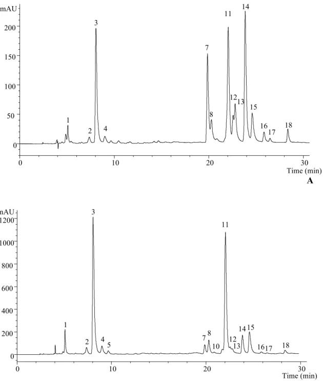
A
B
Figure 1. Phenolic compounds profile of oven-dried Stevia rebaudiana leaves recorded
at 370 (A) and 280 (B) nm.
Time (min)
0 10 20 30
mAU 0 50 100 150 200 2 3 4 7 8 11 12 14 15 13 1 17 18 16
0 10 20 30
mAU 0 200 400 600 800 1000 1200 Time (min) 1 2 3 4 5 78 10 11 12 13 1415
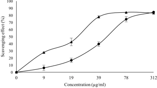
Figure 2. Percentage of scavenging activity on DPPH radicals in extracts of frozen fresh ( ) and
oven-dried samples ( ) of Stevia rebaudiana. EC50: extract concentration corresponding to 50% of DPPH
scavenging activity. Frozen fresh samples: EC50 = 50.66 ± 3.64 µg/mL; oven-dried samples: EC50 =22.87
± 2.17 µg/mL. 0 10 20 30 40 50 60 70 80 90 100
0 9 19 39 78 312
S
ca
ve
ngi
ng
ef
fe
ct
(
%
)
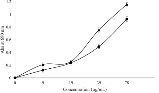
Figure 3. Reducing power in extracts of frozen fresh ( ) and oven-dried samples ( ) of Stevia
rebaudiana (higher absorbance indicates higher reducing power). EC50: extract concentration
corresponding to 0.5 of absorbance. Frozen fresh samples: EC50 = 39.73 ± 0.10 µg/mL; oven-dried
samples: EC50 =28.79 ± 0.09 µg/mL. 0
0.2 0.4 0.6 0.8 1 1.2
0 9 19 39 78
A
b
s
at
6
9
0
n
m