DISSERTAÇÃO DE MESTRADO
TOXICOLOGIA E CONTAMINAÇÃO AMBIENTAIS
Bioactive compounds from seaweed
with in vitro anti-leukemia activity
Tânia Patrícia Teixeira Almeida
M
TÂNIA PATRÍCIA TEIXEIRA ALMEIDA
BIOACTIVE COMPOUNDS FROM SEAWEED
WITH IN VITRO ANTI-LEUKEMIA ACTIVITY
Dissertation for the degree of Master in
Environmental Contamination and Toxicology, presented to
Institute of Biomedical Sciences Abel Salazar
of the University of Porto
Supervisor – Alice Fernanda Abreu Ramos
Category – Post-Doc Researcher
Affiliation – Interdisciplinary Centre of Marine and
Environmental Research, University of Porto
Co-supervisor – Eduardo Jorge Sousa da Rocha
Category – Full Professor
Affiliation – Institute of Biomedical Sciences Abel Salazar,
University of Porto
v
ACKNOWLEDGEMENTS
I would like to thank the following people that made this Masters dissertation possible:
Doctor Alice Ramos, my supervisor, for all the help, guidance and devotion to this work. In a personal way, I would also like to thank her for her friendship and for being such an inspiration. Professor Eduardo Rocha, my co-supervisor, for giving me the opportunity to work on this project with an amazing team, and for the support provided throughout the year.
All members of the PATH group, particularly Professor Daniela Gargiulo and my colleague Mariana Abreu.
Also, I would like to thank the Laboratory of Toxicology of the Department Pharmacology and Toxicology, University of Navarra for receive me, and with special attention to my mentor Doctor Amaya Azqueta, and colleges Violeta Gomez, Tamara Iglesias, and Emma Atkinson.
More than a friend, Joana Ferreira, was a very important person who participated throughout this work, helping both with the practical work and discussions.
Finally, I would like to thank my family (parents and grandparents) for giving me the opportunity to complete this dissertation.
vi
This work was partially funded by the Research Line NOVELMAR – Novel Marine Products with Biotechnological Applications, integrated in the Structured Program of R&D&I INNOVMAR - Innovation and Sustainability in the Management and Exploitation of Marine Resources (reference NORTE-01-0145-FEDER-000035), funded by the Northern Regional Operational Programe (NORTE2020) through the European Regional Development Fund (ERDF).
vii
ABSTRACT
Nowadays, cancer is a major, worldwide problem, affecting both developed and developing countries. Despite the incredible amount of research spanning this field, the statistics on cancer are not optimistic. Leukemia is a type of cancer with a worldwide incidence of 2.5% and a mortality rate of 3.4% (out of all cancers). Chronic myeloid leukemia (CML) represents 15 - 20% of all new cases of leukemia and is characterized by an uncontrolled proliferation of myeloid cells. Currently, the first-line of treatment involves tyrosine kinase inhibitors (TKI), which act in a specific way to inhibit the activity of BCR-ABL. However, resistance, mainly due to mutations, can occur. When the disease reaches a more advanced stage – the blast crisis – more aggressive chemotherapeutics are applied, such as anthracyclines. Doxorubicin is an example that is used in several types of cancers, including leukemia, yet the action of this drug can also cause resistance and other important issues such as non-targeted cytotoxicity. In the attempt to find more effective and less toxic therapies, two in vitro approaches were explored in this work: (i) the study of the anticancer activity of natural compounds; and (ii) the (less explored) improvement of current therapy combined with natural compounds.
Marine organisms are a rich source of bioactive compounds with diverse biological activities. Several compounds isolated from seaweed, namely carotenoids and phlorotannins present anticancer activity against numerous cancer cell lines. In the current work, the in vitro anticancer activity of fucoxanthin and phloroglucinol alone and co-incubated with anticancer drugs (imatinib and doxorubicin) was assessed on two human cancer cell lines (K562 and TK6) derived from blast crisis of CML. For this (i) cytotoxicity at short time; (ii) proliferation capability; (iii) induction of DNA damage; and (iv) induction of cell death by apoptosis, were evaluated. Our results showed that doxorubicin was more cytotoxic than imatinib in both cell lines. Both anticancer drugs decreased cell proliferation in a dose-dependent manner in K562 cell line without induction of DNA damage or apoptosis. Proliferation inhibition of TK6 cell line induced by doxorubicin was accompanied by an increase of DNA damage and apoptosis. Furthermore, fucoxanthin decreased cell proliferation without effects on DNA damage or apoptosis in both cell lines. When co-incubated with the anticancer drugs the inhibition was not improved when compared with fucoxanthin alone for both cell lines. Phloroglucinol did not show cytotoxic effect at short time in both cell lines, however, in TK6 cells, phloroglucinol alone and co-incubated with imatinib inhibited cell proliferation, but without induction of DNA damage and apoptosis.
viii
This work demonstrated that both the natural compounds exhibit antiproliferative effects against K562 (fucoxanthin) and TK6 (fucoxanthin and phloroglucinol) cell lines. When co-incubated with the drugs (doxorubicin and imatinib) their efficacy was enhanced in certain conditions. The findings warrant more studies to understand the mechanisms of action involved in the antiproliferative effects of fucoxanthin and phloroglucinol in the cell lines tested. This knowledge could be interesting to explore the natural compounds as new adjuvant strategy for the treatment of cancer cells that share the same molecular characteristics that the cell lines evaluated in this work.
ix
RESUMO
Atualmente o cancro é um grande problema ao nível global, afetando tanto países em desenvolvimento bem como desenvolvidos. Apesar da incrível quantidade de pesquisa neste campo os dados estatísticos existentes são pouco otimistas. Em particular, a leucemia é um cancro com uma incidência de 2.5% e um índice de mortalidade de 3.4% em todo o mundo. Leucemia mielóide crónica representa entre 15 – 20% de todos os tipos de leucemia e é caracterizada pela proliferação descontrolada de células mieloides. Atualmente a primeira linha de tratamento para esta doença são os inibidores de tirosina quinase, estes atuam de uma forma bastante especifica inibindo a atividade do BCR-ABL, contudo pode ocorrer o aparecimento de resistências devido ao desenvolvimento de mutações. Quando a doença atinge um estado mais avançado – a fase ou crise blástica - quimioterapêuticos mais agressivos são aplicados, como é o exemplo dos antraciclinas. Doxorrubicina é uma antraciclina muito usada para tratar vários tipos de cancro, incluindo a leucemia, contudo a ação desta droga tem vindo a ser relacionada com o aparecimento de resistência e também a sua ação não específica torna-a tóxica para outras células (células normais). De maneira a tornar os tratamentos mais efetivos e menos tóxicos, duas abordagens in vitro foram exploradas neste trabalho: (i) o estudo da atividade anticancerígena dos compostos naturais; e (ii) o (menos estudado) melhoramento dos atuais tratamentos utilizados através da combinação com compostos naturais.
Os organismos marinhos são uma fonte de compostos bioativos com diversas atividades biológicas. Alguns compostos isolados das algas, nomeadamente carotenóides e florotaninas, apresentam atividade anticancerígena contra diversas linhas celulares de cancro. No presente trabalho, a atividade anticancerígena in vitro da fucoxantina e floroglucinol sozinhos ou co-incubados com os fármacos anticancerígenos (imatinib e doxorrubicina) foi avaliada em duas linhas celulares cancerígenas (linha celular K562 e TK6) provenientes da fase blástica da leucemia crónica mielóide. Para isso, (i) a citotoxicidade de curta exposição; (ii) a capacidade de proliferação; (iii) a indução de danos no ADN; e (iv) a indução de morte celular por apoptose foram avaliados. Os nossos resultados mostraram que a doxorrubicina foi mais citotóxica do que o imatinib para as duas linhagens celulares. Ambos os fármacos, imatinib e doxorrubicina, diminuem a proliferação celular de uma forma dose-dependente nas K562 sem indução de danos no ADN e apoptose. A inibição da proliferação induzida pela doxorrubicina nas TK6 foi acompanhada por um aumento dos danos no ADN e da apoptose. Além disso, fucoxantina demonstrou uma redução da proliferação celular sem efeitos ao nível
x
dos danos no ADN ou na apoptose para ambas as linhas celulares. Quando co-incubada com os fármacos a inibição não aumenta quando comparado com o composto sozinho. Floroglucinol não mostrou citotoxicidade para curtos períodos de exposição em ambas as linhas celulares, contudo nas TK6 floroglucinol sozinho ou em co-incubação com imatinib inibiu a proliferação celular, mas sem induzir danos ao nível do ADN e apoptose.
Com este trabalho demonstramos que ambos os compostos naturais exibem um efeito antiproliferativo contra as K562 (fucoxantina) e TK6 (fucoxantina e floroglucinol). Quando co-incubados com os fármacos (doxorrubicina e imatinib) a eficiência dos mesmos é melhorada em certas condições. Os achados justificam que se implementem mais estudos de modo a compreender quais os mecanismos de ação envolvidos nestes efeitos antiproliferativos da fucoxantina e do floroglucinol nas linhagens testadas. Poderá ser de interesse explorar este efeito de modo a poder-se utilizar estes compostos em novas estratégias como adjuvantes para o tratamento de células cancerígenas que apresentem características moleculares semelhantes às utilizadas neste estudo.
xi
INDEX
Acknowledgements ...v Abstract ... vii Resumo ...ix Index ...xiList of figures and tables ...xiii
Abbreviations ...xvii
List of publication and communications………xxi
CHAPTER I – General introduction ... 1
1. Cancer ... 3
1.1. Cancer statistics ... 3
1.2. Risk factors ... 4
1.3. Cancer cell biology ... 5
1.3.1. Genetic instability ... 5
1.3.2. Proliferation and cell cycle ... 6
1.3.3. Apoptosis ... 7
2. Leukemia ... 8
2.1. Chronic myeloid leukemia ... 8
2.1.1. Molecular approach ... 9
2.1.2. Downstream signaling pathways of BCR-ABL ... 10
2.2. Treatment of CML ... 11
2.2.1. Tyrosine kinase inhibitors ... 12
2.2.1.1. Current strategies to enhance the therapeutic effect of imatinib ... 13
2.2.2. Anthracyclines ... 14
2.2.2.1. Current strategies to enhance the therapeutic effect of doxorubicin ... 15
xii
3.1. Carotenoids ... 16
3.1.1. Fucoxanthin ... 17
3.1.2. Anticancer effect of fucoxanthin in leukemia ... 18
3.2. Phlorotannins ... 18
3.2.1. Anticancer effect of phloroglucinol in leukemia ... 19
4. References ... 21
CHAPTER II - Objective ... 29
Objective ... 31
CHAPTER III - Bioactive compounds from seaweed with in vitro anti-leukemia activity ... 33
CHAPTER IV – Conclusions and future perspectives ... 67
APPENDICES ... 71
P1. Cell culture ... 73
P2. Viability assay ... 777
P3. Comet assay ... 79
xiii
LIST OF FIGURES AND TABLES
CHAPTER I:Figure 1 – The role of environmental factors in the development of cancer, with the
percentage contribution of each factor. Adapted from Anand et al. (2008). ... 4
Figure 2 – Cell cycle and respective checkpoints. Adapted from Garrett (2001). ... 7 Figure 3 – Mechanisms of drug resistance induced by BCR-ABL. In a normal cell
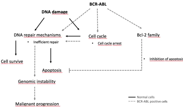
(represented as black) DNA repair mechanisms are activated and DNA damage can be completely repaired – the cell will survive. In the case of inefficient repair, and if the cell cycle checkpoints are not strong enough to allow more time for repair, death signals are activated with induction of apoptosis. In a BCR-ABL expressed cell (represented as gray) the global levels of DNA repair mechanisms are increased. This also affects the checkpoints, providing more time for repair, however, the efficiency is compromised. In addition, in case of some unrepair or misrepair lesion escape, the apoptotic signals are inhibited by the regulation of Bcl-2 protein family. Overall, this process leads to an accumulation of DNA damage, increasing the genomic instability. Adapted from Skorski (2002).. ... 11
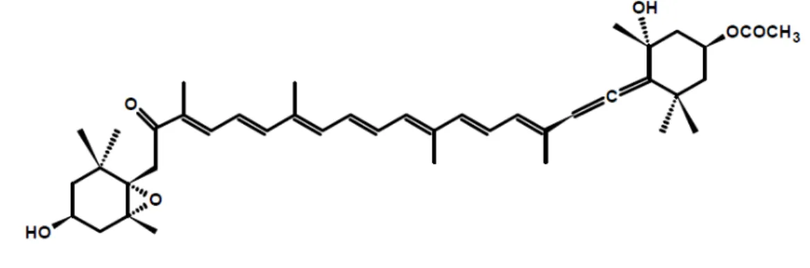
Figure 4 – Fucoxanthin structure. ... 17 Figure 5 – Phloroglucinol structure. ... 19
CHAPTER III:
Figure 1 – Dose-response effects on cell survival, after 24 h of incubation with (a)
doxorubicin (Dox), (b) imatinib (Imat), in K562 cell line, (c) Dox and (d) Imat, in TK6 cell line, evaluated by cell counting with a Neubauer chamber. Results are expressed as mean ± standard deviation (SD) of at least four independent experiments. Significant differences (* p ≤ 0.05; ** p ≤ 0.01; **** p ≤ 0.0001), when compared with those of the control, were tested by one-way ANOVA, followed by post-hoc Dunnett’s tests. ... 45
Figure 2 – Dose-response effects on cell proliferation via Relative Suspension Growth
(RSG), after 24 h of incubation plus 48 h in fresh new medium with (a) doxorubicin (Dox) and (b) imatinib (Imat), in K562 cell line; (c) dox and (d) imat in TK6 cell line, evaluated by cell counting with a Neubauer chamber. Results are expressed as mean ± standard deviation (SD) of at least four independent experiments. Significant differences (*** p ≤ 0.001; **** p ≤ 0.0001) when compared with the control were tested by one-way ANOVA, followed by post-hoc Dunnett’s tests. ... 46
xiv
Figure 3 – Effect of fucoxanthin (Fx) alone (black bars) or in combination (gray bars)
with doxorubicin (Dox) and imatinib (Imat) (at IC30) on cell cytotoxicity after 24 h of treatment and assessed by cell counting. Fx combined with (a) Dox or (b) Imat in K562 cell line; Fx combined with (c) Dox or (d) Imat in TK6 cell line. Results are expressed as mean ± standard deviation (SD) of at least four independent experiments. Significant differences (* p ≤ 0.05) among groups per situation of exposure were tested by two-way ANOVA, followed by post-hoc Bonferroni multiple comparison test. Percentages in brackets showed the decrease on cell viability in relation to respective control. ... 47
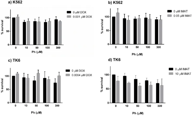
Figure 4 - Effect of phloroglucinol (Ph) alone (black bars) or in combination (gray bars)
with doxorubicin (Dox) and imatinib (Imat) (at IC30) on cell cytotoxicity after 24 h of treatment and assessed by cell counting. Ph combined with (a) Dox or (b) Imat in K562 cell line; Ph combined with (c) Dox or (d) Imat in TK6 cell line. Results are expressed as mean ± standard deviation (SD) of at least four independent experiments. ... 48
Figure 5 - Effect of fucoxanthin (Fx) alone (black bars) or in combination (gray bars)
with doxorubicin (Dox) and imatinib (Imat) (at IC30) on cell proliferation via Relative Suspension Growth (RSG) after 24 h of treatment plus 48 h in fresh medium, and assessed by cell counting. Fx combined with (a) Dox or (b) Imat in K562 cell line; Fx combined with (c) Dox or (d) Imat in TK6 cell line. Results are expressed as mean ± standard deviation (SD) of at least four independent experiments. Significant differences (* p ≤ 0.05; *** p ≤ 0.001) among groups per situation of exposure were tested by two-way ANOVA, followed by post-hoc Bonferroni multiple comparison test. Percentages in brackets showed the decrease on cell proliferation in relation to respective control. ... 49
Figure 6 - Effect of phloroglucinol (Ph) alone (black bars) or in combination (gray bars)
with doxorubicin (Dox) and imatinib (Imat) (at IC30) on cell proliferation via Relative Suspension Growth (RSG) after 24 h of treatment plus 48 h in fresh medium and assessed by cell counting. Ph combined with (a) Dox or (b) Imat in K562 cell line; Ph combined with (c) Dox or (d) Imat in TK6 cell line. Results are expressed as mean ± standard deviation (SD) of at least four independent experiments. Significant differences (* p ≤ 0.05; ** p ≤ 0.01; *** p ≤ 0.001; **** p ≤ 0.0001) among groups per situation of exposure were tested by two-way ANOVA, followed by post-hoc Bonferroni multiple comparison test. Percentages in brackets show the decrease or increase on cell proliferation in relation to respective control. ... 50
Figure 7 - Effect of natural compounds, fucoxanthin (Fx) and/or phloroglucinol (Ph)
xv
breaks – SBs; FPG-sensitive sites) after 24 h in (a) K562 cell line and (b) TK6 cell line, assessed by comet assay, (c) Images from comet assay in TK6 cell line: 1 – SBs of negative control; 2 – FPG-sensitive sites of positive control; and 3 – SBs and 4 – FPG-sensitive sites in 0.0004 μM of Dox. Results are expressed as mean ± standard deviation (SD) of at least two independent experiments. Significant differences (*** p ≤ 0.001) among groups per situation of exposure were tested by one- way ANOVA, followed by post-hoc Bonferroni multiple comparison test. Percentages in brackets are show the increase in relation to respective control. In the case of K562 cells, the results are exploratory as they correspond at only one independent experiment (ongoing work). Scale bar – 100 μm………..52 Figure 8 - Effect of fucoxanthin alone or co-incubated with doxorubicin (Dox) or imatinib
(Imat) (IC30) on the induction of nuclear chromatin condensation in (a) K562 cell line and (b) TK6 cell line, and (c) phloroglucinol (Ph) in TK6 cell line, evaluated by the nuclear condensation assay after 48 h of incubation. Results are expressed as mean ± standard deviation (SD) of at least four independent experiments………...54
xvii
ABBREVIATIONS
∙OH – Hydroxyl radicalABL1 – Abelson murine leukemia viral oncogene homolog 1 AGP – Alpha-1-acid glycoprotein
ALL - Acute lymphoid leukemia AP – Accelerated phase
ATCC - American type culture collection ATL - Adult T cell leukemia
ATP – Adenosine triphosphate BC – Blast crisis
BCR – Breakpoint cluster region CBC – Complete blood count
CCyR – Complete cytogenetic response Cdk – Cyclin-dependent kinases
CHF – Congestive heart failure CML – Chronic myeloid leukemia CP – Chronic phase
DAPI – 4’,6-diamidino-2-phenylindole DISC – Death inducing signaling complex DMSO – Dimethyl sulfoxide
xviii
Dox – Doxorubicin
EDTA – Ethyleneadiaminetetraacetic acid FBS – Fetal bovine serum
FGF-2 – Fibroblast growth factor 2
FGFR-1 – Fibroblast growth factor receptor 1 FPG - Formamidopyrimidine-DNA glycosylase Fx – Fucoxanthin
GIST – Gastrointestinal stromal turmors H2O2 – Hydrogen peroxide
HEPES – 4-(2-hydroxyethyl)-1-piperazineethanesulfonic acid HL-60 – Human promyelocytic leukemia cell line
hOCT1 – Human organic cation transporter 1 HSCT – Hematopoietic stem cell transplantation HTLV-1 – Human T-cell leukemia virus type 1 IARC – International agency for research on cancer IC50 – Half maximal inhibitory concentration IFN-α – Interferon-α
Imat - Imatinib
JNK – c-Jun N-terminal kinase
K562 – Human erythromyeloblastoid leukemia cell line LMP – Low melting point agarose
xix
MDR – Multidrug resistance
MKK4 – Mitogen-activated protein kinase kinase-4 MMP – Mitochondrial membrane potential
NF-κB – Nuclear factor kappa B NMP – Normal melting point agarose O2∙ – Superoxide anion
P-loop – Phosphate-binding loop P-gp – P-glycoprotein
PARP – Poly (ADP-ribose) polymerase PBS – Phosphate buffered saline PFA – Paraformaldehyde
Ph – Phloroglucinol
PP2A – Protein phosphatase 2A
RanGAP1 – RanGTPase activating protein 1 RNA – Ribonucleic acid
ROS – Reactive oxygen species
RPMI – Roswell Park Memorial Institute Medium RSG - Relative suspension growth
SAHA – Suberoylanilide hydroxamic acid SCGE - Single-cell gel electrophoresis STI-571 – Imatinib mesylate
xx
TK6 – Human lymphoblastoid cell line, derived from blast crisis of CML TKD – Tyrosine kinase domain
TKI – Tyrosine kinase inhibitor TNF – Tumor necrosis factor TSG – Total suspension growth UV – Ultraviolet
xxi
LIST OF PUBLICATIONS AND COMMUNICATIONS
The elaboration of this Master thesis, and collaboration with other ongoing related works rendered data sets that were included both in presentations in international meetings, conferences, and in papers, as is follows mentioned:
1. Articles under submission or to be submitted to international peer-reviewed journals
- Almeida, T., Ferreira, J., Azqueta, A., Rocha E., Ramos A.A. (2016) Bioactive compounds from seaweed with anti-leukemia activity: Carotenoids and phlorotannins – a mini-review. To be submited
- Almeida, T., Ferreira, J., Azqueta, A., Rocha E., Ramos A.A. (2016) Bioactivity of fucoxanthin and phloroglucinol from seaweed with anti-leukemia activity. To be submited
- Ferreira, J., Almeida, T., Azqueta, A., Rocha E., Ramos A.A. (2016) Anticancer activity against gliobastoma cell lines by compounds present in algae, alone in combination with anticancer drugs – a mini-review. To be submited
- Ferreira, J., Almeida, T., Azqueta, A., Rocha E., Ramos A.A. (2016) Anticancer effects of fucoxanthin and phloroglucinol, alone and in combination with temozolomide, on the U251 and T98G glioblastoma cell lines. To be submited
2. Communications in conferences
- Tânia Almeida, J. Ferreira, V. Gomez-Rodriguez, E. Rocha, A. Ramos, A. Azqueta. Bioactive compounds from seaweed with anti-leukemia activity: Carotenoids and phlorotannins. ICOETOX | IBAMTOX 2016 (Poster).
CHAPTER I – GENERAL INTRODUCTION
3
1. Cancer
The term “cancer” originates from Greek physician Hippocrates, who noted the similarity between crabs and the cut surface of a solid tumor (Hajdu 2011). In medicine, the earliest report of cancerous diseases, refers to breast cancer, and dates back to, approximately, 3000 B.C. However, paleopathological findings show that tumors existed in animals long before men appeared on Earth (Hajdu 2011).
Our body is made up of trillions of living cells. Every cell has the ability to grow, divide and die in a well-ordered way. During the first years of a person’s life, the normal cells divide faster allowing the person to grow. Upon reaching adulthood, most cells divide only to replace worn-out or dying cells or to repair injuries. Normal cells become cancer cells as a result of alterations in cell DNA. If these alterations are not repaired and if they confer a survival advantage, the cell will not die as it should. Instead, the altered cells proliferate faster than normal cells and make new cells that the body does not need. Most types of cancer cells form a solid tumor (a mass), but in the case of leukemia, tumors are rare. In leukemia, neoplasia cells are mixed in blood and blood-forming organs (American Cancer Society, 2015).
1.1. Cancer statistics
According to worldwide GLOBOCAN estimates, produced by the International Agency for Research on Cancer (IARC) in 2012, there were 14.1 million new cases and 8.2 million deaths due to cancer (Ferlay et al. 2015). The overall data showed that lung and breast cancer are the most frequently diagnosed and caused the highest number of death in men and women, respectively in both developed and developing countries. In developed countries, colorectal and prostate cancer were also at the top of the list for the highest number of cases. The problem becomes more tragic in less developed countries where the cancer incidence has been increasing in the last decade and incurs lower survival rates, representing 57% of all cancer cases and 65% of cancer deaths worldwide (Sloan and Gelband 2007, Torre et al. 2015). Currently, cancer continues to be a worldwide killer disease, despite the large amount of research undertaken. By 2020, it is estimated that there will be a word population of 7.5 billion, with approximately 15 million of new cases of cancer and 12 million cancer deaths (Anand et al. 2008).
4
1.2. Risk factors
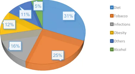
Several risk factors have been associated with the appearance of cancer. These include internal factors, which represent 5 - 10% of all cases (e.g. inherited mutations, hormones and immune conditions) and environmental/acquired factors, which represent 90 - 95% (e.g. smoking tobacco, alcohol consumption, lack of physical activity, bad eating habits, infectious agents, environmental pollution and radiation) (Figure 1) (Anand et al. 2008, Jemal et al. 2011).
Figure 1 – The role of environmental factors in the development of cancer, with the percentage contribution of
each factor. Adapted from Anand et al. (2008).
Several agents from environmental or cellular (endogenous) sources are able to induce DNA damage directly, or in an indirect way that may contribute to mutagenesis (Loeb and Loeb 2000, Helleday et al. 2014). As mentioned earlier, just 5-10% of all cancers are related with inherited gene defects, meaning that lifestyle factors have a great importance in cancer development. Changes in environmental and lifestyle factors seem a promising strategy for the prevention of cancer. There are some environmental factors that can promote the appearance of leukemia, such as viral infections, exposure to polycyclic aromatic hydrocarbons, pesticides, chlorinated drinking water, the presence also of nitrates in the water, radiation, usually from radioactive substances and ultraviolet (UV) light, and low-frequency electromagnetic fields (Belpomme et al. 2007). 31% 25% 16% 12% 11% 5% Diet Tobacco Infections Obesity Others Alcohol
5
1.3. Cancer cell biology
Cancer is a heterogeneous group of diseases, the main feature is the production of abnormal cells that grow beyond the natural boundaries. Nowadays, several hallmarks of cancer have been proposed, forming the fundamental principles of this malignant transformation. Tumor formation is a multistep process, where the normal cells evolve progressively into the neoplastic stage by acquiring particular capacities that enable them to become tumorigenic. These hallmarks are self-sufficiency in growth signals, insensitivity to antigrowth signals, genomic instability, avoiding apoptosis, limitless replicative potential, sustained angiogenesis, tissue invasion, metastasis, tumor-promoting inflammation, metabolic reprogramming, and evasion of the immune system (Hanahan and Weinberg 2011, Floor et al. 2012). Each cancer has its own way of action. The following sections will highlight those characteristics particularly important for the development of leukemia, including genetic instability, proliferation and cell cycle arrest, and apoptosis.
1.3.1. Genetic instability
The greatest differences between cancerous and normal cells are their abilities to divide, survive, invade, metastasize and destroy the host. Genetic instability is one of the hallmarks found in cancer cells, including genetic changes such as mutations in specific genes and structural and numerical changes at the chromosomal level (Shen 2011). Mutations can appear due to DNA damage inflicted by environmental (e.g. radiations, industrial chemicals, natural carcinogens) or cellular sources (e.g. depurination, free radicals, DNA polymerase errors). In normal cells, DNA is replicated with high fidelity where the repair mechanisms are normally able to resolve the damage. However, due to the frequency of occurrence of DNA damage, the inaccessible structure of human chromatin and defects with DNA repair mechanisms, it is possible that some lesions escape the DNA repair mechanisms and produce mutations, contributing to genomic instability (Loeb and Loeb 2000, Lord and Ashworth 2012, Ferguson et al. 2015). Some of these changes will give the cell the capacity to ignore the regulatory processes needed to control cell division, expression, adaptation and even cell death, contributing to cancer initiation and progression. Besides that, this lack of stability generates cancer cells which are widely heterogenic, resulting in the development of cells resistant to certain chemotherapeutics (Loeb and Loeb 2000).
6
1.3.2. Proliferation and cell cycle
The cell cycle is a process that allows the cell to grow, replicate its DNA and divide. It is divided into four sequential phases (as shown in Figure 2): S phase is when the DNA replicates, M phase is responsible for the cell division producing two daughter cells, and two gaps (G1 and G2). G1 appears after M phase, a time where the cell is responsive to positive and negative growth signals, and G2 follows S phase when the cell prepares to start mitosis. There is also a fifth state, G0 or quiescence, in the case of deprivation of the growth-promoting signals in the G1 gap (Garrett 2001, Williams and Stoeber 2012).
The control of each phase and the transitions between them are controlled by sensor mechanisms. These monitor the cellular environment, mainly the genomic integrity, and determine if the cell must go on in the cell cycle progress. The main cell cycle checkpoints occur in the: G1/S phase transition, the biggest sensor of DNA damage; G2/M to monitor the fidelity of DNA replications and finally a mitotic checkpoint where the fidelity of chromosome segregation in mitosis is controlled (Garrett 2001). If some abnormality is detected, such as DNA damage, signaling pathways are activated resulting in cell cycle arrest in the attempt to “solve the problem” (Williams and Stoeber 2012). Mammalian cell cycle progression is controlled by cyclin-dependent kinases (Cdk), a family of serine/threonine kinases, through the phosphorylation of certain proteins (Garrett 2001). In the case of failure at cell cycle checkpoints, uncontrolled proliferation may occur - a typical characteristic for malignant phenotypes (Williams and Stoeber 2012).
7
Figure 2 – Cell cycle and respective checkpoints. Adapted from Garrett (2001).
1.3.3. Apoptosis
Apoptosis is a mechanism of programmed cell death that presents several morphological and biochemical modifications such as rounding-up of the cell, membrane blebbing, externalization of phosphatidylserine, mitochondrial fragmentation, protein cleavage, retraction of pseudopods, pyknosis (reduction of cellular volume), condensation of chromatin, karyorrhexis (nuclear fragmentation) and formation of apoptotic bodies, amongst others. It is important to consider that other types of programmed cell death exist and others may yet be discovered (Elmore 2007, Kroemer et al. 2009, Fuchs and Steller 2015).
There are two main apoptotic pathways: the extrinsic or death receptor pathway and the intrinsic signals or mitochondrial pathway. The intrinsic pathway is initiated by several stress signals, such as radiation, drugs, free radicals, absence of growth factors, viral infections, amongst others that induce changes in mitochondrial permeability. Consequently, cause the release of cytochrome c, activation of caspase-9, -3 and other caspases that culminate with cell death (Kalimuthu and Se-Kwon 2013). The involvement of the Bcl-2 family proteins, responsible for mitochondrial outer membrane permeability, and the p53 tumor suppressor protein, which regulates the Bcl-2 proteins is crucial for the occurrence of this pathway. The Bcl-2 family includes anti- and pro- apoptotic proteins. The anti-apoptotic members are: Bcl-2,
G1 S G2 M G2 Checkpoint G1/S Checkpoint Go Mitotic Checkpoint
8
Bcl-xL and Mcl-1 and the pro-apoptotic members are: Bax and Bak amongst others (Burke 2010). In the extrinsic pathway, apoptosis is initiated by transmembrane receptor-mediated pathways, where cytokine ligands, such as TNF, bind to death receptors presents on cell surface, forming a death inducing signaling complex (DISC) that activates the caspase8 and -10, followed by the rest of the caspase cascade - once caspase-8 is activated, apoptosis is triggered. Disabling the apoptosis is one of the pathogenic occurrences which contributes to cancer initiation, promotion, and progression (Elmore 2007, Kalimuthu and Se-Kwon 2013).
2. Leukemia
Leukemia is one cancer that affects the blood-forming cells present in bone marrow and the hematopoietic process (Gibson et al. 2013). According to the GLOBOCAN project 2012, leukemia has a worldwide incidence of 351 965 cases which correspond to 2.5% of all cancers, and a mortality of 255 471 cases corresponding to 3.4% of all cancer deaths (Torre et al., 2015). This disease can affect the lymphoid or the myeloid stem cells, both resulting in the production of many white blood cells that are abnormal and do not mature normally (Vardiman et al. 2009). Referring to morphology, genetics, and clinical features, leukemia could be classified into four main groups: Acute lymphoid leukemia, chronic lymphoid leukemia, acute myeloid leukemia and chronic myeloid leukemia. In lymphoid leukemia, also known as lymphoblastic or lymphocytic leukemia, the abnormalities start in the cells that become lymphocytes. In myeloid leukemia, the cancer cells come from granulocytic, monocytic/ macrophagic, erythroid, megakaryocytic and mast cell lineages. The terms acute and chronic differ in the maturity of the leukemic cells (Vardiman et al. 2009).
2.1. Chronic myeloid leukemia
Chronic myeloid leukemia (CML) represents 15% of all cases of leukemia and the median age for this disease is 64 years (Gibson et al. 2013). The myeloid abnormally starts in the blood-forming cells of the bone marrow and is characterized by uncontrolled proliferation of neoplastic hematopoietic precursor cells and weakened production of normal hematopoiesis, causing several abnormalities in the blood such as neutropenia, anemia and thrombocytopenia (Ghosh et al. 2014).
9
CML can occur in two or three stages: the first one, known as chronic phase (CP), is normally asymptomatic and presents as a myeloid hyperplasia in the bone marrow and peripheral blood (< 20 %). The intermediate stage, called accelerated phase (AP), occurs with few symptoms. The second phase – the blast crisis (BC) – appears 3-5 years after diagnosis of untreated CML-CP patients, as a rapid progression, and the number of undifferentiated myeloblasts is higher than 20 %. In some cases the transition from CP into BC phase occurs without AP signals (O'Brien et al. 2009, Burke 2010, Jabbour and Kantarjian 2014).
Some patients with CML have no symptoms at all, however, some of the symptoms commonly associated with this disease are the non-specifics (lethargy, fatigue, fevers, night sweats) or splenomegaly (such abdominal pain) (Gibson et al. 2013). Facing these symptoms, the main tests to diagnose this disease are a complete blood count (CBC) and bone marrow biopsy. Bone marrow cytogenetics is recommended to help choose the best treatment, taking into consideration the karyotypic abnormalities (O'Brien et al. 2009).
2.1.1. Molecular approach
Chronic myeloid leukemia is a hematopoietic stem cell disease which presents as a chromosome translocation between chromosomes 9 and 22 during cell division, resulting in a shorter chromosome 22 called Philadelphia chromosome. This translocation t(9;22)(q34;11) of DNA means that part of chromosome 22 breakpoint cluster region (BCR) gene at band q11 fusion with chromosome 9 Abelson 1 (ABL) gene at band q34, leading to the formation of a new oncogene BCR-ABL (Rowley 1973). ABL is a proto-oncogene that encodes for a protein tyrosine kinase found mainly in the nucleus, which regulates the cell cycle, differentiation, migration, invasion, genomic instability and the response to genotoxic stress (Deininger et al. 2000, Burke and Carroll 2010, Greuber et al. 2013). The BCR gene encodes for a serine/threonine kinase that can act as a GTPase-activating protein for members of the Rho family of guanine nucleotide exchange factors, and can also phosphorylate histones and casein (Burke 2010).
BCR-ABL encodes a new protein, p210BCR-ABL which shows deregulated tyrosine kinase activity and contains the NH2-terminal domains of BCR and the COOH-terminal domains of ABL. The critical functional changes found with the expression of this new protein are: ABL protein becomes constitutively active as a protein tyrosine kinase enzyme; attenuation of DNA
10
protein-binding activity of ABL; and the improvement of the binding of ABL to cytoskeletal actin microfilaments (O'Brien et al. 2009). Animal models have confirmed that this protein plays a crucial role in the pathogenesis of CML, increasing cell proliferation, blocking apoptosis and stromal interactions of the cell. Hence, the development of targeted therapy with a specific action on tyrosine kinase was a breakthrough in the treatment of myeloid leukemia (Gibson et al. 2013).
In very few cases, less than 10%, the leukemia cells present the BCR-ABL oncogene, but not the Philadelphia chromosome, and so it is possible that the BCR-ABL gene can be formed in a different way. There are also very rare cases where neither the oncogene nor the Philadelphia chromosome are found, which means that other oncogenes may be causing this disease (Onida et al. 2002). It is also possible to express another fusion protein (p190) however this is more common in cases of acute lymphoid leukemia (ALL) (O'Brien et al. 2009, Cutler et al. 2015).
2.1.2. Downstream signaling pathways of BCR-ABL
The BCR-ABL chimeric oncogene encodes for a constitutively active tyrosine kinase that modulates different signaling pathways, such as PI3K/AKT/mTOR, JAK-STAT, Wnt/β-catenin and autophagy. This brings benefits in terms of cell survival, proliferation, differentiation, and migration, allowing cell proliferation, protection against cell death in the absence of external factors and promotion of invasion and metastasis (Sinclair et al. 2013). Modulation of survival pathways by BCR-ABL activation (Figure 3) has also been related to resistance to genotoxic therapeutics since BCR-ABL-positive cells can repair DNA damage more quickly through the up-regulation of RAD51 (a protein of homolog recombination repair system), decreasing its degradation and activating it through post-translational modification. The expression of RAD51 in the cells seems to be positively correlated with the appearance of a resistance phenotype (Collis et al. 2001). The capacity to repair DNA is not only influenced by the efficiency of the repair mechanisms but also by the time that the cell has to carry out the mechanism. BCR-ABL-positive cells are able to activate DNA damage-dependent cell cycle checkpoints faster by displaying a pronounced G2/M delay (Bedi et al. 1995). Another mechanism involved in drug
11
resistance is the modulation of Bcl-2 family members, namely the up-regulation of anti-apoptotic proteins (e.g. Bcl-xl and Bcl-2) (Skorski 2002).
2.2.
Treatment of CML
With advances in the knowledge of cancer biology, CML treatments have changed. Conventional chemotherapy treatment was widely used allowing the destruction of the cells. Examples of the drugs used in CML were busulfan followed by hydroxyurea. Both chemotherapeutics showed an improvement regarding the symptoms and hematology, but the cytogenetic remission was not significant (Henkes et al. 2008, Zhang et al. 2008). Allogeneic hematopoietic stem cell transplantation (HSCT) seems to be the type of treatment that proves
Figure 3 – Mechanisms of drug resistance induced by BCR-ABL. In a normal cell (represented as black) DNA
repair mechanisms are activated and DNA damage can be completely repaired – the cell will survive. In the case of inefficient repair, and if the cell cycle checkpoints are not strong enough to allow more time for repair, death signals are activated with induction of apoptosis. In a BCR-ABL expressed cell (represented as gray) the global levels of DNA repair mechanisms are increased. This also affects the checkpoints, providing more time for repair, however, the efficiency is compromised. In addition, in case of some unrepair or misrepair lesion escape, the apoptotic signals are inhibited by the regulation of Bcl-2 protein family. Overall, this process leads to an accumulation of DNA damage, increasing the genomic instability. Adapted from Skorski (2002).
Normal cells BCR-ABL positive cells
12
to cure more patients. Yet this method presents some disadvantages, such as the need for a suitable donor and the high toxicity of the procedure, which generates some side-effects such as immunodeficiency, infections, organ toxicity and graft versus host disease (Henkes et al. 2008). Another treatment is the Interferon-α (IFN-α) therapy, introduced in the 1980s, this presents some positive final results regarding remission and disease-free survival. However, the toxicity levels in the patients proved to be a problem, because of their ability to induce fatigue, myalgias, arthralgias, headaches, weight loss, depression, diarrhea, neurological symptoms, memory changes, hair thinning, autoimmune diseases, and cardiomyopathy (Henkes et al. 2008, Burke 2010). The need to find more potent and specific agents combined with the better understanding of molecular mechanisms underlying CML led, in the early 1990s, to the development of tyrosine kinase inhibitors (e.g. imatinib). Imatinib results in the inhibition of proliferation, restoration of the cell cycle, apoptosis induction and reversal of genetic instability in BCR-ABL dependent cells. However, even with the exceptional efficiency of TKI, problems may occur, such as drug resistance, loss of response, kinase domain mutations and transformation of the disease (e.g. evolution to an accelerated or blast phase). The BC is typically lethal, at this point more aggressive treatments are applied such anthracyclines (O'Brien et al. 2009). The previous two treatments (TKI and anthracyclines) will now be explored in further detail.
2.2.1. Tyrosine kinase inhibitors
Nowadays, the first-line of treatment for myeloid leukemia in the chronic phase is imatinib mesylate (STI-571), a potent BCR-ABL tyrosine kinase inhibitor (TKI) (Druker et al. 2006). TKI will bind competitively to the adenosine triphosphate (ATP) binding site of the BCR-ABL protein and consequently inhibits the phosphorylation of proteins related to BCR-BCR-ABL signal transduction (Mahon et al. 2000, Gibson et al. 2013). The final outcome is the induction of apoptosis in hematopoietic cells expressing BCR-ABL without affecting normal cells (Druker et al. 1996). Imatinib proved to be an efficient treatment for myeloid leukemia since patients showed 85% of overall survival (Gibson et al. 2013).
Despite the specific mechanisms of action, around 33% of patients started to fail to achieve a complete cytogenetic response (CCyR), either because of the toxicity or (mostly) due to the appearance of a resistant phenotype over time (O'Brien et al. 2009, Bhamidipati et al.
13
2013). The hematologic and nonhematologic toxicities caused by imatinib include neutropenia, thrombocytopenia, gastrointestinal disturbances, edema, skin rashes and musculoskeletal complaints (Gibson et al. 2013). Furthermore, some patients present, after a few years with imatinib treatment, signals of cardiotoxicity with congestive heart failure (CHF) (O'Brien et al. 2009). This resistance may be due to a range of mechanisms, some are BCR-ABL-dependent and others are BCR-ABL-independent mechanisms. The BCR-ABL-dependent mechanisms include, most frequently, point mutations in the ABL tyrosine kinase domain (TKD) and gene amplification of ABL (Bhamidipati et al. 2013). An important mutation is one that results in the formation of amino acid substitutions in imatinib binding sites, frequently called “gatekeeper” mutations (e.g. T315I) (Gibson et al. 2013). The BCR-ABL-independent mechanisms of resistance involve: (1) decrease in drug uptake by expression of the human Organic Cation Transporter 1 (hOCT1) (Thomas et al. 2004); (2) improved drug efflux by overexpression of P-glycoprotein (P-gp) efflux pumps, reducing the amount of intracellular drug (Mahon et al. 2000); (3) increase of plasma protein α1 acid glycoprotein (AGP), which binds to imatinib preventing the ABL kinase inhibition (Gambacorti-Passerini et al. 2000); (4) increase of prostaglandin-endoperoxide synthase 1 (cyclooxygenase 1), which plays an important role in imatinib metabolism (Villuendas et al. 2006).
In response to the emergence of imatinib resistance, the development of second-generation TKI began. These include nilotinib (Tasigna TM) and dasatinib (Sprycel TM). Both drugs achieved positive molecular responses, even activing with imatinib-associated kinase domain mutations, yet each drug induced its own mutations, and neither could inhibit BCR-ABL T315I (Gibson et al. 2013, Zhou and Xu 2015). A mutation in the ATP phosphate-binding loop (P-loop) is associated with poor prognosis and a high risk for progression (O'Brien et al. 2009, Mughal et al. 2013). Other types of TKI are also in development, such bosutinib and ponatinib, the latter presenting activity against the T351I mutation (Gibson et al. 2013).
14
Resistance development in leukemia cells and the potential toxicity have been great limitations for imatinib application (Hu et al. 2009). The future of CML therapy passes to the development of new potent agents, perhaps in combinations with existing treatments in lower doses.
Nimmanapalli et al. (2003) showed that imatinib, when combined with suberoylanilide hydroxamic acid (SAHA), known as an inhibitor of histone deacetylases, can enhance the cytotoxicity effects in leukemia cells (K562 cell line) by up-regulation of p21 and p27 and down-regulation of BCR-ABL levels with induction of apoptosis in BCR-ABL-expressed cells. A decrease in phopho-AKT and Bcl-xL levels was also observed. After experiments in two CML murine models, Hu et al. (2009) concluded that a low dose of imatinib combined with bortezomib (proteasome inhibitor) or proteasome inhibitor might optimize the CML treatment. The results showed inhibition in Bcl-2, increase of cytochrome c and activation of caspases, along with inhibition of proteasomal degradation of protein phosphatase 2A (PP2A). The combination was proven to have an inhibitory effect on tyrosine kinase via suppression of NFκB. Lin et al. (2016) demonstrated that by down-regulating RanGTPase and activating protein 1 (RanGAP1) it is also possible to improve the imatinib efficiency because RanGAP1 can mediate BCR-ABL nuclear entrapment to activate the P73-dependent apoptosis pathway.
2.2.2. Anthracyclines
Anthracyclines are one of the most effective classes of anti-tumor antibiotics used for the treatment of numerous cancers, including breast, ovarian (Bellarosa et al. 2001), prostate, lung (Pratesi et al. 1998), and leukemia, amongst others (Hehlmann 2012). One of the first anthracyclines to be isolated was doxorubicin, isolated from Streptomyces peucetius var. caesius in the 1960s (Malla et al. 2010). Despite the wide use of this drug, its mechanism of action is not completely understood. However, several studies demonstrated its involvement in the inhibition of DNA and RNA synthesis, in the inhibition of topoisomerase II with subsequent formation of DNA double-strand breaks and the induction of cell death (Laroche-Clary et al. 2000, Park et al. 2005), and in the synthesis of free radicals (Lebrecht et al. 2004, Thorn et al. 2011).
Beside the broad anticancer activity, the action of doxorubicin has been related with several side effects such as non-targeted cytotoxicity and acquisition of multidrug resistance
15
(MDR) phenotype. In CML the main mechanisms that contribute for MDR involve the P-gp by sequestration of the drug into cytoplasmic vesicles, the expression of antiapoptotic protein Bcl-2 and the gene BCR-ABL (Misra and Sahoo Bcl-2011).
2.2.2.1. Current strategies to enhance the therapeutic effect of doxorubicin
Referring to the limitations of doxorubicin, such as drug resistance of cancer cells and the side-effects on normal cells, several strategies have been made to overcome these problems. One possibility to potentiate the action of doxorubicin is to combine this drug with synthetic or natural agents (Ghosh et al. 2014).
According to Misra and Sahoo (2011), the combination of doxorubicin with curcumin (a natural polyphenol) exhibited a synergistic inhibitory effect on growth of K562 cells. The synergistic growth inhibition was mediated through different mechanisms that involved the inhibition of p210BCR-ABL. The authors suggest that the synergistic effect is clinically important and may provide combinatorial strategies in cancer therapy. Jang et al. (2013) reported the synergetic effect of the combination treatment of decursin (a natural compound present in Angelica gigas) and doxorubicin, in multiple myeloma cells, by enhancing apoptotic activity via mTOR. Also observed was the activation of caspase -9 and -3, the cleavage of poly (ADP-ribose) polymerase (PARP), increase of sub-G1 population, and decrease in the expression of cyclind-D1 and survivin amongst other effects in U266 cells. Ghosh et al. (2014) showed that extracts from the hot water in black tea have protective effects against chemotherapeutic drugs, such as daunorubicin (an anthracycline), in normal lymphocytes. This extract is able to inhibit drug-induced ROS generation and renew the mitochondrial membrane potential (MMP). It increases cellular viability, up-regulates endogenous antioxidants enzymes, inhibits mRNA expression of apoptotic genes, prevents caspase-3 activation and reduces DNA fragmentation.
3. Seaweed as a source of bioactive compounds
Several epidemiological studies have shown that some phytochemicals present in vegetables and fruits are able to reduce the risk of degenerative processes (Abubakar et al. 2012, Anand et al. 2008, Kim et al. 2010, Rengarajan et al. 2013). Marine organisms are a rich
16
source of bioactive compounds with several biological activities, and the interest by the scientific community has grown rapidly. In particular, seaweed, as photosynthetic organisms, are exposed to high amounts of light and oxygen which favors the formations of free radicals and other oxidative reagents. However, the absence of any serious photodynamic damage means that marine algae have the ability to generate bioactive components to protect themselves (Heo et al., 2008).
Marine algae can be divided into four main groups, according to the type of pigments, morphology, anatomy and reproductive structures: Chlorophyceae (green algae), Phaeophyceae (brown algae), Rhodophyceae (red algae) and Cyanophyceae (blue-green algae) (Kolanjinathan et al. 2014). Seaweed are multicellular eukaryotic and macroscopic organisms living in salty water and mostly belong to the green, brown and red algae. They have been integrated into the human diet in some countries, such as Japan, since ancient times. In line with such traditions, the consumption of this natural product around the world is increasing nowadays (Kolanjinathan et al. 2014). Seaweed are a rich source of polysaccharides, minerals, protein, vitamins, and low-fat carbohydrate (Cornish and Garbary 2010). High levels of bioactive compounds can be produced by a variety of marine macro and microalgae which provide several properties such as antibacterial, antiviral, immunosuppressant, antioxidant, antiproliferative and antitumor activity (Sharif et al. 2014).
The use of bioactive compounds in combination with conventional anticancer drugs is poorly explored but the strategy may improve the clinical performance in conventional chemotherapy by altering the biological disposition to minimize toxicity, maximize efficacy and possibly reduce the dose of therapy (Yamamoto et al. 2011, Ghosh et al. 2014).
3.1. Carotenoids
Seaweed pigments can be divided into three main groups: chlorophylls, carotenoids, and phycobiliproteins. Carotenoids, organic pigments found in chloroplasts and chromoplasts, exhibit a purple, red, orange and yellow color — actually, carotenoids are a class of tetraterpene pigments found in bacteria, fungi, algae, as well as in higher plants and animals (Chojnacka et al. 2012). Nowadays, more than 700 carotenoids have been found in nature (Dembitsky and Maoka 2007, Maoka 2011, Takaichi 2011). The intake of dietary carotenoids has been correlated with a lower incidence of cardiovascular and neurodegenerative diseases, cataract
17
formation and cancer (Barros et al. 2014, Saini et al. 2015). Diverse biological functions such as provitamin A activity, antioxidant (quench singlet oxygen activity, scavenge free radicals), antiproliferative, and proapoptotic activity and enhancement of the immune system has been attributed to the carotenoids (Ishikawa et al. 2008, Fernández-García et al. 2012, Tanaka et al. 2012). The antioxidant activity of carotenoids is related to their structure; the number of double bonds and the presence of functional groups influence their ability to interact with different radicals (Sachindra et al. 2007).
3.1.1. Fucoxanthin
Fucoxanthin is one of the most abundant carotenoids found in nature, particularly in marine environments. This xanthophyll represents more than 10% of the total carotenoid production. Fucoxanthin is widely distributed in brown algae (Phaeophyceae) and diatoms (Bacillariophyta) and has an unusual structure that includes an allenic bond and 5,6-monoepoxide moiety (Figure 4) (Rengarajan et al. 2013).
Fucoxanthin has preventive effects against several types of cancer, including leukemia, due to its mechanisms of action, which include antiproliferation, cell cycle arrest, apoptosis induction, and suppression of angiogenesis (Anand et al. 2008, Rengarajan et al. 2013, Wang et al. 2012). Furthermore, the anticancer effect of fucoxanthin seems to be in some cases specific for cancer cells without cytotoxic effects against normal cell lines (Ishikawa et al. 2008). In leukemia, the effects of fucoxanthin are poorly understood. Here we tried to summarize the current knowledge of the effects of fucoxanthin in leukemia.
18
3.1.2. Anticancer effect of fucoxanthin in leukemia
In vitro and in vivo research has shown the anticancer effects of fucoxanthin in leukemia. Fucoxanthin and fucoxanthinol (its metabolite) decrease the viability of HTLV-1 infected T-cell lines and ATL cells through the induction of G1 cell cycle arrest, associated with down-regulation of cyclin D1, cyclinD2, CDK4 and CDK6 expression, and up-regulation of GADD45α, which will inhibit the entry of cells into S phase (Ishikawa et al. 2008). These authors also showed induction of apoptosis with activation of caspase-3, -8 and -9, and down-regulation of antiapoptotic protein expression, such as XIAP, cIAP2, Bcl-2 and survivin. In mice, fucoxanthinol suppresses tumor growth without adverse effects (Ishikawa et al. 2008).
Nakazawa et al. (2009) observed that fucoxanthin induces apoptosis in HL-60 leukemia cells and other cell lines with down-regulation of anti-apoptotic Bcl-2 proteins. Kim et al. (2010) also showed that the inhibitory growth of fucoxanthin in leukemia cell lines was due to apoptosis induction caused by the generation of ROS, along with the indication of cell cycle arrest in G1 stage. However, Kotake-Nara et al. (2005) demonstrated that apoptotic activity of fucoxanthin in promyelocytic cell lines is not caused by ROS. Besides the antiproliferative and proapoptotic effects of fucoxanthin in HL-60 leukemia cells, Ganesan et al. (2011) showed the anti-angiogenesis effects of fucoxanthin mediated by down-regulation of signal transduction by fibroblast growth factor 2 (FGF-2) and its receptor (FGFR-1) in human umbilical vein endothelial cells (Ganesan et al. 2013). However, information about the effects of fucoxanthin in CML is clearly missing.
3.2. Phlorotannins
Phlorotannins are tannin derivatives composed of phloroglucinol units linked to each other in different ways (Cornish and Garbary 2010, Thomas and Kim 2011). Phloroglucinol (1,3,5-trihydroxybenzene) is a phenolic compound isolated from Ecklonia cava that has three hydroxyl groups in the benzene ring (Figure 5). This class of polyphenols is unique in brown seaweed and several biological activities have been attributed to it, such as antioxidant, anti-inflammatory, antibacterial, anti-allergic and anticancer properties (Pádua et al. 2015, Corona et al. 2016).
19
Figure 5 – Phloroglucinol structure.
3.2.1. Anticancer effect of phloroglucinol in leukemia
Phloroglucinol has demonstrated cytoprotective effects in normal cells from the toxicity found in cancer treatments, proving to be a good candidate to improve the quality of these treatments. The protective effect in mice intestinal cells was accomplished by blocking the activation of p53, downregulation of Bax and Bak-dependent pathway and up-regulation of the levels of Bcl-2 and Bcl-XS/L (Ha et al. 2013).According to Kang et al. (2010), phloroglucinol efficiently protected cells against radiation and extended the survival of mice exposed to lethal doses of radiation through the inhibition of mitogen-activated protein kinase kinase-4 (MKK4/SEK1), c-Jun NH2-terminal kinase (JNK), activator protein-1 (AP-1) cascades and protected the depletion of reduced glutathione. Phloroglucinol also has a protective effect against lipid peroxidation and DNA damage induced by radiation (Kang et al. 2010).
Besides the cytoprotective effects in normal cells, phloroglucinol also showed cytotoxic effects against cancer cells. Such effects in leukemia cell lines are poorly explored, however, some studies showed in vitro antiproliferative activity of phloroglucinol and of its derivatives in a variety of cancer cell lines including K562 cells. According to Liu et al. (2011) hyperforin, an abundant phloroglucinol-type, induced, in K562 cells, the dissipation of mitochondrial transmembrane potential caused by radiation, through the release of cytochrome c. Phloroglucinol induced the activation of the caspase-3, -8 and -9 cascade and consequently PARP cleavage. Cell cycle arrest was also observed with the increased expression levels of both p53 and p27kip1. Quiney et al. (2006) presented the same results for other leukemia cells, e.g. B-cell chronic lymphocytic leukemia (B-CLL), where hyperforin induced apoptosis by disruption of the mitochondrial transmembrane potential, activation of caspase-3 and cleavage of the PARP. Furthermore, downregulation of Bcl-2 proteins, Mcl-1 (anti-apoptotic proteins) and nitric oxide (NO) synthase of type 2 were also observed. In this study, downregulation of the cell cycle inhibitor, p27kip1 was detected in contrast with the results from Liu et al. (2011). Using
20
chronic lymphocytic leukemia cells in ex vivo studies Merhi et al. (2012) showed that the mitochondrial pathway of caspase-dependent apoptosis was induced by up-regulation of Noxa (protein of Bcl-2 family). More studies are needed to properly understand the effects of phloroglucinol against leukemia and information about the effects in CML is clearly missing.
21
4. References
Abubakar, M. B., W. Z. Abdullah, S. A. Sulaiman and A. B. Suen (2012). "A review of molecular mechanisms of the anti-leukemic effects of phenolic compounds in honey." International Journal of Molecular Sciences 13(11): 15054-15073.
Anand, P., A. B. Kunnumakara, C. Sundaram, K. B. Harikumar, S. T. Tharakan, O. S. Lai, B. Sung and B. B. Aggarwal (2008). "Cancer is a preventable disease that requires major lifestyle changes." Pharmaceutical Research 25(9): 2097-2116.
Barros, M. P., S. C. Poppe and E. F. Bondan (2014). "Neuroprotective properties of the marine carotenoid astaxanthin and omega-3 fatty acids, and perspectives for the natural combination of both in krill oil." Nutrients 6(3): 1293-1317.
Bedi, A., J. Barber, G. Bedi, W. El-Deiry, D. Sidransky, M. Vala, A. Akhtar, J. Hilton and R. Jones (1995). "BCR-ABL-mediated inhibition of apoptosis with delay of G2/M transition after DNA damage: a mechanism of resistance to multiple anticancer agents." Blood 86(3): 1148-1158.
Bellarosa, D., A. Ciucci, A. Bullo, F. Nardelli, S. Manzini, C. A. Maggi and C. Goso (2001). "Apoptotic events in a human ovarian cancer cell line exposed to anthracyclines." Journal of Pharmacology and Experimental Therapeutics 296(2): 276-283.
Belpomme, D., P. Irigaray, L. Hardell, R. Clapp, L. Montagnier, S. Epstein and A. Sasco (2007). "The multitude and diversity of environmental carcinogens." Environmental research 105(3): 414-429. Bhamidipati, P. K., H. Kantarjian, J. Cortes, A. M. Cornelison and E. Jabbour (2013). "Management of imatinib-resistant patients with chronic myeloid leukemia." Therapeutic Advances in Hematology 4(2): 103-117.
Burke, B. and M. Carroll (2010). "BCR–ABL: a multi-faceted promoter of DNA mutation in chronic myelogeneous leukemia." Leukemia 24(6): 1105-1112.
Burke, B. A. (2010). "Effects of the BCR-ABL oncogene on DNA damage and repair." Dissertation Paper 196
Chojnacka, K., A. Saeid, Z. Witkowska and L. Tuhy (2012). “Biologically active compounds in seaweed extracts-the prospects for the application.” The Open Conference Proceedings Journal 3: 20-28.
Collis, S., A. Tighe, S. Scott, S. A. Roberts, J. H. Hendry and G. P. Margison (2001). "Ribozyme minigene-mediated RAD51 down-regulation increases radiosensitivity of human prostate cancer cells." Nucleic Acids Research 29(7): 1534-1538.
22
Cornish, M. L. and D. J. Garbary (2010). "Antioxidants from macroalgae: potential applications in human health and nutrition." Algae 25(4): 155-171.
Corona, G., Y. Ji, P. Anegboonlap, S. Hotchkiss, C. Gill, P. Yaqoob, J. P. Spencer and I. Rowland (2016). "Gastrointestinal modifications and bioavailability of brown seaweed phlorotannins and effects on inflammatory markers." British Journal of Nutrition 115(07): 1240-1253.
Cutler, J., R. Tahir, J. Han, R. S. Nirujogi, T.-C. Huang, X. Wong, S. Mallampati, X. Sun, P. Brown and K. Reddy (2015). "Differential Signaling through p190 and p210 Forms of BCR-ABL Fusion Proteins Revealed By Proteomic Analysis." Blood 126(23): 3651-3651.
Czyz, M., J. Jakubowska and M. Sztiller-Sikorska (2008). "STI571/doxorubicin concentration-dependent switch for diverse caspase actions in CML cell line K562." Biochemical Pharmacology 75(9): 1761-1773.
Deininger, M. W., J. M. Goldman and J. V. Melo (2000). "The molecular biology of chronic myeloid leukemia." Blood 96(10): 3343-3356.
Dembitsky, V. M. and T. Maoka (2007). "Allenic and cumulenic lipids." Progress in Lipid Research 46(6): 328-375.
Druker, B. J., F. Guilhot, S. G. O'Brien, I. Gathmann, H. Kantarjian, N. Gattermann, M. W. Deininger, R. T. Silver, J. M. Goldman, R. M. Stone, F. Cervantes, A. Hochhaus, B. L. Powell, J. L. Gabrilove, P. Rousselot, J. Reiffers, J. J. Cornelissen, T. Hughes, H. Agis, T. Fischer, G. Verhoef, J. Shepherd, G. Saglio, A. Gratwohl, J. L. Nielsen, J. P. Radich, B. Simonsson, K. Taylor, M. Baccarani, C. So, L. Letvak and R. A. Larson (2006). "Five-year follow-up of patients receiving imatinib for chronic myeloid leukemia." New England Journal of Medicine 355(23): 2408-2417. Druker, B. J., S. Tamura, E. Buchdunger, S. Ohno, G. M. Segal, S. Fanning, J. Zimmermann and N. B.
Lydon (1996). "Effects of a selective inhibitor of the Abl tyrosine kinase on the growth of Bcr-Abl positive cells." Nature Medicine 2(5): 561-566.
Elmore, S. (2007). "Apoptosis: a review of programmed cell death." Toxicologic Pathology 35(4): 495-516.
Ferguson, L. R., H. Chen, A. R. Collins, M. Connell, G. Damia, S. Dasgupta, M. Malhotra, A. K. Meeker, A. Amedei and A. Amin (2015). “Genomic instability in human cancer: Molecular insights and opportunities for therapeutic attack and prevention through diet and nutrition.” Seminars in Cancer Biology 35: 55-524.
Ferlay J, Soerjomataram I, Ervik M, Dikshit R, Eser S, Mathers C, Rebelo M, Parkin DM, Forman D, Bray, F. GLOBOCAN 2012 v1.0, Cancer Incidence and Mortality Worldwide: IARC CancerBase No. 11 [Internet]. Lyon, France: International Agency for Research on Cancer; 2013. Available from: http://globocan.iarc.fr, accessed on 20/April/2016.
23
Fernández-García, E., I. Carvajal-Lérida, M. Jarén-Galán, J. Garrido-Fernández, A. Pérez-Gálvez and D. Hornero-Méndez (2012). "Carotenoids bioavailability from foods: From plant pigments to efficient biological activities." Food Research International 46(2): 438-450.
Floor, S. L., J. E. Dumont, C. Maenhaut and E. Raspe (2012). "Hallmarks of cancer: of all cancer cells, all the time?" Trends in Molecular Medicine 18(9): 509-515.
Fuchs, Y. and H. Steller (2015). "Live to die another way: modes of programmed cell death and the signals emanating from dying cells." Nature Reviews Molecular Cell Biology 16(6): 329-344. Gambacorti-Passerini, C., R. Barni, P. le Coutre, M. Zucchetti, G. Cabrita, L. Cleris, F. Rossi, E.
Gianazza, J. Brueggen, R. Cozens, P. Pioltelli, E. Pogliani, G. Corneo, F. Formelli and M. D'Incalci (2000). "Role of alpha1 acid glycoprotein in the in vivo resistance of human BCR-ABL(+) leukemic cells to the abl inhibitor STI571." Journal of the National Cancer Institute 92(20): 1641-1650.
Ganesan, P., K. Matsubara, T. Sugawara and T. Hirata (2013). "Marine algal carotenoids inhibit angiogenesis by down-regulating FGF-2-mediated intracellular signals in vascular endothelial cells." Molecular and Cellular Biochemistry 380(1-2): 1-9.
Ganesan, P., K. Noda, Y. Manabe, T. Ohkubo, Y. Tanaka, T. Maoka, T. Sugawara and T. Hirata (2011). "Siphonaxanthin, a marine carotenoid from green algae, effectively induces apoptosis in human leukemia (HL-60) cells." Biochimica et Biophysica Acta 1810(5): 497-503.
Garrett, M. D. (2001). "Cell cycle control and cancer." Current Science 81(5): 515-522.
Ghosh, D., S. K. Dey and C. Saha (2014). "Protective effect of black tea extract during chemotherapeutic drug induced oxidative damage on normal lymphocytes in comparison with cancerous K562 cells." International Journal of Scientific and Engineering Research 5(2): 437-447.
Gibson, J., H. Iland, S. Larsen, C. Brown and D. Joshua (2013). "Leukaemias into the 21st century–part 2: the chronic leukaemias." Internal Medicine Journal 43(5): 484-494.
Greuber, E. K., P. Smith-Pearson, J. Wang and A. M. Pendergast (2013). "Role of ABL family kinases in cancer: from leukaemia to solid tumours." Nature Reviews Cancer 13(8): 559-571.
Ha, D., S. J. Bing, J. Cho, G. Ahn, D. S. Kim, M. Al-Amin, S. J. Park and Y. Jee (2013). "Phloroglucinol protects small intestines of mice from ionizing radiation by regulating apoptosis-related molecules: a comparative immunohistochemical study." Journal of Histochemistry and Cytochemistry 61(1): 63-74.
Hajdu, S. I. (2011). "A note from history: landmarks in history of cancer, part 1." Cancer 117(5): 1097-1102.