UNIVERSIDADE DA BEIRA INTERIOR
Ciências da Saúde
Breast Adenomyoepithelioma:
Case Report and Review of the Literature
Ana Margarida Catarino Gomes
Dissertação para obtenção do Grau de Mestre em
Medicina
(ciclo de estudos integrado)
Orientador: Doutor Carlos Alberto Martins Gomes
ii
Dedication
In memory of my grandmother Maria Amália, who was always tremendously supportive throughout the course. Your prayers were heard.
iii
Acknowledgments
First and foremost, I would like to express my sincere gratitude to my advisor Doctor Carlos Alberto Martins Gomes, for his continuous support of my Master’s study and research, for his patience, motivation, enthusiasm and knowledge and for being open to new ideas as well as for helping me to shape my interest and thoughts. His guidance helped me throughout my research and the writing process of this thesis. I could not have imagined having a better advisor and mentor for my Master’s study.
After my advisor, I would like to express my deep gratitude to the Beira Interior University for the opportunity given to me to complete my degree at this educational institution, recognized both nationally and internationally.
I would like to thank Professor Moutinho, who kindly provided all the data of the case report.
A big thank you to CEDAP – Centro de Diagnóstico Anatomo-Patológico for providing the original microscope slides from the patient for a better documentation of the case report. I would like to also thank Mrs. Rosa Farate from the Anatomo-Pathology Department of Cova da Beira Hospital for the help in obtaining the tumor microscopic image photographs and Miss Carla Bernardo from the Library and Documentation Service of Cova da Beira Hospital for the prompt help on the articles research.
My sincere thanks also go to Professor Ondřej Daum, professor of the Faculty of Medicine of Charles University in Pilsen, who kindly sent an article published by Dr. Kinkor on the subject of my thesis some years ago.
Last but not the least, I would like to thank all my friends but especially my family: my parents, Alfredo Gomes and Isabel Catarino, and my sister, Carolina Gomes, for the unconditional support throughout my life, and specially during my degree. Special thanks to my grandfather José Catarino as well, who always believed in my capacities and encouraged me.
iv
Resumo
O adenomioepitelioma da mama é uma proliferação neoplásica bifásica de células luminais e mioepiteliais. É normalmente assintomático, sendo diagnosticado como massa palpável ou achado imagiológico ocasional(1). A punção aspirativa por agulha fina é o melhor
método de diagnóstico(2) e a imuno-histoquímica é muito útil na sua confirmação(3) Os
adenomioepiteliomas são geralmente benignos embora possam recorrer localmente(4) sendo a
variante tubular, os tumores lobulares com alta atividade mitótica ou os tumores com atipia citológica particularmente propensos à recorrência local. O tratamento é a excisão local completa(5-7) e em caso de reincidência a excisão ampla(5).
Caso clínico: Paciente de 47 anos com uma massa no quadrante superior-externo da mama esquerda. A ecografia revelou um nódulo com 8 milímetros de maior eixo, homogéneo, bem delimitado, de contornos regulares e a mamografia mostrou uma massa opaca, bem definida, com bordos irregulares. Os estudos anátomo-patológico e imunohistoquímico da amostra da microbiópsia foram inconclusivos pelo que foi realizada uma tumorectomia cuja análise anatomo-patologica revelou um adenomioepitelioma tubular. Não foi realizado tratamento adicional e o follow-up ao fim de 20 meses não teve recorrência.
Avaliação dos casos publicados: dos 159 casos de adenomioepitelioma descritos, 158 são em mulheres e maioritariamente acima dos 45 anos de idade. Apresenta-se como uma massa, em média com 30 milímetros de diâmetro, sendo o padrão lobular o mais frequente. Demonstrou malignidade limítrofe especialmente em mulheres entre 15 a 40 anos ou com mais de 80 anos. O padrão lobular parece ser o mais benigno enquanto o padrão spindle o mais maligno e o padrão tubular é o que mais apresenta recorrências locais. Taxas mitóticas baixas estão associadas a menos recorrências e as altas a grande potencial de metastização.
Conclusões: O adenomioepitelioma afeta predominantemente mulheres entre os 16 e os 92 anos de idade, sem predomínio mamário. Pode exibir um padrão heterogéneo e uma malignidade limítrofe. A ecografia é importante como uma primeira abordagem e a punção aspirativa por agulha fina combinada com a imunohistoquímica definem o diagnóstico. Adenomioepiteliomas com potencial maligno estão associados a alta atividade mitótica, atipia citológica(6), necrose e limites imprecisos(4). As metástases foram documentadas em tumores
de 20 milímetros de diâmetro ou maiores(8) e os locais de metástases distantes incluem
pulmão, cérebro, mandíbula, tecidos moles, tiroide, gânglios linfáticos (mediastino e axilar) e fígado(6, 9-13). O Índice mitótico parece ser um ótimo parâmetro para o prognóstico e para
prever o comportamento do tumor.
Palavras-chave
v
Abstract
Adenomyoepithelioma is a biphasic neoplastic proliferation of luminal and myoepithelial cells. Patients are 22 to 92 years old, normally asymptomatic. It is either diagnosed as a palpable mass or as an occasional mammographic finding(1). Fine-needle
aspiration cytology is the investigation of choice to diagnose(2) and immunohistochemistry is
very useful in confirming the diagnosis(3). Adenomyoepithelioma is usually benign, although it
may recur locally(4). Tubular variants and some lobular tumors with high mitotic activity or
cytological atypia are particularly prone to local recurrence. Its treatment is complete local excision(5-7) and, in case of recurrence, a wider excision(5).
Case Report: 47-year-old female with a breast mass on the external upper quadrant of the left breast. Ultrasound revealed an 8 millimeter homogeneous, well delimitated nodule with regular borders and medio-lateral mammography showed a well defined opaque mass with irregular borders. The anatomo-pathological and immunohistochemisty of the specimen from the needle core biopsy was inconclusive so an excisional biopsy was performed diagnosing a tubular adenomyoepithelioma. No additional treatment was performed with a follow-up of 20 months without recurrence.
Review of published cases: 159 cases of adenomyoepithelioma almost all women mostly older than 45 years old. Adenomyoepithelioma is presented as a mass of on average 30 millimeters in either breast of lobular pattern most of the times. Adenomyoepithelioma demonstrated a borderline malignancy especially in women from 15 to 40 years of age or older than 80 years. Lobular pattern proved to be the most benign, while spindle pattern the most malignant variant and tubular pattern is the commonest one to recur. Low mitotic rates are associated with less recurrence or malignancy and high mitotic rates accompany adenomyoepitheliomas with great potential to metastasize.
Conclusions: Adenomyoepithelioma affects women aged between 16 and 92 years in either breast. It may display a heterogeneous pattern and a borderline malignancy. Ultrasonographic images may play an important role as a first approach and fine-needle aspiration cytology combined with immunohistochemistry should define the diagnosis. Potential malignant adenomyoepitheliomas are associated with high mitotic activity, cytologic atypia(6), necrosis and infiltrative borders(4). Metastases have only been documented
in tumors 20 millimeter in diameter or larger(8) and distant metastases locations include lung,
brain, jaw, soft tissues, thyroid, lymph nodes (mediastinal and axillary) and liver(6, 9-13).
Mitotic rate appears to be a good parameter for prognosis and to predict the tumor behavior.
Keywords
vi
Index
1. Introduction ... 1 2. Objectives ... 2 3. Methodology ... 3 4. Epidemiology ... 4 5. Clinical Features ... 5 6. Diagnosis ... 6 Ultrasonography ... 6Magnetic Resonance Imaging ... 6
Mammography ... 6 Macroscopic Pathology ... 7 Microscopic Pathology ... 7 Immunohistochemistry ... 8 Cytogenetics ... 10 7. Differential Diagnosis ... 12
8. Biologic Behavior and Prognosis... 14
9. Treatment ... 16
10. Case Report ... 17
11. Review of the Published Cases ... 22
Results ... 22
12. Discussion ... 27
13. Study Limitations ... 31
14. Advantages of the Study ... 32
15. Conclusion ... 33
vii
List of Figures
Fig.1: Ultrasound showing a homogeneous, well delimitated nodule with regular borders. ... 17
Fig.2: Mammography showing a well defined opaque mass on the superior external quadrant of the left breast. ... 18
Fig.3: Histological appearance of the specimen excised by quadrantectomy showing a lobulated well-defined mass (HE staining). ... 19
Fig.4: HE staining of the lesion showing a confluent multinodular injury with central tubular glandular structures. ... 19
Fig.5: Immunohistochemistry for PAS showing lumina filled with fibrillar material. ... 20
Fig.6: Immunohistochemistry for p63 antibody showing myoepithelial hyperplasia. ... 20
viii
List of Charts
Chart 1: Total number of diagnosed adenomyoepitheliomas by age groups. ... 22
Chart 2: Distribution of the breast affected by adenomyoepithelioma on the studied cases. . 22
Chart 3: Distribution of the different patterns of adenomyoepithelioma on the studied cases. ... 23
Chart 4: Recurrent adenomyoepitheliomas distribution per age groups. ... 23
Chart 5: Metastatic adenomyoepitheliomas distribution per age groups. ... 24
Chart 6: Recurrence and metastasis according dimensions (in millimeters) of the adenomyoepithelioma. ... 24
Chart 7: Metastasizing adenomyoepithelioma distribution by side of primary tumor. ... 25
Chart 8: Recurrences of the different patterns of adenomyoepithelioma. ... 25
Chart 9: Metastases of the different patterns of adenomyoepithelioma. ... 26
Chart 10: Total number of adenomyoepitheliomas diagnosis per age groups. ... 27
Chart 11: Rates of recurrence and metastases of adenomyoepithelioma. ... 28
Chart 12: Recurrence of adenomyoepithelioma per age groups. ... 28
Chart 13: Metastases of adenomyoepithelioma per age groups. ... 28
ix
List of Tables
Table 1: Immunohistochemical characterization of adenomyoepithelioma (positive result (+); negative result (-))(34). ... 10
Table 2: Overview of prognosis of the different patterns of adenomyoepithelioma. ... 26 Table 3: Overview of prognosis of adenomyoepithelioma according to the mitotic rate of the tumor. ... 26
x
Lista de Acrónimos
AME – adenomyoepitheliomaAMPA - α-amino-3-hydroxy-5-methyl-4-isoxazolepropionic acid BI-RADS - breast imaging-reporting and data system
BRCA-1 – breast cancer 1, early onset CEA – carcinoembryonic antigen
CGH – comparative genomic hybridization CK – cytokeratin
Cm – centimeter(s)
DLC-1 – deleted in liver cancer 1 DNA - deoxyribonucleic acid EMA – epithelial membrane antigen ER – estrogen receptor
FGF2 – fibroblast growth factor 2 FNAC – fine-needle aspiration cytology GH – growth hormone
GluR2 – AMPA-selective glutamate receptor 2 HE - hematoxylin and eosin
HER2/neu - human epidermal growth factor receptor 2 HMW - high molecular weight
HPC-1 – hereditary prostate cancer 1 Id - once-daily dosing
LOH – loss of heterozygosity Mcg – micro centigram Mm – millimeter(s) MR – mitotic rate
mRNA – messenger ribonucleic acid PAS - periodic acid–Schiff
PAS-diastase - periodic acid–Schiff–diastase PR – progesterone receptor
PTHrP- parathyroid hormone-related protein SMA – smooth muscle actin
UBI – Universidade da Beira Interior US - ultrasonography
1
1. Introduction
Myoepithelial cells are a normal component of the breast tissue(8). They are present in
the normal mammary duct system and they are often prominent in benign lesions, such as usual ductal hyperplasia, sclerosing adenosis, and intraductal papilloma(14). Their presence in
neoplastic lesions has been considered a hallmark of benignity. Recently, however, breast neoplasms have been described that are entirely or partially composed of myoepithelial cells(5). In these tumors, myoepithelial cells can demonstrate squamous, chondromyxoid,
plasmacytoid, clear cell and myoid spindle cell differentiation. Myoepithelial lesions were classified by Tavassoli(5) into three types: benign myoepithelioma neoplasm
(adenomyoepithelioma), hyperplasia (myoepitheliosis), and malignant neoplasm with myoepithelial differentiation (malignant myoepithelioma). Myoepitheliomas are neoplasms of purely myoepithelial origin and may be benign or malignant in approximately equal proportions. Tumors with bicellular proliferation of both epithelial and myoepithelial cells are the so called adenomyoepitheliomas.
Adenomyoepithelioma (AME), first described by Hamperl(14), is a biphasic neoplastic
proliferation of luminal and myoepithelial cells. It is a neoplasm arising from progenitor cells likely from the suprabasal site(15) that can differentiate into both luminal epithelium and
basal myoepithelium(15). As Hamperl noted, this tumor may display a heterogeneous pattern
because of the variable proliferation of epithelial and myoepithelial cells. Papillary architecture is seen in most tumors, and, therefore, AME is considered to be a variant of intraductal papilloma(16). Striking morphologic heterogeneity, such as papillary configuration
or multinodularity, especially in a limited biopsy sample(17-19), may not be appreciated, which
may lead to erroneous diagnoses of carcinoma(16). So far, other than case reports, only
comprehensive series studieshave been reported(5, 6, 16, 17, 19). Although most AMEs have been
benign, sporadic malignant AMEs with distant metastases have also been reported(5, 6, 8, 9).
Recognition of this entity, accurate diagnosis, and knowledge of the expected behavior are important in guiding the most appropriate patient management.
2
2. Objectives
This thesis focuses on a rare breast tumor. The main objectives of this review are: - To characterize pathophysiologically breast AME;
- To reflect upon the most accurate methods to diagnose AME; - To know more about the biological behavior of AME.
3
3. Methodology
Data collection for the present literature review was done through a research on PubMed database using the sentence “breast adenomyoepithelioma” OR “breast AND adenomyoepithelioma”. It was considered any relevant publication written in English and no limits were set on the date of publication.
The case reports where identified on PubMed database and through a systematic review of cases documented in the internet using the search platform from Google. The research was done using the sentence “case report AND breast adenomyoepithelioma OR case report AND breast AND adenomyoepithelioma”. No limits were set on the date of publication or the language in which the articles were written in order to obtain the greatest possible number of cases. This review focuses on 7 aspects: patient age, tumor side, tumor size, variant of the tumor, metastasis, recurrence and mitotic rate.
The information was read and the most relevant data were collected for further analysis in order to characterize adenomyoepithelioma pathophysiological patterns and prognosis.
4
4. Epidemiology
Approximately 150 cases of AME of the breast have been reported in the literature(20).
Patient age ranged from 22 to 92 years old (average age, 59 years)(19). Nearly all of
5
5. Clinical Features
Patients are normally asymptomatic and without previous personal history of cancer or high-risk breast lesions(23) and the adenomyoepithelioma is either diagnosed as a palpable
mass or as an occasional mammographic finding(1).
Patients usually present a solitary, well-circumscribed, sometimes palpable nodule (depending on location and size), measuring on average 20-30mm(15). Presentation as an
intracystic tumor is rare(15). The duration of symptoms varies from several weeks to several
months(5). The tumors are usually located in a peripheral portion of the breast(16) and there is
no predilection for either breast(4). Tenderness and serous nipple discharge have been found
6
6. Diagnosis
These lesions can be diagnostically challenging, because of the heterogeneity of adenomyoepitheliomas, especially when a core needle biopsy is performed even though this is considered the most accurate mechanism of diagnosis. Recognition of the biphasic cellular elements and the characteristic overall architecture of the tumors in combination with immunohistochemistry are essential to establish the correct diagnosis. Therefore, adequate sampling of the tumor to identify these features is necessary(24).
Ultrasonography
The most frequent US features for adenomyoepithelioma are lesions with small, solid, irregular or oval, non-parallel orientation, microlobulated or indistinct margin, hipoechogenicity and increased peripheral vascularity(23, 25).
Magnetic Resonance Imaging
Adenomyoepithelioma appearance in magnetic resonance has been described by some authors as an oval, smooth mass with persistent pattern on kinetic curve; irregular speculated mass with washout pattern on kinetic curve; round, speculated mass with a plateau pattern on kinetic curve or ill-defined solid tumor in cysts with demonstrated heterogeneous enhancement. Magnetic resonance images lack a pattern of specificity for this tumor(23).
Mammography
Mammographic findings of benign adenomyoepithelioma are non-specific(23, 26). On
mammography, AME appears as a round or lobulated, dense, mostly circumscribed mass, sometimes with partially indistinct margins(27). Calcifications and cystic appearance are not
7
Macroscopic Pathology
The size of AME ranges from 3 mm to 70 mm, with an average size of 25 mm(5, 19). The
tumors have been described as round to lobulated, well circumscribed or discrete, and firm(5, 16). Multinodular or papillary configurations and focal cystic changes have also been
described(19, 28, 29). The recurrent tumors have irregular borders and range from 2 cm to 6
cm(6). The cut surface reveals pink-white to gray-tan tissue with focal to diffuse translucency (5, 6). Hemorrhage and focal necrosis have also been reported(16, 18, 30). Satellite nodules may
appear in AME.
Microscopic Pathology
FNAC is the investigation of choice to diagnose breast lumps with reported diagnostic accuracy of 96,9% and a positive predictive value of 98,4%. Wide-bore needle biopsy is performed in case of inconclusive cytology results or if suspicion of unusual pathology(2).
The AME tumor has a biphasic nature, composed of cuboidal to columnar, epithelial-lined tubules surrounded by myoepithelial cells(19). It is a well circumscribed tumor either
encapsulated or multinodular(4).
A spectrum of histological patterns, however, has been observed among various examples of these tumors and even in different areas of individual tumors(16). These variations
were based on the distribution of proliferating glandular and myoepithelial cells, the extent of spindle or polygonal configuration of myoepithelial cells, the prominence of papillary component, and the degree of fibrosis(16).
Most AMEs have papillary configuration and, therefore, have been considered a variant of intraductal papilloma by some authors or a morphologic evolution from intraductal papilloma(6, 7, 16). It may appear as aggregated lobules of glands with tall lining epithelium
with scant cytoplasm and hyperchromatic nuclei surrounded by myoepithelial cells with clear cytoplasm(4). Spindle-shaped, clear or polygonal myoepithelial cells can be dominant(4)
forming nests or sheets of cells with clear cytoplasm.
Three variants of AMEs were described by Tavassoli(5). The first variant is the tubular
pattern, which is characterized by a balanced proliferation of rounded tubules, as well as unusually prominent hyperplastic myoepithelial cells(5). This pattern has ill-defined margins
and it may resemble a tubular adenoma(4). The second variant is the spindle cell type, which
is composed of a predominantly spindled myoepithelial cell proliferation admixed with a few columnar, epithelial-lined tubules(5). It resembles leiomyoma(4). Finally, the third variant
exhibits a lobular pattern composed of solid nests of myoepithelial cells proliferating around compressed tubules; the solid nests of tumor are then surrounded by fibrous connective tissue septa of varying thicknesses(5). Combinations of growth patterns sometimes exist.
8
Adenomyoepitheliomas are moderate to highly cellular. They are composed of clusters of epithelium and myoepithelium either in small clusters or dispersed cells(1, 26).
Adenomyoepitheliomas may displace, compress, or obliterate the epithelial gland, resulting in a zone nearly devoid of glands(5, 16, 19). These areas, if pronounced, may lead to a differential
diagnosis of myoepithelioma. The myoid areas may exhibit myoepithelial cells with pink to amphophilic cytoplasm or a plasmacytoid appearance with dense, hyaline-like, glassy eosinophilic cytoplasm and eccentric nuclei(5, 7, 27). The myoepithelial cells usually appear as
small, comma-shaped or ovoid, dark nuclei, devoid of cytoplasm(31) or with clear cytoplasm
and often a naked bipolar nuclei(1, 26). Cells present mild to moderate nuclear atypia and
occasionally a metachromatic fibrillary stroma can be found. Myoepithelial cells have an epithelioid morphology, polygonal shape, intranuclear inclusions or intracytoplasmic vacuoles, clear cytoplasm and often a naked bipolar nuclei(1, 26). Myxochondroid matrices produced by
the myoepithelial cells may also be noted, as seen in pleomorphic adenomas(19, 27). Dense,
collagenous, hyaline-like matrix materials can be seen in thick basement membranes(32).
Epithelial cells that usually form glandular spaces(4) tend to have hyperchromatic nuclei and
dense eosinophilic to amphophilic cytoplasm, when compared with myoepithelial cells(7).
Epithelial cells can display nuclear grooves(26). Apocrine metaplasia of epithelial cell
component, as well as squamous and sebaceous metaplasia or foam cells may be variably present(5, 7, 33) and it can be mistaken with fibroadenoma, a lesion suspicious for malignancy or
even with a malignant lesion(34).
Iyengar et al. after a study of twelve fine-needle aspiration cytologies considered intracytoplasmic vacuoles and intranuclear inclusions helpful in the identification of myoepithelial component of adenomyoepithelioma(26).
Atypical features, including increased mitotic activity, cytologic atypia with nuclear pleomorphism, prominent nucleoli, hyperchromasia, and necrosis, if observed, are usually associated with cases that either recurred or had a malignant clinical outcome(5, 6, 8).
Collagenous spherulosis in histologic sections has been reported in a single patient(26).
Descriptions of electron microscopy of the tumor refer classic myoepithelial features – myofibrils with dense bodies, pinocytic vesicles, desmososmes, tight junctions and patchy basement membrane.
Immunohistochemistry
Immunohistochemistry is very useful in confirming the diagnosis of AME(3) specially in
uncertain specimens. The interplay between epithelial and myoepithelial cell elements is highlighted by immunohistochemical staining with antibodies specific for these two components.
9
The cytoplasm of epithelial cells uniformly reacts with antibodies to cytokeratins, such as cytokeratin AE1/ AE3, CK 5, 7, 14 and 17, CAM 5.2(6, 19) or carcinoembryonic antigen
(CEA)(4). The luminal surfaces of the glandular cells are positive for the epithelial membrane
antigen (EMA)(7).
Polygonal and spindle myoepithelial cells are not reactive to epithelial membrane antigen (EMA) and are often only subtly reactive or weakly reactive to cytokeratin AE1/3(6, 19).
The myoepithelial component is highlighted by p63, smooth muscle myosin heavy chains, CK5, CD10, calponin, actin and S100(6, 7, 19, 35), adhesion molecules (CD44) and host active molecules
like: maspin, glutathionine-S-transpherase and metallothioneins.
The distribution and intensity of staining for anti-actin antibodies is heterogeneous(7).
Myoepithelial cells stain with actin depending on their level of differentiation(8). It is more
conspicuous in spindle cells than in clear polygonal cells. However, no reactivity to actin is seen in epithelial cells(7).
According to Bassler and Katzer(36), cells staining with antibodies against S100 and
cytokeratins but not with antibodies against smooth muscle actin (SMA) express incomplete features of the fully differentiated myoepithelial cells and can be considered transitional, intermediate, or precursor cells.
McLaren et al.(19) reported that p63 produced the best results with consistent, intense
nuclear staining, whereas some studies have reported discontinuous staining patterns(35).
Smooth muscle myosin heavy-chain is the most sensitive marker, which is easier to interpret than smooth muscle actin and muscle-specific actin because of its low cross-reactivity with myofibroblasts(7, 35, 37). Calponin is also highly sensitive for detecting
myoepithelial cells with cytoplasmic staining, but calponin-positive myofibroblasts can be detected in up to 74% of breast proliferations by light, patchy staining patterns(35). S100 is
expressed in virtually all of the myoepithelial cells, with variable intensity and uniformity of reactivity(6, 7, 35). Most of the luminal epithelial cells are S100 negative, but some reports have
noted strong expression in the luminal epithelial cells. In these cases, its diagnostic utility as a myoepithelial marker may be limited(7, 38-40). The myoepithelial-specific antibodies display
various cross-reactivity patterns and variable protein expression, especially in the neoplastic myoepithelial cells compared with their normal counterparts(7, 35). Therefore, a panel-based
approach of two or more markers should be used to minimize the chance of failure in detecting the myoepithelial cells(7, 35).
Proliferative indices of Ki-67 immunostaining are present in both compartments of the tumor but may be higher in the myoepithelial cells than it is in the ductal cells(41). Ki-67 is a
nuclear protein associated with and necessary for cellular proliferation. Immunostain for estrogen is either negative or weakly positive in a patchy pattern(4, 27). Progesterone receptor
and ERBB2 (formerly Her2/neu) have, however, been consistently reported to be negative in all the published studies (16, 27).
10
Some studies also showed that myoepithelial cells secret growth factors important in morphogenesis and maintenance of normally mammary gland – PTHrP, basic fibroblast growth factor FGF2(34).
Table 1: Immunohistochemical characterization of adenomyoepithelioma (positive result (+); negative result (-))(34).
Antigen Smooth muscle actin (+)
S-100 (+) Maspin (+) Vimentin (+) Pan-Cytokeratin (+) HMW Cytokeratin (+) EMA (-) ER (+ in 20%) PR (-) HER-2 (-)
Cytogenetics
Involvement of chromosome regions 8p and 16q have shown to be involved in breast tumors, frequently reported as acquired clonal abnormalities, and thus might represent genomic hot spots in the pathogenesis of such tumors. The evidence suggests a possible clustering of 8p breakpoints in breast carcinomas, but none of the studies have identified any possible candidate gene(s) within this region. The chromosomal translocation seen in Gatalica
et al.(34) case report is distinct, as it appears at the cytogenetic level to be balanced with no
loss of material either on 8p or on 16q, although it is possible for some DNA material to be lost at a molecular level. 8p23 breakpoint map important genes to oncogenesis like a tumor suppressor gene – DLC1 - in 8p22 region which was found down regulated in Gatalica et al.(34)
patient suggesting a rule of DLC1 in adenomyoepithelioma. There is a case report of a tumor with t(8;16)(p23;q21) mutation and a case of instability for the BRCA1 microsatellite marker (17q11.2)(4, 42).
Cadherin 5 or vascular endothelial cell adhesion molecule (VECAM) mapped to 16q22.1 was also down regulated in the same patient. Cadherins are suggested to play a role in development and maintenance of tissue and could be involved in invasion and metastasis of malignant tumors including breast carcinoma. VECAM may indeed have a role in the pathogenesis of adenomyoepithelioma(34).
GH mRNA and GH itself were found to be strongly expressed in adenomyoepithelioma as well as an ionotropic AMPA receptor GluR2 which mRNA levels are increased in adenomyoeptihelioma indicating that glutamate signaling may be involved in growth
11
stimulation for the tumor. Also ninjurin-1 mRNA and protein were found elevated indicating a potential role in growth stimulation of myoepithelium(34).
In Salto-Tellez(42) study, one adenomyoepithelioma was found with microsatellite
instability for BRCA1 microsatellite marker and lost of heterozygosity of HPC1 gene. This finding highlights the importance of 17p chromosomal loss where TP53 and BRCA1 genes are relatively close.
Trojani et al.(10) described a malignant AME with lung metastases and identified a DNA
diploid epithelial population and DNA tetraploid myoepithelial cells.
Jones et al.(43) performed comparative genomic hybridization (CGH) analysis on a
malignant AME case. They found a loss of heterozygosity (LOH) at 11q23-q24 and 16q22-q23. A liver metastasis for the same case was showing exclusively myoepithelial differentiation with additional LOH at 12q24.
12
7. Differential Diagnosis
Most AMEs appear to represent variants of intraductal papilloma(16). The
differentiation of papilloma with prominent myoepithelial cells from AMEs can be made based on architecture, pattern, and degree of myoepithelial proliferation(19). Those cases with only
myoepithelial cells lining the papillae and forming the basal layer below the epithelial elements and without nests, nodules, or an increased proportion of myoepithelial cells are categorized as papillomas with prominent myoepithelial cells(19). Myoepithelial cell markers
highlight the presence of prominent myoepithelial cells within the papillae and at the periphery(35). When the stroma associated with papillary lesions are sclerotic or exuberant,
nuclear stains, such as p63 and maspin, should be included in a myoepithelial marker panel to avoid cross-reactivity with myofibroblasts within the sclerotic stroma(35). A diagnosis of AME is
favored if myoepithelial proliferation is extensive and involves the lesion diffusely(27). Nipple
adenoma may mimic AME, but the presence of florid ductal hyperplasia and the pseudoinfiltrative pattern of stromal sclerosis entrapping glandular epithelium without an entrapped fibrous tissue or supporting fibrovascular cores are useful features to help differentiate the former(19). Again, prominent mioepithelial component differentiates nipple
adenoma from AME. Clear cell carcinoma may also mimic AMEs, but that may be differentiated by the presence of both epithelial and myoepithelial cell types, and confirmed with immunohistochemical stains, if necessary(19). Metaplastic tumors associated with
papilloma are also included in the differential diagnoses(44). The rarest adenosis variant of
AME has an infiltrative growth pattern that resembles microglandular adenosis. Microglandular adenosis is characterized by an absence of myoepithelial layer and S100 positivity(7).
Breast malignancies such as neuroendocrine, glycogen-rich, clear-cell and secretory carcinomas of the breast should be considered if the aspirate comprises a majority of cells suggesting such cell spectra(7).
Fibroadenoma also makes differential diagnosis with adenomyoepithelioma. Adenomyoepithelioma can be distinguished from fibroadenoma because it presents a more tridimensional and crowded groups and fewer sheets of ductal epithelium than the ones seen in fibroadenoma(7).
When the tumor predominantly displays a spindle cell component, it may morphologically be mistaken for a myoid hamartoma or leiomyoma(5). Strong reactivity for
S100 and p63, and minimal staining for actin and cytokeratin in AME are helpful in differentiating the two lesions(5, 7). Myoid hamartomas also strongly express CD34, unlike AME,
which is negative. Lesions composed exclusively of benign myoepithelial cells suggest a diagnosis of myoepithelioma(5, 7, 14). Through sampling of the tumor to identify the luminal
13
Some areas of an AME may resemble adenoid cystic carcinoma of the breast, but that has infiltrative borders and a characteristic cribriform architecture in most cases. The myoepithelial cells of an adenoid cystic carcinoma tend to be smaller, more hyperchromatic, and basaloid appearing and have much less cytoplasm than do those of an AME(27). In addition,
adenoid cystic carcinoma may be excluded by the absence of the two types of mucins and the presence of apocrine epithelia(19). Pleomorphic adenomas have features that overlap with
AMEs, but a hyaline matrix with chondroid areas and distinct encapsulation are more prominent in pleomorphic adenoma(7, 19, 27).
The diagnosis of AME on a needle core biopsy can be challenging because of morphologic heterogeneity. In limited biopsy material, the sampled tissue may even be mistaken for invasive carcinoma, especially in tumors that have compact glandular structures with clear cell epithelioid myoepithelial proliferation(7, 15-17). The presence of regularly
spaced, rounded or ovoid glands, unidirectional streaming of the glands and prominent clear cell or spindle cell myoepithelium are some morphologic clues to the diagnosis of AME(17).
Cytologic differential diagnosis for adenomyoepithelioma includes: cellular fibroadenoma, phyllodes tumor, myofibroblastoma, breast carcinomas including adenoid cystic carcinoma, metaplastic carcinoma, and extramammary metastases(31). Immunostains
for myoepithelial markers, especially p63, are useful for highlighting the abundant myoepithelial components(17). Atypical features, such as pronounced nuclear pleomorphism,
mitotic activity, necrosis, invasive growth, and the overgrowth of one or the two components of the lesion may not be evident in the needle core biopsy. Therefore, excisional biopsy is recommended to rule out a carcinoma arising in an AME(17).
14
8. Biologic Behavior and Prognosis
AME are usually benign although they may recur locally(4). The best predictor for local
recurrence is an initial incomplete or narrow excision margin(11). Tubular variants and some
lobular tumors with high mitotic activity are particularly prone to local recurrence(43).
Most AMEs can be treated by local excision, but local recurrences have occurred 8 months to 5 years after initial excision(5, 6, 45). In the Tavassoli(5) study, most tumors with
recurrence were of the tubular type of lesions, extending into, and blending with, the adjacent normal ducts. The recurrent lesions lacked an aggressive morphologic appearance or noticeable mitotic activity(5). The recurrent tumor with a lobulated variant displayed only
cytologic atypia and had an increased mitotic activity with 8/10 high-power fields (as compared with 3/10 high-power fields in the original tumor)(5). Cytologic atypia and mitotic
rates were found to be variable in other studies(6). Peripheral intraductal extension,
incomplete excision, and variable cytologic atypia may be linked to local recurrence(5, 6, 16).
Although most AMEs are benign, malignant transformations of tumors have been reported in the literature(5, 6, 8-10, 12) but they have not yet been described in male patients(23).
Malignant transformation may take the form of myoepithelial carcinoma (low to high grade), leiomyosarcoma or undifferentiated overgrowth(1). Increase mitoses, marked atypia and
spindle cell overgrowth are hallmarks of malignancy(1).
Loose et al.(6) suggested using the term malignant AME to identify tumors with
metastatic potential for the lesions associated with more-aggressive histologic features that include high mitotic activity and marked cytologic atypia. Necrosis and infiltrative borders favor malignancy(4). Axillary lymph node metastasis was reported in the Tavassoli(5) study, but
the possibility of the direct extension of the tumor to the lymph node was taken into. Malignant adenomyoepithelioma is normally heralded by a long history of a stable mass followed by a rapid growth phase(27). The rapid enlargement of the tumor is always
highly suggestive of malignant change(43). The tumor is usually nodular and the nodules
normally show cystic changes, necrosis and foci of calcification(13). Most of these
demonstrated malignant transformations are only of one cellular component - epithelial more often than myoepithelial. Only eleven adenomyoepitheliomas were reported in which both the epithelial and myoepithelial cells were malignant(8). The malignant lesions reported to
arise from a benign adenomyoepithelioma, however, were all carcinomas(46). Because of the
biphasic nature of the tumor, carcinomas may arise from ductal epithelial cells, myoepithelial cells, or both(8, 21, 47).
This tumor has potent metastatic characteristics(8). Only two cases of axillary node
involvement have been reported(5, 48) one of which could not be determined whether lymph
node involvement was due to true metastases or direct extension from the primary tumor(5).
Metastases have only been documented in tumors 20 mm in diameter or larger(8) and consist
15
soft tissues, thyroid, lymph nodes (mediastinal and axillary) and liver(6, 9-13). It appears to have
hematogenous rather than lymphatic spread(49).
A myoepithelial carcinoma arising in an adenomyoepithelioma with a high Ki-67 labeling index in myoepithelial cells has also been reported(47).
16
9. Treatment
Complete local excision with appropriate margins is recommended to prevent local recurrence or potential metastasis(5-7). If the lesion recurs, a wider excision would be
required(5). Mastectomy or breast-conserving surgery with radiation and axillary dissection are
17
10. Case Report
A 47-year-old female patient was sent for a referral consultation due to the presence of a breast nodule.
There was no family history of breast cancer and the patient was healthy, with no previous diseases, non-smoker, denied allergies or regular consumption of alcohol.
Menarche at the age of 13, first sexual intercourse when she was 18 years old. History of one gestation and one parturition at the age of 27, delivery by a caesarean section. The baby was breastfed for 16 months. She denied dysmenorrhea, dyspareunia and cyclic mastalgia. Presently on oral contraception with desogestrel 75 mcg id.
Physical examination revealed a non-adherent and movable slightly papular paracentral mass with regular borders on the external superior quadrant of the left breast measuring approximately 1cm in diameter. Axillary lymph nodes were not palpable.
Ultrasound revealed, on the superior external quadrant of the left breast, an 8 mm homogeneous, well delimitated nodule with regular borders (see fig.1).
Fig.1: Ultrasound showing a homogeneous, well delimitated nodule with regular borders.
Medio-lateral mammography showed a well defined opaque mass on the superior external quadrant of the left breast with slightly irregular borders, less than 1cm in length (BIRADS R4a) (see fig.2). This nodule was not present on a previous mammogram. Therefore, fine needle cytology or nodule biopsy recommended.
18
Fig.2: Mammography showing a well defined opaque mass on the superior external quadrant of the left breast.
Needle core biopsy was performed and the anatomo-pathological examination of five cylindrical fragments, the larger measuring 12mm long, was analyzed. It revealed a non-capsulated lesion, with relatively well-defined boundaries, composed of juxtaposed masses of various sizes with occasional glandular differentiation in the form of small dispersed acini within it. There were also apparently pseudoglandular areas forming rounded spaces filled with acellular basophil to discrete eosinophil, PAS-diastase positive and Alcien Blue positive material without polarized cells around them. This staining also showed the presence of basement membrane type material around the cell masses.
Imunohistochemisty examination of the specimen revealed predominant myoepithelial cells with myoepithelial phenotype, with immunostaining for p63 antibody, with dispersed areas of ductal differentiation confirmed by immunostaining for EMA and CAM 5.2 antibodies. S100 staining was diffusely positive but heterogeneous as well as for C-Kit proto-oncogene antibody. There was no staining for estrogens receptors, progesterone receptors and C-erb B2. Neoplastic cells were relatively monotonous, without mitotic activity (MR=0) and immunostaining for ki67 revealed a heterogeneous proliferative activity achieving 20% in some areas and activity inferior to 2% in other areas. In conclusion, the examination revealed a non-specific epithelial and myoepithelial lesion that did not allow the differential diagnosis between nodular adenosis area with marked myoepithelial proliferation and myoepithelioma in the studied fragments. Neither could the remotest chance of well differentiated adenoid cystic carcinoma be excluded.
Because of the inconclusive results of the needle core biopsy and the possibility of it being a carcinoma, quadrantectomy with sentinel lymph node was performed.
Gross examination of the surgical specimen, from the left breast quadrantectomy, with 52g and 80x20x50mm included a 80x18mm cutaneous flap with a paracentral cutaneous lesion quietly popular of 8x4mm.
On the medial zone of the specimen, 50 mm away from the axillary vertex there was a tumor lesion of 9x8mm with lobulated, whitish, firm edges and a small satellite-nodule with
19
2mm and bluish color, 2 mm distally from the main nodule. The lesion was 23mm from the cutaneous surface, 15mm from the deep margin, 3mm from the superior-internal margin and 9mm from the inferior-external margin. Remaining mammary tissue was yellow and hard-elastic and between the injury and the mammillary apex, firmer whitish areas outlining ill-defined septa could be observed (see fig.3).
Fig.3: Histological appearance of the specimen excised by quadrantectomy showing a lobulated well-defined mass (HE staining).
The specimen was stained with hematoxylin-eosin and immunohistochemically by MNF116 antibody (which marks tissues with epithelial origin), p63 antibody and PAS and PAS-diastase. On the light microscopy examination, the tumor was seen as a confluent multinodular injury with a central area revealing papillary differentiation, consisting of small and tubular glandular structures proliferation, frequently collapsed with hardly identified lumen, surrounded by myoepithelial hyperplasia positive to p63 antibody immunostaining. There were other areas with patent lumina, some of which were cleft with occasional microcalcifications and in other areas lumina were filled with fibrillar PAS-positive material. On the central part of the lesion fibrosis could be observed, and ducts were slightly papillary (see fig. 4 - 7).
Fig.4: HE staining of the lesion showing a confluent multinodular injury with central tubular glandular structures.
20
Fig.5: Immunohistochemistry for PAS showing lumina filled with fibrillar material.
Fig.6: Immunohistochemistry for p63 antibody showing myoepithelial hyperplasia.
21
The lesion was totally excised with a minimal lateral margin of 6mm. The discontinuous nodular lesion described macroscopically corresponded to a thrombosed vessel, a sequel of the previous biopsy puncture, having exuberant citoesteatonecrotic lesions around. Remaining breast tissue was without pathological meaningful alterations.
The macroscopic anatomo-pathological examination of the sentinel lymph node disclosed an adipose tissue fragment with the larger axis measuring 40mm. Two juxtaposed ganglia could be isolated (25x10mm and 15x11mm). The biggest one showed a bluish peripheral area and the smallest one had a lipomatosus aspect.
The two sentinel nodes were stained with HE method and immune-staining techniques with MNF116 antibody, p63 antibody, PAS and PAS-diasterase. 10:12 cutoff levels of the nodes stained with HE were studied and also 8:10 alternating sections were studied by immunostaining with the antibody MNF116, revealing no metastasis.
The results further supported the existence of myoepithelial cells and glandular cells on the lesion. The final diagnoses was benign nodular lesion of tubular adenomyoepithelioma type on the superior external quadrant of the breast with 8mm in diameter, totally excised with free resection margins and two axillary sentinel nodules without metastases.
Post operation course was uneventful with a good clinical outcome. No additional treatment was performed. The patient was well and without any signs of recurrence of the disease 20 months after surgery.
Despite all the immunohistochemical studies, it was relevant to find that surgery was still necessary for the correct diagnosis of an adenomyoepithelioma.
22
0 5 10 15 20 0 - 5 5 - 10 10 1 5 15 2 0 20 2 5 25 3 0 30 3 5 35 4 0 40 4 5 45 5 0 50 5 5 55 6 0 60 6 5 65 7 0 70 7 5 75 8 0 80 8 5 85 9 0 90 9 5 0 0 0 1 1 3 7 7 12 12 11 16 13 15 13 13 5 3 0
AME diagnosed per age
Total number of cases
11. Review of the Published Cases
Results
This research provided 159 cases of adenomyoepithelioma with sufficient information to be considered in this meta-analysis.
The tumor was found to be almost exclusive for the female gender (only one male patient has been reported) and the average age of the patients was 57 years old with the youngest patient reported being just 16 years old at the time of diagnosis and the oldest 87. The number of cases reported increase after the age of 40 (see chart 1).
Chart 1: Total number of diagnosed adenomyoepitheliomas by age groups.
The tumor size varied from 5mm to 313mm with the average size of approximately 30mm.
No significant predilection was found for either breast with 36% (58 cases) of the tumors located on the right breast and 31% (49 cases) appearing on the left side, but in 32% (51 cases) there was no data available. There is one case of bilateral disease (see chart 2).
Chart 2: Distribution of the breast affected by adenomyoepithelioma on the studied cases.
31%
36% 1%
32%
Breast affected by AME
Left Breast Right Breast Left and Right Breast
23
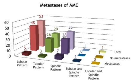
The most common pattern found was the lobular pattern accounting for 33% (53 cases) of the cases described, followed by the spindle pattern, accounting for 22% (35 cases) and finally, by the tubular pattern in 18% (29 cases). Two patterns on the same tumor was rare (1%; 2 cases) (see chart 3).
Chart 3: Distribution of the different patterns of adenomyoepithelioma on the studied cases.
Adenomyoepitheliomas with local recurrence or with metastasis are more common in the age group 15 to 40 and after the age of 80. Both metastasis and recurrence cases exceed the number of non-metastasizing and non-recurred cases at these ages (see chart 4 - 5).
Chart 4: Recurrent adenomyoepitheliomas distribution per age groups.
25% 33% 18% 22% 1% 1% Patterns of AME Not defined Lobular Pattern Tubular Pattern Spindle Pattern
Tubular and Spindle Pattern Lobular and Spindle Pattern
0 5 10 15 20 0 - 5 5 - 10 10 1 5 15 2 0 20 2 5 25 3 0 30 3 5 35 4 0 40 4 5 45 5 0 50 5 5 55 6 0 60 6 5 65 7 0 70 7 5 75 8 0 80 8 5 85 9 0 90 9 5
Recurrence of AME per age
24
Chart 5: Metastatic adenomyoepitheliomas distribution per age groups.
Analyzing the tumor dimensions impact on prognosis, no pattern of behavior dependent on the size of the tumor could be found. It appears that tumor dimensions are not correlated with the tumor potential to metastasize or recur (see chart 6).
Chart 6: Recurrence and metastasis according dimensions (in millimeters) of the adenomyoepithelioma.
Metastasizing tumors revealed no predilection for either breast. Metastatic rate was 20% for the left breast (10 cases of metastases in 49 on the left breast) and 17% for the right
0 5 10 15 20 0 - 5 10 1 5 20 2 5 30 3 5 40 4 5 50 5 5 60 6 5 70 7 5 80 8 5 90 9 5
Metastatic AME per age
With Without Total number of cases
0,0% 10,0% 20,0% 30,0% 40,0% 50,0% 60,0% 70,0% 80,0% 90,0% 100,0% 0 5 10 15 20 25 0 - 5 5 - 10 10 1 5 15 2 0 20 2 5 25 3 0 30 3 5 35 4 0 40 4 5 45 5 0 50 6 0 60 7 0 70 8 0 80 9 0 90 1 00 10 0 - 15 0 15 0 - 20 0 20 0 - 40 0
Recurrence and metastases according AME dimention
Total of cases Number of recurrencies Number of metastizations % Recurrence % Metasteses
25
breast (10 cases of metastases in 58 on the right breast). In 9 cases of metastases (31%), it wasn’t described from which side the primary tumor was (see chart 7).
Chart 7: Metastasizing adenomyoepithelioma distribution by side of primary tumor.
Considering the recurrence distribution for the different patterns, tubular pattern has the highest recurrence rate of 17% (5 cases) followed by lobular pattern with a recurrence of 15% (8 cases) and spindle pattern presenting only 9% of recurrent cases (3 cases) (see chart 8).
Chart 8: Recurrences of the different patterns of adenomyoepithelioma.
Comparing the metastatic rates of the different tumor patterns, spindle pattern appears to be the most prompt to metastasize (20% metastasizing rate). Tubular pattern metastasized in 10% of the cases and lobular pattern only metastasized in 9% of the cases.
Finally, analyzing the patterns of dual appearance tumors, the tumors with tubular and spindle patterns metastasized, but it didn’t recur, and the tumor with lobular and spindle pattern recurred, but it didn’t metastasize (see chart 9 and table 2).
0 20 40 60 Left Breast Right Breast Not defined 10 10 49 58 9
Side of metastasising AME
Metastasizing AME Total 0 10 20 30 40 50 60 Lobular Pattern Tubular
Pattern SpindlePattern Tubular and Spindle Pattern Lobular and Spindle Pattern 8 5 3 1 0 45 24 32 0 1 53 29 35 1 1 Recurrence of AME Total Recurence No recurence
26
Chart 9: Metastases of the different patterns of adenomyoepithelioma.
Table 2: Overview of prognosis of the different patterns of adenomyoepithelioma.
Recurrences and metastases were also analyzed taking into account the mitotic rate presented by the adenomyoepitheliomas. Mitotic rate was considered low for values lower than 4 mitoses per high powered field, medial for values between 4 and 9 and high for values equal to or higher than 10 mitoses per high powered field. Low mitotic rates were associated with less probability to recur (4% against 20% for tumors with medial or high mitotic rates). High mitotic rates were associated with metastases (31% of the cases against 13% for the tumors with low or mean mitotic rate) (see table 3).
Table 3: Overview of prognosis of adenomyoepithelioma according to the mitotic rate of the tumor.
0 10 20 30 40 50 60 Lobular Pattern Tubular Pattern Spindle
Pattern Tubular and Spindle Pattern Lobular and Spindle Pattern 5 3 7 1 0 48 26 28 0 1 53 29 35 1 1 Metastases of AME Total Metastases No metastases
Metastases metastases No Recurrences Recurrences No Total of Cases
Lobular Pattern 5 9% 48 91% 8 15% 45 85% 53
Tubular Pattern 3 10% 26 90% 5 17% 24 83% 29
Spindle Pattern 7 20% 28 80% 3 9% 32 91% 35
Tubular and Spindle Pattern 1 100% 0 0% 1 1% 0 100% 1
Lobular and Spindle Pattern 0 0% 1 100% 0 100% 1 0% 1
Recurrence Metastases
Total Percentage Total Percentage
Low MR 0 0% 2 17%
Mean MR 8 31% 8 31%
27
12. Discussion
The last article published about adenomyoepithelioma (Bajpai, 2013(20)) referred
approximately 150 cases of AME. The review includes 159 described cases of AME worldwide. Adenomyoepithelioma is known for being a women’s tumor rarely occurring in the male population(21, 22). This is verified in the present study – only one male patient was found
compared to 158 women. According to American Cancer Society(50) breast cancer is about 100
times more frequent in women than in men. It seems that adenomyoepithelioma follows this tendency.
According to McLaren(19), the average age for adenomyoepithelioma diagnosis is 59
and the patients’ age range from 22 to 92 years, embracing all ages. The same could be concluded with the present study (range of ages: 16-87 and a mean age of 57 years old) (see chart 10). There is an increased incidence of adenomyoepithelioma after the age of 45 (see chart 10). This finding is not surprising since, according to Canadian Cancer Society and
American Cancer Society(50), breast carcinomas tend to occur more commonly between the
ages of 50-69.
Chart 10: Total number of adenomyoepitheliomas diagnosis per age groups.
Adenomyoepithelioma size varied from 5 to 313mm among the cases studied showing a mean size of 30mm, a close value to Hikino’s findings in a study of 2007 (mean size value: 25mm)(15).
Nassar(4) stated that adenomyoepithelioma has no predilection for either breast,
which is also true for this review. Lobular pattern prevailed as the most common histological pattern of adenomyoepithelioma – accounting for 33% of all cases. The same prevalence was found in 2003 by Howlet(43). 0 0 0 1 1 3 7 7 12 12 11 16 13 15 13 13 8 0 2 4 6 8 10 12 14 16 18 0 5 10 15 20 25 30 35 40 45 50 55 60 65 70 75 80 85 90
28
Adenomyoepithelioma is described in literature as a benign tumor, although there are clinical cases published where a malignant behavior is revealed(5, 6, 8-10, 12). Analyzing the
present data, it was found that from all 159 cases of AME, 26% either recurred or metastasized, with 5% of them metastasizing (see chart 11).
Chart 11: Rates of recurrence and metastases of adenomyoepithelioma.
This behavior seems more common in younger women (under 40) and in the age group above 80. Both metastatic and recurrent cases exceeded the number of non-metastasized and non-recurred tumors within these periods (see chart 12, 13).
Chart 12: Recurrence of adenomyoepithelioma per age groups.
Chart 13: Metastases of adenomyoepithelioma per age groups.
73,60%
5,60%
20,80% 26,40%
AME recurence and metastasis
No metastasis nor recurrence Metastasis and recurrence Only Metastasis or only recurrence 0 5 10 15 20 0 - 5 5 - 10 10 1 5 15 2 0 20 2 5 25 3 0 30 3 5 35 4 0 40 4 5 45 5 0 50 5 5 55 6 0 60 6 5 65 7 0 70 7 5 75 8 0 80 8 5 85 9 0 90 9 5 95 1 00
Recurrence of AME per age
Recurrence No recurrence 0 5 10 15 20 0 - 5 5 - 10 10 1 5 15 2 0 20 2 5 25 3 0 30 3 5 35 4 0 40 4 5 45 5 0 50 5 5 55 6 0 60 6 5 65 7 0 70 7 5 75 8 0 80 8 5 85 9 0 90 9 5 95 1 00
Metasteses of AME per age
Mestases No metastases
29
These findings follow the tendency of breast cancer, described in 2011 by the
Canadian Cancer Society, Canadian Cancer Statistics and American Cancer Society(50) (For
breast cancer, survival is significantly worse for those aged 15–39 and 80–99 at the time of diagnosis) (see chart 14).
Chart 14: Recurrence and metastases of adenomyoepithelioma per age groups.
Analyzing the recurrence and metastatic rates of the different AME patterns, it can be noticed that lobular, although representing the most prevalent pattern, shows a lower grade of metastization.
On the contrary, spindle pattern, although having the lowest prevalence, revealed the highest metastasizing rate (20%). This fact has previously been referred by Dabbs(1). Tubular
pattern is the most frequently associated with recurrence among literature(5) fact also
demonstrated in the present review.
Mitotic rate is expectedly shown to be associated with recurrence and metastatic rates. Low mitotic rates (MR≤3) were associated with less probability for the tumor to recur (4% against 20% for tumors with average or high MR), confirming Dabbs’(1) and FA’s(5)
conclusions. High mitotic rate (≥10), in accordance with Dabbs(1) was associated with
metastases (31% of metastases against 13% for the tumors with low or average MR).
Breast adenomyoepithelioma is characterized by proliferation of epithelial and myoepithelial cells. The recognition of this entity, accurate diagnosis, and knowledge of the expected behavior are important in guiding the most appropriate patient management. From
0 2 4 6 8 10 12 14 16 0 - 5 5 - 10 10 1 5 15 2 0 20 2 5 25 3 0 30 3 5 35 4 0 40 4 5 45 5 0 50 5 5 55 6 0 60 6 5 65 7 0 70 7 5 75 8 0 80 8 5 85 9 0 90 9 5 95 1 00
AME metasteses and recurrence per age
30
all the cases found not all of them were followed after surgery. A correct follow-up is necessary not only to diagnose any change of behavior suspicious for malignant transformation but also to learn more about its pathophysiology.
The review demonstrated that adenomyoepithelioma is more common for females after 45 years old and the clinical case patient, who is 47 years old, proves this predominance.
Our clinical case revealed a typical case of adenomyoepithelioma: an asymptomatic patient, without previous history of cancer, a solitary breast mass on the peripheral portion of the breast found and an inconclusive or suspicious mammogram(1). The lesions are usually
diagnostic challenging even with a core needle biopsy and the morphological heterogeneity may lead to the erroneous diagnosis of carcinoma(24). Adequate sampling of the tumor is
necessary to identify the overall architecture correctly, and our patient, after being submitted to needle core biopsy (imunohistochemistry revealed an epithelial and myoepithelial lesion), was later submitted to tumorectomy, which permitted classifying the tumor as an adenomyoepithelioma. Mitotic rate was low. No additional treatment was needed because adenomyoepithelioma is recommended to be treated only with local excision with appropriate margins(5-7). The tumor revealed a tubular pattern – the most common
31
13. Study Limitations
Adenomyoepithelioma of the breast is a rare medical condition. Perhaps because of this rarity, the 159 articles evaluated did not provide the needed information for a clearer comprehension of this pathology. Some articles did not refer to the age of the patient, breast side or location, nodule dimensions, AME patterns, mitotic rates, recurrence or metastasis. This lack of a standard of information limits a more adequate characterization of the tumor.
32
14. Advantages of the Study
It is important for every diagnosed AME to be compared with the previous cases so that a solid pathophysiological pattern can be found to achieve a padronized diagnostic procedure and treatment.
Our clinical case follows what seems to be typical for adenomyoepithelioma. To highlight: grade 4 mammogram, inconclusive biopsy and the need of tumorectomy to achieve the final diagnose.
33
15. Conclusion
After an exhaustive study of selected literature and analysis of the case report and the published cases, some considerations about adenomyoepithelioma may be done.
Adenomyoepithelioma is a biphasic neoplastic proliferation of luminal and myoepithelial cells(14). Around 160 cases of AME of the breast have been reported in the
literature, nearly all women. The tumor may display a heterogeneous pattern because of the variable proliferation of epithelial and myoepithelial cells and striking morphologic heterogeneity may lead to the erroneous diagnoses of carcinoma(16). So far, other than case
reports, only comprehensive series of studies have been reported (5, 6, 16, 17, 19). Recognition of
this entity, accurate diagnosis, and knowledge of the expected behavior are important in guiding the most appropriate patient management.
Patients’ age ranged from 16 to 92 years, with the average age near 60. They are normally asymptomatic and without previous personal history of cancer or high-risk breast lesions(23) and the adenomyoepithelioma is either diagnosed as a palpable mass or as an
occasional mammographic finding(1). Patients usually present with a solitary,
well-circumscribed, sometimes palpable, firm nodule, measuring on average 30mm present for several weeks to several months(5). The tumors are usually located in a peripheral portion of
the breast(16), having most commonly a tubular pattern and there is no predilection for either
breast(4).
These lesions can be diagnostically challenging, even when a core needle biopsy is performed because of heterogeneity of adenomyoepitheliomas. Recognition of the biphasic cellular elements and the characteristic overall architecture of the tumors in combination with immunohistochemistry are essential to establish the correct diagnosis. Therefore, adequate sampling of the tumor to identify these features is crucial(24).
On US images adenomyoepithelioma are small, solid, irregular or oval, non-parallel orientation lesions, microlobulated or with indistinct margin characterized by hipoechogenicity and increased peripheral vascularity(23, 25). Magnetic resonance imaging and
mammography lack a pattern of specificity for this tumor(23). Although all imaging methods
are non-specific for adenomyoepithelioma, US can be done after a physical examination positive for a mammary mass in order to look for a well defined mass and, if positive, the nature of the mass should be done and confirmed by FNAC, combined with immunohistochemistry.
FNAC shows of displace, compress, or obliterate the epithelial gland, myoid areas composed by myoepithelial cells with pink to amphophilic cytoplasm(7) or a plasmacytoid
appearance with dense, hyaline-like, glassy eosinophilic cytoplasm and dark, eccentric, small, comma-shaped or ovoid nuclei(5, 7, 27, 31).
Immunohistochemistry is very useful in confirming the diagnosis of AME(3). The