Cop
yright
© ABE&M t
odos os dir
eit
os r
eser
vados
.
MicroRNAs
miR-146-5p
and
let-7f
as prognostic tools for
aggressive papillary thyroid
carcinoma: a case report
MicroRNAs miR-146-5p e let-7f como ferramenta de prognóstico para o carcinoma papilífero de tiroide: relato de caso
Murilo Vieira Geraldo1*, Cesar Seigi Fuziwara1*, Celso Ubirajara Moretto Friguglieti2, Ricardo Borges Costa3, Marco Aurélio Vamondes Kulcsar2, Alex Shimura Yamashita1, Edna Teruko Kimura1
SUMMARY
Papillary thyroid cancer (PTC) is the most incident histotype of thyroid cancer. A certain frac-tion of PTC cases (5%) are irresponsive to convenfrac-tional treatment, and refractory to radioio-dine therapy. The current prognostic factors for aggressiveness are mainly based on tumor size, the presence of lymph node metastasis, extrathyroidal invasion and, more recently, the presence of the BRAFT1799Amutation. MicroRNAs (miRNAs) have been described as
promis-ing molecular markers for cancer as their deregulation is observed in a wide range of tumors. Recent studies indicate that the over-expression of miR-146b-5p is associated with aggressive-ness and BRAFT1799Amutation. Furthermore, down-regulation of let-7f is observed in several
types of tumors, including PTC. In this study, we evaluated the miR146b-5p and let-7f status in a young male patient with aggressive, BRAFT1799A-positive papillary thyroid carcinoma, with
extensive lymph node metastases and short-time recurrence. The analysis of miR-146b-5p and let-7f expression revealed a distinct pattern from a cohort of PTC patients, suggesting caution in evaluating miRNA expression data as molecular markers of PTC diagnosis and prognosis. Arq Bras Endocrinol Metab. 2012;56(8):552-7
SUMÁRIO
O carcinoma papilífero (PTC) é o histotipo mais prevalente de câncer de tiroide. Cerca de 5% dos casos são refratários ao tratamento convencional e à radioiodoterapia. Os fatores prognós-ticos para agressividade mais utilizados atualmente são o tamanho do tumor, a presença de metástases linfonodais ao diagnóstico, a presença de invasão extratiroideana e, mais recen-temente, a presença da mutação BRAFT1799A. A análise de peril de expressão de microRNAs
(miRNA) mostra que esses pequenos RNAs são marcadores moleculares promissores para o câncer, por apresentarem desregulação de sua expressão em uma ampla gama de tumores, includindo o PTC. Estudos recentes revelam a associação entre o aumento da expressão do miRNA e miR-146b-5p e a presença da mutação BRAFT1799A como um fator de pior prognóstico no
PTC. Além disso, observa-se a diminuição da expressão de let-7f em diversos tipos de tumores, incluindo tumores tiroideanos. Neste relato de caso, realizamos a quantiicação da expressão de miR-146b-5p e let-7f em um paciente jovem, de sexo masculino, apresentando PTC positivo para a mutação BRAFT1799A com extensas metástases linfonodais ao diagnóstico e recidiva precoce.
A análise da expressão de miR-146b-5p e let-7f mostrou um padrão diferente do observado em um grupo de pacientes PTC, sugerindo a necessidade de cautela na interpretação da expressão de miRNAs como marcador molecular no diagnóstico e prognóstico de PTC. Arq Bras Endocrinol Metab. 2012;56(8):552-7
1 Department of Cell and
Developmental Biology, Institute of Biomedical Sciences, University of Sao Paulo, Sao Paulo, SP, Brazil
2 Santa Catarina Hospital,
Sao Paulo, SP, Brazil
3 Ferdinando Costa Laboratory,
Sao Paulo, SP, Brazil
* These authors contributed
equally to this work.
Correspondence to:
Edna Teruko Kimura
Av. Prof. Lineu Prestes, 1524, sala 414 Instituto de Ciências Biomédicas, Universidade de São Paulo 05508-900 – São Paulo, SP, Brazil [email protected]
Cop
yright
© ABE&M t
odos os dir
eit
os r
eser
vados
.
INTRODUCTION
P
apillary thyroid cancer (PTC) is the most incidenthistotype of thyroid cancer, accounting for more than 80% of the 35,000 annual cases in the USA. To date, prediction of PTC outcome is mainly based on tumor size, gender, age of patient at time of diagnosis, presence lymph node metastasis, capsular invasion, and extra-thyroidal extension. Although most PTCs cases display overall good prognosis, a certain fraction of PTC cases (5%) are irresponsive to conventional treat-ment and refractory to radioiodine therapy (1). Thus, a number of cases diagnosed as low-risk PTCs eventu-ally recur loceventu-ally and as lymph node metastases, lead-ing to death (2), indicatlead-ing the lack of current reliable outcome predictors. Indeed, no consensus has been achieved regarding the clinical application of molecular markers to predict the outcome of PTC patients (3).
More recently, several studies suggest that the pres-ence of mutation in BRAF oncogene is associated with a poor outcome due to frequent extra-thyroidal inva-sion, recurrence, and lower survival rate (4-6). In this context, microRNAs (miRNAs) arise as interesting mar kers for cancer, as their deregulation affects innu-merous cell processes, such as proliferation and apop-tosis (7). MiRNAs negatively regulate protein levels by binding to 3’UTR of target mRNA and impairing pro-tein translation (8). The potential of miRNAs as PTC molecular markers is highlighted, once their expres-sion may be detected from formalin-ixed parafin-em-bedded (FFPE) tissue, ine needle aspiration cytology (FNAC), as well as serum samples (9).
Recent studies have explored the use of several miRNAs as cancer diagnostic tools (10). In the last 4 years, several studies have demonstrated the potential
of miRNAs in PTC diagnosis. Although miR-146b-5p
is down-regulated in other types of cancer (11-13), this miRNA is highly overexpressed in PTCs (14,15), re-liably distinguishing this type of cancer from follicular carcinoma (FTC), and from benign lesions (16,17). Moreover, Schwertheim and cols. have shown that a set of miRNAs that includes miR-146b-5p, distinguish-es well-differentiated from undifferentiated thyroid tumors (18). Recent studies suggested that miRNA expression may be used as a thyroid cancer prognos-tic tool. The expression levels of miR-146b-5p, along with other two miRNAs, miR-221 and -222, were sig-niicantly higher in high-risk PTC patients who showed extra-thyroidal invasion (19). Moreover, among
BRAFT1799A-positive PTCs, the aggressive subset
pres-ents high miR-146b-5p expression levels (20). Let-7f
down-regulation was irstly described by Takamizawa and cols. (21) in lung cancer with poor prognosis, and rapidly arose as a promising cancer molecular marker
(22). Although let-7f down-regulation has been
de-scribed in PTC, its usefulness as a diagnostic and prog-nostic marker for PTC is uncertain.
In this study, we present a case report of a young male patient presenting an aggressive, BRAFT1799A
-posi-tive papillary thyroid carcinoma, displaying lymph node metastases at diagnosis and short-term recurrence after surgery, in whom we evaluated the expression pattern of the miRNAs miR-146b-5p and let-7f.
SUBJECT AND METHODS
Case report
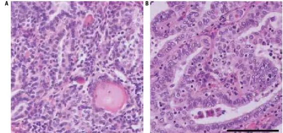
A 23-year-old caucasian male (named here ABC) with parents bearing Hashimoto’s thyroiditis, was submitted to a health checkup, and was diagnosed with hypothy-roidism, positive for anti-TPO and anti-TG antibodies. At the ultrasound examination, calciied thyroid no-dules of 12 mm and 8 mm were found at the isthmus and right lobe, respectively. The patient ABC also had lymphadenopathy of 33 mm, with calciication at level III of right side, and 9 mm at level III of left side. Fine needle aspiration citology (FNAC) pattern was com-patible with PTC in the thyroid and in the right lateral lymph nodes, but not in the left side. The patient un-derwent total thyroidectomy, and central compartment and right lateral lymph node dissection (levels II to V). Histopathological examination of the primary tumor conirmed the diagnosis of PTC (Figure 1A) with lym-phatic invasion. Metastases were found in right lymph nodes at levels III (2 out of 7), IV (6 out of 6) (Figure 1B), V (2 out of 3), and at the central compartment (3 out of 4). Neither vascular nor peri-neural invasion were detected. According to the American Joint Com-mittee on Cancer Staging System, the tumor was classi-ied as T3N1bM0 and MACIS score was 3.37 (23,24).
Three months after surgery, the patient received
200 mCi of 131I under Thyrogen® (Genzyme,
Cop
yright
© ABE&M t
odos os dir
eit
os r
eser
vados
.
largest one with 19 mm, and elevated anti-Tg antibody levels (11,580 IU/m). FNAC revealed PTC and PET-CT examination showed FDG uptake at level II. At the second surgery, lateral lymph node dissection was per-formed on the left side (levels II to V). Histopathologi-cal examination of lymph nodes revealed metastases at levels II (2 out 4), III (3 out 17), IV (2 out of 8) and V (9 out of 11). The patient received 200 mCi of 131I
under Thyrogen® stimulation. Three months after the
second surgery, radioiodine uptake was negative.
Patient population
Human PTC and non-tumor paired thyroid tissue sam-ples were collected from patients, including ABC, after the informed consent was signed. This samples belong to the thyroid tumor tissue bank, approved by the ethi-cal committee of the Institute of Biomediethi-cal Sciences (no. 1030/CEP), Universidade de Sao Paulo. Thyroid carcinomas were classiied according to the last WHO classiication (25).
BRAF mutation detection
Genomic DNA was extracted from thyroid tissue
us-ing the saltus-ing-out protocol. Briely, after digestion
with proteinase K, protein was precipitated with 5 M NaCl, and DNA was isolated with isopropanol. Exon 15 of BRAF, containing the hotspot T1799A muta-tion, was ampliied by PCR using speciic primers (FW: 5´- AAACTCTTCATAATGCTTGCTCTG-3’; RV: 5’-GGCCAAAAATTTAATCAGTGGA-3’), and the
product was puriied in QIAquick® PCR puriication
Figure 1. Histopathological slides of the resected differentiated PTC and lymph node metastasis. (A) Primary PTC. (B) Level IV lymph node metastasis. Hematoxylin-eosin, the black bar represents 100 µm.
kit (Qiagen, Valencia, CA, USA). For direct sequenc-ing reaction, the puriied product was ampliied ussequenc-ing
BigDye® Terminator v3.1 Cycle Sequencing Kit (Life
Technologies), forward primer, and injected in multi-capillary sequencer MegaBace1000 (GE Healthcare, Little Chalfont, UK) (26). The similarity of the result-ing sequence was analyzed and conirmed usresult-ing BLAST software (http://www.ncbi.nlm.nih.gov/BLAST/).
MicroRNA expression
Excised tumor tissues were immediately stored in RNAlater®. Total RNA was extracted by
phenol-chloroform using TRIzol® reagent (LifeTechnologies,
Carlsbad, CA, USA), according to the manufacturer’s instructions, and stored at -80oC. Detection of miRNAs miR-146b-5p, let-7f and RNU6B was performed using commercial kits (Life Technologies) according to the manufacturer’s speciications. Briely, cDNA was synthesized from 10 ng of total RNA using speciic pri mers in a stem-loop based technology with TaqMan MicroRNA Reverse Transcription Kit (Life Technologies). Expression was analyzed by qPCR using speciic Taqman miRNA Assay (Life Technologies)
and TaqMan® Master Mix no AmpErase® UNG (Life
Technologies).
Statistical analysis
Quantiication of miRNA expression is presented as mean ± S.E., and data were submitted to Student’s t-test to compare results between the two groups. Differen ces were considered signiicant at p < 0.05.
Cop
yright
© ABE&M t
odos os dir
eit
os r
eser
vados
.
RESULTS
Analysis of genomic DNA from a set of PTC sam-ples and paired contra-lateral thyroid tissue revealed
BRAFT1799A mutation in 41% of patients, including the
patient ABC (7/17).
MiR-146b-5p is substantially up-regulated in the cohort of PTCs from our tissue bank when compared with their matched contra-lateral samples, irrespective of their BRAFT1799A status (Figure 2A). Interestingly,
both the primary tumor and metastatic tissue of ABC
exhibited lower miR-146b-5p fold-change levels
com-pared with the cohort of PTC samples (Figure 2A), and no statistical differences compared with its paired nor-mal thyroid tissue (P = 0.773).
On the other hand, the BRAFT1799A tumors showed
no signiicant changes in let-7f expression levels, while
BRAF-wild type tumors exhibit increased let-7f
lev-els in comparison with matched contra-lateral thyroid tissue (Figure 2B). ABC primary tumor, which was
BRAFT1799A-positive, exhibited a slight increase in let-7f
levels (31%, P < 0.01) (Figure 2B). However, the meta-static tissues, also positive for BRAFT1799A-, displayed
decreased let-7f levels (Fig 3B).
in distinguishing malignant from benign tumors, and that different thyroid cancer histotypes display distinct miRNA expression (17,27). However, in patient ABC, the expression pattern of miR-146b-5p and let-7f levels could not be associated with aggressiveness and poor prognosis, as previously described in the literature.
It is not clear whether the overexpression of miR-146b-5p is associated to BRAFT1799A status. While initial
studies showed no correlation between this miRNA and oncogenic activation (9), recent studies have demon-strated a positive correlation between the overexpres-sion of miR-146b-5p and aggressive BRAFT1799A tumors
(19). Moreover, in the literature, high expression of
miR-146b-5p isassociatedwith BRAF-mutated tumors that display more aggressive phenotypes (20). Con-versely, our cohort of patients did not show any corre-lation regarding BRAF status and miR-146b-5p expres-sion. MiR-146b-5p is associated with the deregulation of NFκB and TGFβ pathways, while the blockage of
miR-146b-5p action restores TGFβ signal transduction in thyroid cells in vitro (28). Non-thyroid tumors fre-quently exhibit reduced levels of miR-146b-5p (11,29). We have previously shown that activation of MAPkinase pathway in normal thyroid cells increases miR-146b-5p
levels in vitro (28). Therefore, the overexpression of
miR-146b-5p, only observed in thyroid tumors, could be related with a thyroid-speciic oncogenic activation, which might include the MAPK pathway.
The association between BRAFT1799A and let-7f
ex-pression also remains poorly understood. Classically,
the main target of let-7 is RAS oncogene, an
impor-tant transducer for the MAPK pathway. Interestingly, a functional study revealed that the reinforced expression of let-7f in a PTC cell line blocks ERK phosphoryla-tion without affecting RAS protein levels (30). On the other hand, we have previously shown that activation of RET/PTC rearrangement in rat normal thyroid cell line, but not of BRAF mutation, decreases let-7f levels. In our cohort of patients, BRAFT1799A-positive tumors
showed no change in let-7f levels, in comparison with contra-lateral thyroid tissue, while BRAFwild type tu-mors displayed increased levels of this miRNA. Inter-estingly, the patient ABC, which was BRAFT1799A
-posi-tive, showed slightly increased let-7f expression, even though his metastatic tissue, also BRAFT1799A-positive,
showed decreased levels of let-7f. Little is known about the targets regulated by let-7f in PTC. Different mRNA targets might be regulated by let-7f to promote matrix remodeling, vascular iniltration, and the colonization
Figure 2. MiR-146b and let-7f levels in PTC samples. Expression of
miR-146b-5p(A) and let-7f (B) in PTC samples in comparison with
contra-lateral thyroid tissue. Values are represented as mean ± SE of expression fold-change matched to contra-lateral thyroid tissue. RNU6B gene expression was used as an internal control. ABC, patient ABC; Meta IV, lymph node metastasis at level IV; Meta VI, lymph node metastasis at level VI. (*) P < 0.01, against matched thyroid contra-lateral tissue.
A B
100 4
3
2
1
0
Let-7f/RNU6B (a.u.)
miR-146b-5p Let-7f
*
BRAFwt
BRAFT1799A
Primar
y
Meta IV Meta VI
80
60
40
20
0
Patient ABC
miR-146b/RNU6B (a.u)
BRAFwt
BRAFT1799A
Primar
y
Meta IV Meta VI
Patient ABC
DISCUSSION
Cop
yright
© ABE&M t
odos os dir
eit
os r
eser
vados
.
of the metastatic site. Functional studies are needed to elucidate whether the loss of let-7f by primary tumor is implicated in thyroid tumor progression and what is the clinical signiicance of this data.
To date, clinical and histological features, such as age, gender, extra-thyroidal invasion, and, vascular in-vasion, histotype, and more recently, BRAFT1799A status,
are still the indicated parameters for accurate PTC out-come prediction. Furthermore, recent lines of evidence have shown that the number of lymph node metasta-ses is also important to predict the patient’s outcome, and may be used as an independent prognostic factor for aggressiveness (31), which corroborate the clinical characteristics of patient ABC.
This case showed to be interesting because of the discordance of two widely studied PTC miRNA mar kers in this index patient and our cohort of patients. Our data indicate that the expression pattern of miR-146b-5p and
let-7f genes may not reproduce the clinical features of the tumor, clearly illustrating that outcome prediction based on the analysis of the expression of a single or few miRNA genes should be viewed with caution.
Acknowlegments: the authors would like to thank Dr. Kelly Cris-tina Saito for her technical assistance, and Fundação de Amparo à Pesquisa do Estado de São Paulo (Fapesp), Conselho Nacio-nal de Desenvolvimento Cientíico e Tecnológico (CNPq), and Coordenação de Aperfeiçoamento de Pessoal de Nível Superior (Capes) for the inancial support.
Disclosure: no potential conlict of interest relevant to this article was reported.
REFERENCES
1. Mazzaferri EL, Jhiang SM. Long-term impact of initial surgical and medical therapy on papillary and follicular thyroid cancer. Am J Med. 1994;97(5):418-28.
2. Mazzaferri EL, Kloos RT. Clinical review 128: current approaches to primary therapy for papillary and follicular thyroid cancer. J Clin Endocrinol Metab. 2001;86(4):1447-63.
3. Cooper DS, Doherty GM, Haugen BR, Hauger BR, Kloos RT, Lee SL, et al. Revised American Thyroid Association management guidelines for patients with thyroid nodules and differentiated thyroid cancer. Thyroid. 2009;19(11):1167-214.
4. Nikiforova MN, Kimura ET, Gandhi M, Biddinger PW, Knauf JA, Basolo F, et al. BRAF mutations in thyroid tumors are restricted to papillary carcinomas and anaplastic or poorly differentiated carcinomas arising from papillary carcinomas. J Clin Endocrinol Metab. 2003;88(11):5399-404.
5. Xing M. BRAF mutation in papillary thyroid cancer: pathogenic role, molecular bases, and clinical implications. Endocr Rev. 2007;28(7):742-62.
6. Lupi C, Giannini R, Ugolini C, Proietti A, Berti P, Minuto M, et al. Association of BRAF V600E mutation with poor clinicopathologi-cal outcomes in 500 consecutive cases of papillary thyroid carci-noma. J Clin Endocrinol Metab. 2007;92(11):4085-90.
7. Esquela-Kerscher A, Slack FJ. Oncomirs – microRNAs with a role in cancer. Nat Rev Cancer. 2006;6(4):259-69.
8. Bartel DP. MicroRNAs: genomics, biogenesis, mechanism, and function. Cell. 2004;23;116(2):281-97.
9. Nikiforova MN, Tseng GC, Steward D, Diorio D, Nikiforov YE. MicroRNA expression proiling of thyroid tumors: biological signiicance and diagnostic utility. J Clin Endocrinol Metab. 2008;93(5):1600-8.
10. Lu J, Getz G, Miska EA, Alvarez-Saavedra E, Lamb J, Peck D, et al. MicroRNA expression proiles classify human cancers. Nature. 2005;435(7043):834-8.
11. Bhaumik D, Scott GK, Schokrpur S, Patil CK, Campisi J, Benz CC. Expression of microRNA-146 suppresses NF-kappaB activity with reduction of metastatic potential in breast cancer cells. Onco-gene. 2008;27(42):5643-7.
12. Kanaan Z, Rai SN, Eichenberger MR, Barnes C, Dworkin AM, Weller C, et al. Differential microRNA expression tracks neo-plastic progression in inlammatory bowel disease-associated colorectal cancer. Hum Mutat. 2012;33(3):551-60.
13. Man YG, Fu SW, Liu AJ, Stojadinovic A, Izadjoo MJ, Chen L, et al. Aberrant expression of chromogranin A, 146a, and miR-146b-5p in prostate structures with focally disrupted basal cell layers: an early sign of invasion and hormone-refractory cancer? Cancer Genomics Proteomics. 2011;8(5):235-44.
14. He H, Jazdzewski K, Li W, Liyanarachchi S, Nagy R, Volinia S, et al. The role of microRNA genes in papillary thyroid carcinoma. Proc Natl Acad Sci U S A. 2005;102(52):19075-80.
15. Pallante P, Visone R, Ferracin M, Ferraro A, Berlingieri MT, Tron-cone G, et al. MicroRNA deregulation in human thyroid papillary carcinomas. Endocr Relat Cancer. 2006;13(2):497-508.
16. Chen YT, Kitabayashi N, Zhou XK, Fahey TJ, 3rd, Scognamiglio T. MicroRNA analysis as a potential diagnostic tool for papillary thy-roid carcinoma. Mod Pathol. 2008;21(9):1139-46.
17. Sheu SY, Grabellus F, Schwertheim S, Worm K, Broecker-Preuss M, Schmid KW. Differential miRNA expression proiles in variants of papillary thyroid carcinoma and encapsulated follicular thyroid tumours. Br J Cancer. 2010;102(2):376-82.
18. Schwertheim S, Sheu SY, Worm K, Grabellus F, Schmid KW. Anal-ysis of deregulated miRNAs is helpful to distinguish poorly dif-ferentiated thyroid carcinoma from papillary thyroid carcinoma. Horm Metab Res. 2009;41(6):475-81.
19. Chou CK, Chen RF, Chou FF, Chang HW, Chen YJ, Lee YF, et al. miR-146b is highly expressed in adult papillary thyroid carcinomas with high risk features including extrathyroidal invasion and the BRAF(V600E) mutation. Thyroid. 2010;20(5):489-94.
20. Yip L, Kelly L, Shuai Y, Armstrong MJ, Nikiforov YE, Carty SE, et al. MicroRNA signature distinguishes the degree of aggressiveness of papillary thyroid carcinoma. Ann Surg Oncol. 2011;18(7):2035-41. 21. Takamizawa J, Konishi H, Yanagisawa K, Tomida S, Osada H,
En-doh H, et al. Reduced expression of the let-7 microRNAs in hu-man lung cancers in association with shortened postoperative survival. Cancer Res. 2004;64(11):3753-6.
22. Fuziwara CS, Geraldo MV, Kimura ET. Let-7 and Cancer. MicroRNA let-7: role in human diseases and drug discovery. DNA and RNA: Properties and Modiications, Functions and Interactions, Recombination and Applications Genetics – Research and Issues: Nova Science Publishers; 2012. p. 109-24.
23. Edge SB, Compton CC. The American Joint Committee on Cancer: the 7th edition of the AJCC cancer staging manual and the future of TNM. Ann Surg Oncol. 2010;17(6):1471-4.
Cop
yright
© ABE&M t
odos os dir
eit
os r
eser
vados
.
25. DeLellis RA, Lloyd RV, Heitz PU, Eng C. Pathology and Genetics of Tumours of Endocrine Organs. Lyon: IARC Press; 2004.
26. Kimura ET, Nikiforova MN, Zhu Z, Knauf JA, Nikiforov YE, Fagin JA. High prevalence of BRAF mutations in thyroid cancer: genetic evidence for constitutive activation of the RET/PTC-RAS-BRAF signaling path-way in papillary thyroid carcinoma. Cancer Res. 2003;1;63(7):1454-7. 27. Shen R, Liyanarachchi S, Li W, Wakely PE Jr, Saji M, Huang J, et al.
Mi-croRNA signature in thyroid ine needle aspiration cytology applied to “atypia of undetermined signiicance” cases. Thyroid. 2012;22(1):9-16. 28. Geraldo MV, Yamashita AS, Kimura ET. MicroRNA miR-146b-5p
regulates signal transduction of TGF-beta by repressing SMAD4 in thyroid cancer. Oncogene. 2012;12;31(15):1910-22.
29. Xia H, Qi Y, Ng SS, Chen X, Li D, Chen S, et al. microRNA-146b inhibits glioma cell migration and invasion by targeting MMPs. Brain Res. 2009;7;1269:158-65.
30. Ricarte-Filho JC, Fuziwara CS, Yamashita AS, Rezende E, da-Silva MJ, Kimura ET. Effects of let-7 microRNA on Cell Growth and Differentiation of Papillary Thyroid Cancer. Transl Oncol. 2009;2(4):236-41.