Faculdade de Ciências do Mar e do Ambiente
Transthyretin and thyroid hormone transport in fish and
the effect of endocrine disruptors on this process
(Tese para obtenção do grau de doutor no ramo de Biologia, especialidade de Biologia Molecular)
Isabel Maria Sena Morgado
Orientado: Doutora Deborah Mary Power Co-oreintador: Doutor Eduardo Xavier Pinho e Melo Constituição do Júri:
Presidente: Reitor da Universidade do Algarve Vogais: Doutor Glen Sweeney
Doutor Adelino Vicente Mendonça Canário Doutora Deborah Mary Power
Doutor Eduardo José Xavier Rodrigues de Pinho e Melo Doutora Cecília Reis Alves dos Santos
Faculdade de Ciências do Mar e do Ambiente
Transthyretin and thyroid hormone transport in fish and
the effect of endocrine disruptors on this process
(Tese para obtenção do grau de doutor no ramo de Biologia, especialidade de Biologia Molecular)
Isabel Maria Sena Morgado
Orientador: Doutora Deborah Mary Power Constituição do Júri:
Presidente: Reitor da Universidade do Algarve Vogais: Doutor Glen Sweeney
Doutor Adelino Vicente Mendonça Canário Doutora Deborah Mary Power
Doutor Eduardo José Xavier Rodrigues de Pinho e Melo Doutora Cecília Reis Alves dos Santos
The contents of this dissertation are of exclusive responsibility of the author O conteúdo desta dissertação é da exclusiva responsabilidade da autora
During this PhD I have learned about science and its “pathways” and also about life and its lessons. I found out that I didn’t have a clue about both…but now I’m in a better position to get some. For such big accomplishment I have to acknowledge many people:
I acknowledge the Portuguese National Science Foundation (FCT) for funding this work through the project POCTI/CVT/38703/2001 and the PhD fellowship (SFRH/BD/6091/2001).
I acknowledge the University of Algarve and the Centre of Marine Sciences (CCMAR), Laboratory of Molecular and Comparative Endocrinology where the present work was undertaken, in particularly Professor Adelino Canário for receiving me in his team and for his support and advice.
I sincerely want to express my gratitude to my supervisor, Professor Deborah Power for believing in me for this project, for the constant support, encouragement, wise advice and for always transmitting me the confidence to carry on. I especially admire and thank her for being such a great teacher from whom I learned so much throughout these years.
I’m also very grateful to Prof. Eduardo Melo who co-supervised some of this work. I thank him for his availability, orientation and advice in the biophysical studies.
I greatly thank ALL my friends/colleagues who where obviously involved in this work in many different ways. Thanks to everyone in the lab: Guerreiro, Teresa, Vanessa, Ana Passos, Vitor, Nuno, Laurence and Nick (for their company “fora de horas”), Laura, Bela, João, Ana Gomes, Vânia, Patrícia, Rita, Natália, Rute, Pedro Luis, Vera, Liliana, Beta, Elsa, Alejandro, Natália Moura, Dulcineia, Liana, Marco, Bruno, Nádia, Dulce, Lília, Rita Jacinto (my TTR African sister), Rita Costa, Tânia, Angela, Peter, Mar, Olinda, Rui, Frade, Mira, Xoan, Begoña, Pep..
....special thanks to my friends outside the lab: Lamp, Nati, Fifi, Cotinha, Warrior, Eleni, Marianna, Patri, Joni, David, Graça, Anabela, Carlos, César, Sónia e Poka….and to my parents, brothers and sisters!
NOME: Isabel Maria Sena Morgado FACULDADE: Ciências do Mar e do Ambiente ORIENTADOR: Professora Deborah Power DATA: Março 2007
TÍTULO DA TESE: Transtirretina e o transporte das hormonas tiróides em peixes e o
efeito dos disruptores endócrinos neste processo
RESUMO
Nos vertebrados, a transtirretina (TTR) é uma proteína responsável pelo transporte sanguíneo das hormonas tiróides (HT), tiroxina (T4) e triiodotironina (T3). A evolução estrutural e
funcional da TTR nos vertebrados, a sua capacidade de formar fibras amilóides causando doenças humanas e a sua afinidade para disruptores endócrinos (DEs) são questões importantes pouco exploradas em peixes. Neste estudo produziu-se TTR recombinante de dourada (sbrTTR), antisoro específico anti-sbrTTR e dois mutantes sbrTTR no N-terminal. Ensaios competitivos de ligação demonstraram que sbrTTR liga-se com afinidade semelhante à T3 (Kd=10.6nM) e T4 (Kd=9.8nM) e que o N-terminal influencia a ligação à sbrTTR e a
produção de fibras amilóides. Níveis plasmáticos de TTR (3.8µg/ml) aumentaram após administração de HT apesar da expressão génica hepática não ser alterada, sugerindo que as HT regulam a secreção da TTR mas não a síntese. Os DEs competiram significativamente na ligação sbrTTR-HT in vitro, indicando a utilidade deste ensaio na avaliação da disrupção do eixo da tiróide em peixes. In vivo, os ligandos da TTR, ioxynil e diethylstilbestrol, alteram o eixo da tiróide em dourada; a expressão de TSH e deiodinases no cérebro diminuiu apesar dos níveis plasmáticos das HT e a expressão hepática da TTR não sofrerem quaisquer alterações.
Palavras chave: Transtirretina, proteínas de ligação às hormonas tiróides, hormonas tiróides, peixes teleósteos, sistema da tiróide, disruptores endócrinos.
THESIS TITLE: Transthyretin and thyroid hormone transport in fish and the effect
of endocrine disruptors on this process
ABSTRACT
Transthyretin (TTR) is a homotetrameric protein that transports thyroid hormones (THs) thyroxine (T4) and triiodothyronine (T3) in vertebrate’s blood. TTR features raise important
questions: understanding its structural/functional evolution in vertebrates; its ability to form amyloid fibrils causing human disease and TTR’s high affinity for endocrine disruptor chemicals (EDCs). In fish, TTR features are poorly explored. In this study, sea bream recombinant TTR (sbrTTR), specific anti-sbrTTR antisera and two sbrTTR N-terminal mutants were produced. A [125I-T3] competitive binding assay demonstrated that sbrTTR
binds T3 (Kd=10.6nM) and T4 (Kd=9.8nM) with a similar affinity and the N-terminus was
found to influence binding to sbrTTR but also the formation of fibrils. TTR plasma levels, measured (3.8µg/ml) for the first time in fish using a specific ELISA, increased upon administration of THs, although hepatic gene expression was not modified suggesting THs regulate TTR secretion but not synthesis. Putative EDCs strongly displace in vitro sbrTTR-THs binding indicating the sbrTTR binding assay could be a useful risk assessment tool for thyroid axis disruption in fish. In vivo, TTR-binders ioxynil and diethylstilbestrol altered the thyroid axis in sea bream; brain TSH and deiodinases expression was downregulated although TH plasma levels and TTR hepatic expression remained unaltered.
Key-words: Transthyretin, thyroid hormone binding proteins, thyroid hormones, teleost fish, thyroid system, endocrine disrupting chemicals.
Articles in refereed journals
Morgado, I., Santos, C.R.A., Jacinto, R., Power, D. M. 2007. Regulation of Transthyretin by
thyroid hormones in fish. Gen Comp Endocrinol. 152, 189-197.
Morgado, I., Hamers, T., Van der Ven, L., Power, D.M. 2007. Disruption of thyroid hormone
binding to sea bream recombinant Transthyretin by ioxynil and polybrominated diphenyl ethers. Chemosphere. 69, 155-163.
Conference proceedings
Morgado, I., Anjos, L., Power, D. M. 2005. Transthyretin and thyroid hormone transport in
fish. Avances en Endocrinología Comparada vol.II, 277-79.
Morgado I., Sauer-Eriksson, E., Power, D. M. 2006. Thyroid hormone binding by
recombinant sea bream transthyretin: The role of the N-terminal region Journal of experimental zoology. Part A- comparative experimental biology 305A (2): 158-158 FEB 1 2006
Communications in conferences
Morgado, I., Power, D. M. Thyroid hormone binding by recombinant sea bream
transthyretin: the role of the N-terminal region; Oral communication. 15th International Congress of Comparative Endocrinology, May 2005,Boston, USA.
Morgado, I., Power, D. M. Transthyretin and thyroid hormone transport in fish; Oral
communication. V Mini-Symposium on Comparative and Molecular Endocrinology, Junho 2004, Universidade do Algarve.
Morgado, I., Anjos, L., Power, D. M. Transthyretin (TTR) and thyroid hormone transport in
fish-expression and purification of sea bream TTR; Poster communication 4º Congreso de la Asociación Ibérica de Endocrinología Comparada, Setembro 2003, Universidad de Córdoba, Espanha.
Morgado, I., Power, D. M. Production, purification and thyroid hormone binding affinity of
recombinant sea bream transthyretin; Poster communication. 5th International Symposium on Fish Endocrinology, September 2004, Castellon, Spain,
Morgado, I., Power, D. M. Endocrine disruption of thyroid hormone binding to recombinant
sea bream transthyretin; Poster communication. V Congresso da Associação Ibérica de Endocrinologia Comparativa, September 2005, Faro, Portugal.
C
HAPTER1
- General Introduction 13C
HAPTER2
- General Methods 63C
HAPTER3
- Piscine transthyretin hormone affinity andfibril formation: the role of the N-terminal 81
C
HAPTER4
- Regulation of transthyretin by thyroidhormones in fish 103
C
HAPTER5
- Disruption of thyroid hormone binding to sea bream recombinant transthyretin by ioxynil andbrominated flame retardants 125
C
HAPTER6
- Disruption of the thyroid system by diethystilbestroland ioxynil in the sea bream (Sparus aurata) 145
C
HAPTER7
- General Discussion 167REFERENCES 177
18S 18S ribosomal RNA
ALB Albumin
ANOVA Analysis of variance
BDE Brominated diphenyl ethers
BFR Brominated flame retardants
bp Base pairs
BPG axis Brain-pituitary-gonads axis
BSA Bovine serum albumin
cAMP Cyclic adenosine monophosphate
CD Circular dichroism
cDNA Complementary DNA
cGMP Cyclic guanosine monophosphate
CNSA Central nervous system selective amyloidosis
CSF Cerebrospinal fluid
DAB 3,3-diaminobenzidine hydrochloride
DAG Diacylglycerol DBD DNA-binding domain DEPC Diethylpyrocarbonate DES Diethylstilbestrol DIT Diiodotyrosine DMSO Dimethylsulfoxide
DNA Deoxyribonucleic acid
DNase Deoxyribonuclease
dNTP Deoxynucleotide triphosphate
DTT Dithiothreitol
ECL Enhanced Chemiluminescense
EDC Endocrine disrupting chemicals
EDTA Ethylenediaminetetraacetic acid
ELISA Enzyme-linked immunosorbent assay
FAC Familial amyloid cardiomyopathy
FAP Familial amyloid polyneuropathy
IP3 Inositol triphosphate
IPTG Isopropyl-beta-D-thiogalactopyranoside
IRD Inner-ring deiodination
Kb Kilo bases
Kd Dissociation constant
kDa Kilo Dalton
MCS Multiple cloning site
MIT Monoiodeotyrosine
MMI Methimazol
MMLV-RT Mouse Moloney murine leukemia virus reverse transcriptase
MOPS 3-(N-Morpholino) propanesulfonic acid
mRNA Messenger ribonucleic acid
MW Molecular weight
OD Optical density
ORD Outer-ring deiodination
PBDE Polybrominated diphenyl ethers
PCB Polychlorinated biphenyls
PCR Polymerase chain reaction
PEG Polyethilene glycol
PFA Paraformaldehyde
PMSF Phenylmethylsulphonyl fluoride
Poly(A) Polyadenylated RNA (mRNA)
PTU Propilthyouracil
PVDF Polyvinylidene difluoride
RBP Retinol-binding protein
RIA Radioimmunoassay
RNA Ribonucleic acid
RNase Ribonuclease
rpm Rotations per minute
RT-PCR Reverse transcriptase-polymerase chain reaction
RXR Retinoid X receptor
sbrTTR Sea bream recombinant TTR
sbTTR Sea bream TTR
SDS-PAGE Sodium dodecyl sulphate - polyacrylamide gel electrophoresis
SE Standard error
SSA Senile systemic amyloidosis
T3 Triiodothyronine
T4 Thyroxine
TBBPA Tetrabromobisphenol A
TBG Thyroxine-binding globulin
TBPA Thyroxine-binding prealbumin
TG Thyroglobulin
TH Thyroid hormones
THBP Thyroid hormone binding protein
ThT Thioflavine T
TPO Thyroid peroxidase
TR Thyroid hormone receptors
TRE Thyroid hormone response elements
TRH Thyrotropin-releasing-hormone
TSH Thyrotropin-stimulating-hormone
TTR Transthyretin
U Units
1.1 Thesis context
The present manuscript describes research work developed within the aim of the thesis to give an overview of the thyroid hormone axis and the thyroid hormone binding proteins. It is focused on a quite specific aspect of the vast field of Biology, although many tools and sub-fields of this multidisciplinary area were obviously involved. The research field addressed by the thesis and in particular the introduction would fall within the realms of Fish Thyroidology. This, in its turn, can be included in Comparative Endocrinology, a branch of Endocrinology, and an important sub-discipline of Animal Physiology.
Humankind has been dealing with aspects of endocrine physiology and pathology for thousands of years (e.g., domestic castration of dogs to modify behaviour). For instance, description of dysfunctional thyroid gland symptoms and respective treatment with the iodide rich sea water date back over 3.500 years (Medvei, 1982). However, the concepts of “endocrine” and “hormone” were only established at the beginning of the twentieth century (Bayliss and Starling, 1901/1902; Bayliss and Starling, 1902) and since then the field of Endocrinology has been evolving rapidly. The progress in science and technology allowed hormone identification, characterization and provided means of quantification. It also permitted great advances in the understanding of endocrine glands secretory mechanisms, hormone and receptor structures and interactions and signal transduction. Most of these achievements however, have been mainly focused on mammalian systems and in particular the human endocrine system due to its interest for human medicine. Studies focused on non-mammalian animals have been grouped together and classified as “Comparative Endocrinology”. This new field was initially based upon comparisons of “lower-vertebrates” with the better known mammalian model. However, the limited value of such extrapolations is clear when considered in
the context of the entirely different physiological challenges faced by mammals and lower vertebrates in their environments. Today, the concept of comparative endocrinology mostly relies in the search of “commonalities in the endocrine physiology of different vertebrate and invertebrate organisms with a view to establishing underlying principles and evolutionary relationships of the groups” (Leatherland, 1993).
Over the past 20 years Comparative Endocrinology started to devote increasing attention to a specific taxon such as fish in which the endocrine system regulates many fundamental processes (e.g. growth, development, metabolism and reproduction). This phenomenon is also probably explained by the key position of fish in evolution and the fact that they represent the most abundant vertebrates on the planet (over 25,000, Helfman et al.(1997)). Moreover, the perceived health benefits of consuming fish have made them of increasing commercial importance as a food resource, but there is also a buoyant sport fishing and aquarium hobbyist industry.
The present work is focused on a very specific aspect of fish endocrinology or more precisely fish thyroidology: thyroid hormone transport. All the studies herein described concern Transthyretin (TTR), one of several thyroid hormone binding proteins which have been described and characterised in vertebrates. Human TTR is especially well characterised and is an important subject of study due to its role in severe neurodegenerative diseases (e.g. familial amyloidotic polyneurpathy (FAP), senile systemic amyloidosis (SSA)). In fish, however, relatively little is known about this binding protein. Only recently TTR was identified in teleost fish (Funkenstein, 2001; Kawakami et al., 2006; Santos and Power, 1999; Yamauchi et al., 1999). When the present thesis project was conceived and planned, studies concerning fish TTR were very scarce or almost inexistent although there was evidence (mostly from other
vertebrates) of many interesting aspects to explore: TTR binding to thyroid hormones (THs) and the molecular basis of this process; its role in the thyroid axis and involvement in TH regulation; TTR misfolding and stability a causative factor in human diseases and also the ability of TTR to bind endocrine disrupting chemicals (EDC). Furthermore, the sequence of TTR from a teleost fish, the sea bream, had recently been cloned and preliminary studies of its structure were ongoing which made it the perfect tool. In light of the scarcity of information about fish TTR and taking into consideration its potential importance in a number of important aspects of vertebrate thyroidology the present project was undertaken. In the present thesis a number of tools with which to study fish TTR were developed and applied in structural, biochemical and physiological studies. They have responded to some questions about fish TTR but mostly they have raised many new interesting ones about this proteins evolution and function.
1.2 The Endocrine system
As life evolved in its complexity, multicellularity and differentiation gave rise to the extremely organised forms of life presently found in nature. Such multicellular organisms are in the end no more than an orchestra of cells performing different functions as an integrated whole and for this reason coordination is a key factor. Efficient organization and coordination requires an effective communication network and cells in close connection communicate directly by electrical and chemical interactions. However, as animals became larger networks were required to sense and bring about rapid responses to specific stimulus, two important networks which evolved were the nervous and the endocrine system. In the latter system, information is integrated in a set of chemical messengers known as “hormones” (from the Greek hormao = to excite) which are produced by a number of endocrine glands widely spread
throughout the body. In general, endocrine integration controls long-term processes like growth, development, metabolism, reproduction, etc.
The term hormone was introduced for the first time by Bayliss and Starling in 1902. They demonstrated unequivocally, when trying to identify the factors responsible for pancreatic secretion, that this vital process is regulated by a hormone, secretin, and defined in their study the concept of “endocrine”. The definition of an endocrine hormone is a “factor secreted from a gland into the blood stream which acts at distant
targets”. However, with the advance of the field of endocrinology a number of new concepts have been introduced. Currently, it is established that hormones may circulate in the blood, other body fluids or by diffusion between cells and their action can be endocrine (if the target is a distant organ) but also autocrine (when acting on the same cell type that secreted them or paracrine (if acting on neighbouring cells of different type). In 1989, (Bolander, 1989) defined a hormone as a “chemical, non-nutrient, intercellular messenger carrying information between two or more cells”.
Different chemical compounds can act as hormones. They can be peptides and proteins, steroids or amino acid derivatives. The structural and chemical properties of each hormone greatly determine their mode of action. These chemical messengers are synthesized by the metabolic machinery of the gland cells, stored in vesicles or granules and released, on appropriate stimulation, by exocytosis directly into the blood or other body fluids. The stimulus for release is usually a consequence of alterations in the external environment (e.g. temperature) or in vivo (e.g. nutritional status) and can be transmitted as an electrical or chemical signal like a neurotransmitter, nutrient or other hormone. Once in the circulation (or in the extracellular space) the released hormone travels to its effector sites. Most water-soluble hormones, like peptides, are carried in physical solution while hydrophobic and lipid soluble hormones (like steroid or some
peptide derivatives) are carried bound to plasma proteins. When reaching the target cell the hormone is recognized by specific receptors in the plasma membrane, cytoplasm or nucleus. Hormone-receptor binding gives rise to a cascade of intracellular events that ultimately lead to the hormone´s physiological effect (e.g., changes in enzyme activity, ion levels, transcription of responsive genes, etc). In general, small lipophilic hormones like steroid or thyroid hormones pass through the lipid plasma membrane of receptor cells and bind a cytoplasmic or nuclear receptor. The complex formed binds to acceptor sites on the chromosome regulating the transcription of specific genes, the products of which are usually enzymes. In contrast, peptide or protein hormones which are water-soluble do not cross the plasma membrane and act by binding to transmembrane receptors on the cell surface. The hormone-receptor complex activates an enzyme inside the membrane, which in turn leads to the formation of a second intracellular messenger (e.g. cyclic adenosine monophosphate (cAMP), cyclic guanosine monophosphate (cGMP), diacylglycerol (DAG), inositol triphosphate (IP3) and Ca2+ (Bentley, 1998;
Lodish et al., 2000). The secondary messengers trigger a cascade of intracellular responses, generally involving activation of ion channels or enzymes such as protein kinases that give rise to the hormone’s physiological effect. The response is of a rapid non-genomic kind unlike that generally provoked by hormones binding to nuclear receptors. However, recently membrane receptors have also been shown to control transcription of specific genes through transcription factor activation (Lodish et al., 2000; Neves et al., 2002) and nuclear receptors located in the cytoplasm and cell membrane appear to interact with intracellular signalling factors mediating non-genomic actions (Aranda and Pascual, 2001).
Hormone concentrations in the blood stream are usually very low but cause profound effects as a consequence of their amplification by the cascade of events they activate.
However the triggering of such events is highly dependent on circulating hormone concentrations which are mainly determined by their rate of secretion and clearance. The balance of circulating hormone levels is crucial and is generally regulated by a feedback system. In this way, negative feedback loops, often involving two or more glands, act inducing a gland to raise/decrease hormone secretion in response to lower or higher circulating hormone concentrations. One well established example of this mechanism is the hypothalamus – pituitary - thyroid axis which will now be described as it provides the background for the present thesis.
1.3 Thyroid axis and hormones
The presence of a thyroid gland in mammals has been recognised for several thousand years (Leatherland, 1993); in humans it consists of a small bilobed structure that develops embryonically as an outgrowth from the front of the pharynx and migrates in a posterior direction to the ventral surface of the neck surrounding the trachea (Dorit et al., 1991). This position is generally comparable in all vertebrates. Structurally, the thyroid gland consists of a follicle formed by a single layer of epithelial cells surrounding a central cavity filled with colloid. In an active thyroid gland the epithelial cells of the follicle are columnar and the colloid contains numerous vacuoles while in an inactive gland the epithelial cells are flattened and follicles are distended with colloid (Bentley, 1998). The thyroid appears to have the longest phylogenetic history of any endocrine gland and its basic follicular unit has been conserved in all vertebrates and homologous tissues have been identified in protochordates, including amphioxus (Cephalochordata) and ascidians (Urochordata) (Barrington, 1962).
The thyroid gland exerts its action through the synthesis of two biologically active hormones, Thyroxine (T4) and Triiodothyronine (T3) the production of which is
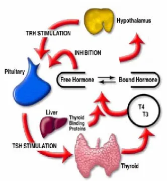
regulated by a feedback inhibition mechanism operating at the level of the hypothalamus - pituitary gland - thyroid gland axis. Thyroid follicles have the remarkable ability to extract iodine from the blood which is essential for thyroid hormone synthesis. Iodine uptake is a critical step for hormone production and by regulating this step the thyroid gland regulates its own activity. Thyroid hormone levels in the blood are primarily controlled by negative feedback at the hypothalamus (fig.1). This system-wide regulatory centre located in the brain, synthesizes and releases thyrotropin-releasing-hormone (TRH) which in turn stimulates the secretion of thyrotropin-stimulating-hormone (TSH) by the pituitary gland. TSH is secreted into the blood by the pituitary gland and acts on the thyroid stimulating the synthesis and release of biologically active TH molecules. Synthesis of TRH by the hypothalamus is regulated by both the concentration of THs and TSH in the blood. A reduction in the concentration of circulating THs is also directly sensed by the pituitary thyrotrophs and stimulates TSH release.
1.3.1 Thyroid hormones
Thyroid hormones have been identified throughout the vertebrates including basal groups such as, cyclostomes, teleosts, amphibians and reptiles (Chiu et al., 1975; Higgs and Eales, 1973; Packard et al., 1976) and iodothyronine active compounds were also found to be present in a number of protochordates (Bentley, 1998). The thyroid hormones are probably the most pluripotent hormones found in vertebrates as they can influence virtually every metabolic activity of an organism (e.g., concentration and function of numerous enzymes; metabolism of fats, carbohydrate, vitamins, proteins; heart rate; stimulation of erythropoiesis or regulation of bone synthesis, etc.).
Figure 1 Hypothalamic - pituitary gland - thyroid gland axis. Thyroid hormone levels are regulated by
a feedback inhibition mechanism which operates along the hypothalamic-pituitary-thyroid axis. The hypothalamus secretes thyrotropin releasing hormone (TRH) which stimulates the pituitary to secrete thyroid stimulating hormone (TSH). TSH, in turn, stimulates the thyroid gland to produce and secrete thyroid hormones (T4 and T3) into the circulation where they bind thyroid binding proteins. The system
attempts to maintain constant the free TH fraction through a negative feedback loop. Alterations on free hormone concentrations are sensed at the level of the hypothalamus and pituitary and TSH production is stimulated/inhibited in order to re-establish adequate TH levels.
Adapted from: http://www.dpcweb.com/medical/thyroid/thyroid_function.html
Physiologically, THs act through two major mechanisms: by increasing protein synthesis and oxygen consumption. Ultimately, their action influences basic life cycle events such as growth, differentiation, metamorphosis, reproduction, hibernation and thermogenesis (Bentley, 1998). Normal thyroid function is essential for both mental and physical development of young mammals. Inadequate production or overproduction of thyroid hormones is strongly associated with two types of thyroid disease states in
THs (hypothyroidism) can compromise normal physical and mental development, a conditions known as cretinism. The same condition in adults results in low metabolism and several associated complication like weight increase, low body temperatures, etc (Dorit et al., 1991). Usually hypothyroidism can be readily treated by oral administration of synthetic THs. Abnormally high TH levels (hyperthyroidism) causes an elevation of the metabolic rate, weight loss, excessive perspiration, nervousness and other complications (Dorit et al., 1991) Hyperthyroidism is commonly treated by administering anti-thyroid drugs (e.g. propylthiourea, methimazole), which suppress synthesis of thyroid hormones primarily by interfering with iodination of thyroglobulin. In other vertebrates the function of THs is still not fully understood. For example they are essential for moulting in amphibians and reptiles, and their essential role in amphibian methamorphosis is widely recognised (Leloup and Buscaglia, 1977). Recent reports also highlight their importance in fish development, growth and metamorphosis (Power et al., 2001; Yamano, 2005). In fact, many vertebrates have a characteristic rise in THs during crucial stages of development (Hulbert, 2000; Leloup and Buscaglia, 1977).
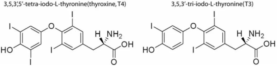
Functional THs are small amino acid derivatives (fig. 2) consisting of a hydrophobic thyronine nucleus, which accounts for their poor water solubility, a hydrophilic hydroxyl group attached to the phenolic ring and four or three iodines at positions 3, 5, 3’, and 5’ in thyroxine (T4) or 3, 5, 3’ in triiodo L-thyronine (T3) (Power et al., 2000a).
Figure 2 The chemical structure of thyroid hormones T4 (left) and T3 (right) (Lundberg, 2006)
1.3.2 THs biosynthesis
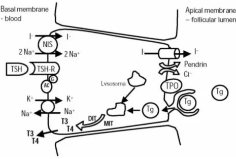
The general principals of thyroid hormonogenesis as presently accepted were first outlined by Gorbman and Bern (1962) and are essentially similar throughout vertebrates (Leatherland, 1993). TH synthesis takes place in the follicular cells of the thyroid and starts with the uptake of extracellular iodide. See fig. 3 for a schematic representation of TH biosynthesis. The thyroid utilizes I-, the elemental form in the blood, which is ultimately derived from the environment. Terrestrial vertebrates obtain I- mostly from food while fish take up iodine from the water via the gills and gut. Iodide is pumped into thyrocytes in an ATP dependent manner by means of a sodium/iodide co-tranporter (symporter, NIS) protein in the plasma membrane. Once inside the follicular cell it is converted by thyroid peroxidase (TPO), most likely at the luminal surface of the cell, to an oxidized species of iodine which is incorporated into tyrosyl groups of thyroglobulin (TG) a very large glycoprotein. Iodination occurs at specific tyrosine sites within thyroglobulin giving rise to diiodotyrosine (DIT) and monoiodeotyrosine (MIT). TPO activity facilitates the intramolecular coupling of either two DIT residues, giving rise to tetraiodothyronine (T4), or a MIT with a DIT residue resulting in the formation of
triiodothyronine (T3). At this stage, iodinated thyronine compounds are an integral part
of thyroglobulin and represent a reserve of THs stored in the lumen of the thyroid follicles. Enzymatic degradation of the thyroglobulin, stimulated by TSH, releases THs into the intracellular compartment. Free T4 and smaller amounts of free T3 are then
secreted into the blood. While T4 is formed exclusively in the thyroid, 80% of T3 found
in circulation is thought to derive from T4 conversion in peripheral non-thyroidal tissues
(especially liver and kidney). The difference between the source of T3 and T4 is
probably related to their mode of action. T4 is considered to be a prohormone as in order
to act it has to be converted to its derivative, T3, the biologically more potent TH
because of its greater affinity for thyroid hormone receptors (TRs) (McNabb, 1992).
Figure 3. Thyroid hormones biosynthesis pathway in a thyroid follicle cell. The first step to hormone
synthesis is the import of iodide into the follicular cells, an active transport process (an ATP-pump pumps K+ in and Na+ out of the cell) that occurs through a sodium/iodide "symporter" (transporting iodide in). Once in the follicular cell, iodide is converted into iodine by the enzyme thyroid peroxidase (TPO), which uses hydrogen peroxide (H2O2) as a cofactor. Thyroid peroxidase catalyzes the incorporation of iodide
molecule onto both the 3 and/or 5 positions of the phenol rings of tyrosines present in the large protein thyroglobulin (TG) originating diiodotyrosine (DIT) and monoiodeotyrosine (MIT). Thyroglobulin contains 140 tyrosines but only two to five of these are converted into either T4 or T3 through TPO
1.3.3 TH mode of action and metabolism
Once released into the circulation, a major part of THs reversibly binds to plasma transport proteins and only a minor fraction (approximately 0.03% for T4 and 0.3% for
T3) remains in the free form (fig.1). According to the “free hormone hypothesis”
formulated by Robbins and Rall in 1960 and further supported by Mendel and colleagues (Mendel, 1989),thyroid hormones enter target cells by diffusion and it is the free hormone and not the protein-bound one that is available to enter cells. Therefore, the free extracellular TH concentration constitutes the driving force that determines the rate at which THs reach their targets eliciting physiological responses. However, an alternative proposal (Beraud et al., 1958 and Ingbar and Freinkel, 1960) suggests that transmembrane diffusion of THs is governed by intra and extra-cellular thyroid-hormone binding proteins. More recently several studies have demonstrated the existence of a cellular uptake mechanism for THs in which binding proteins mediate cell entry through protein receptors on the membrane of the target cell (Benvenga and Robbins, 1990; Divino and Schussler, 1990; Kuchler-Bopp et al., 1998; Vieira et al., 1995). Although this membrane translocation mechanism is not completely clarified it is presently accepted that cellular uptake of THs is a carrier-mediated process important for overall regulation of their bioactivity (for review see Hennemann et al., 2001). TH actions after cellular uptake are mediated by nuclear thyroid hormone receptors (TRs) that can bind T3 with high affinity (for reviews see Wu and Koenig, 2000; Zhang
and Lazar, 2000). TRs belong to the nuclear receptor superfamily that also includes receptors for other small lipophylic hormones (e.g. steroids, retinoids and vitamin D). Two distinct genes (TRα and TRβ) located in different chromosomes (17 and 3, respectively in humans) generate multiple TRs isoforms including TRα1, TRα2, TRα3
and TRβ1, TRβ2 (Lazar, 1993) which have distinct functional roles. TRs act as transcription factors regulating gene expression. They bind to specific regulatory DNA sequences of the target genes known as TH response elements (TREs). Although TR can bind TREs as monomers or homodimers, they preferentially bind many TREs as heterodimers with the retinoid X receptor (RXR), another member of the nuclear receptor superfamily. Binding of unliganded TR generally represses basal transcription while ligand (T3) binding triggers a conformational change in the TR activating
transcription of a target gene. Inhibition and enhancement of gene expression can depend on the nature of the TREs, hormonal status and the cellular environment.
TRs like other nuclear receptors exhibit a modular structure with a variable N-terminal region, a central DNA-binding domain containing two zinc fingers and a large highly conserved ligand-dependent activation domain located towards the C-terminus of the protein (Lazar, 1993). The intracellular T3 concentration largely determines the degree
of occupancy of nuclear receptors thus regulating biological responses to THs (Brent, 1994). The intracellular T3 concentration in its turn is directly influenced by factors such
as, T3 and T4 uptake rates from the extracellular fluid or TH inactivation. The latter can
be achieved by glucuronide or sulphate conjugation, deamination or decarboxylation, but the major metabolic pathway is deiodination which takes place mainly in liver and kidney. T4 can lose iodine atoms in a stepwise manner to create an array of
iodothyronines. The most important deiodination pathway is T4 conversion to T3 which
occurs by removal of one iodide unit from the outer ring of T4 (5’-monodeiodination or
outer-ring deiodination, ORD). Monodeiodination of the inner ring (5-monodeiodination or inner-ring deiodination, IRD) can also take place giving rise to 3,3’,5’-T3 (reverse T3 or rT3). Although, rT3 has no biological activity this alternative
non-active rT3 which can be further deiodinated to T2, T1 and thyronine (T0). Elevated
plasma T3 levels can also reduce 5’-monodeiodination activity (MacLatchy, 1993;
Scott-Thomas et al., 1992) suggesting an autoregulation of T3 production. This dual
control mechanism allows independent regulation of the prohormone T4 and of the
biologically active T3 blood levels (Leatherland, 1993). Deiodination is carried out by a
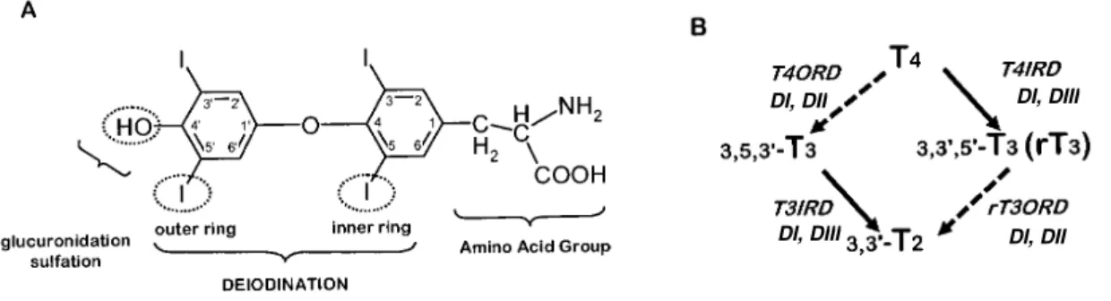
family of selenoprotein enzymes, the deiodinases. At least three different deiodinases are known in homeotherms, type I (DI), type II (DII) and type III (DIII) (Berry et al., 1991; Croteau et al., 1996; Croteau et al., 1995), which vary in tissue location, substrate preference and affinity (fig. 4B). DI, which can deiodinate both inner and outer ring is commonly found in the liver, kidney, muscle, pituitary and thyroid gland. DII can only deiodinate the outer ring and being the major activating enzyme. It is mostly present in the brain but also in the testis, thyroid, muscle, heart and pituitary. DIII can only deoidinate catalyze inner ring deoidination being the major inactivating enzyme. It is found in the brain and skin and also the placenta and fetal tissue.
Figure 4. A- Structure of L-thyroxine indicating the main pathways of metabolism. Deiodination of T4
occurs through removal of one iodide unit form the inner (left) or outer (right) ring of the molecule. B- Representation of the main deiodination pathways: Outer-ring deiodination (ORD, ) and inner-ring deiodination (IRD, ). Active T3 is formed from T4 outer ring deiodination (T4ORD) through the action
of deiodinases DI or DII. Inner-ring deiodination of T4 (T4IRD) by DI or DIII results in the formation of
the inactive reverse T3 (rT3). T3 itself can be degraded by removal of one of a inner (T3IRD) by DI or DIII
or outer-ring iodine (T3ORD) by DI or DII originating the presumed inactive 3, 3’-T2. Adapted from
Brown et al (2004).
DI, DII
DI, DII DI, DIII
These enzymes act as a complex and coordinated system which enables hormonal activation or inactivation\degradation to playing an important role in modulating TH levels in specific tissues and overall THs homeostasis and economy.
1.3.4 Thyroid axis in fish
In higher vertebrates THs have a widespread and diverse action and are essential from early stages of life. Since in lower vertebrates the best known feature of THs is its crucial role in amphibian metamorphosis (Shi et al., 1996), THs are expected to greatly influence embryonic and larval stages of development. In fact, due to its pluripotent action, fish thyroid systems inevitably attract the interest of many fields like fish physiology, biochemistry or aquaculture. The TH axis has also become of considerable concern in environmental toxicology as a number of toxic chemicals may strongly interfere with normal thyroid function, mostly due to their structural resemblance to THs. Throughout the past two decades knowledge about the role of THs in fish has improved and reports mostly relate to their role in regulation of development, growth and aspects of reproduction (for review see Leatherland, 1994; Power et al., 2001; Yamano, 2005) while regulation of basal metabolism, so evident in endotherms, is not so well documented in teleosts.
The existence of a thyroid gland in mammals has been accepted for thousands of years but the recognition of a comparable tissue in fishes only occurred in the mid-late 19th century (Maurer, 1886; Simon, 1844). Unlike tetrapods, which possess a single discrete gland, in fish, thyroid follicles may not be encapsulated by connective tissue. Most orders of teleost fish lack a “glandular” structure and instead thyroid follicles are found scattered throughout the connective tissue of the lower jaw (pharyngeal region) usually aggregated around the ventral aorta (Bentley, 1998; Leatherland, 1994). The essential
components making up the thyroid axis have, however, been largely conserved across vertebrates as has the basic structure and function of fish thyroid tissue. Nevertheless, many aspects of the teleost thyroid axis certainly meet fish physiological adaptations with particular mechanisms of action, some of which are still not completely clarified. The basic steps involved in fish thyroid function from TH biosynthesis to TH action seem to fit the current model. TSH stimulates secretion from the thyroid which secretes mainly T4 which is converted in the periphery to T3 by deiodination. TH transport to
target cells involves carrier proteins and T3 is the active TH form that predominantly
binds TRs.
TH biosynthesis depends on an adequate supply of iodide and fish have the capacity to take up iodide from water across the extensive gill surface (Hunn and Fromm, 1966). In teleosts an adequate plasma iodide level is partly determined by dietary and branchial uptake suggesting different strategies in iodide economy. Owing to the greater iodide availability teleosts may have less efficient mechanisms of recovery and retention of hormonal iodide than homeotherms. TH biosynthesis, thyroglobulin properties and intrathyroidal secretion have received limited attention in teleosts. The few studies which exist report that negligible amounts of T3 (Eales and Brown, 1993) are
synthesized or secreted by thyrocytes in teleost species. Although the TH biosynthesis in fish is assumed to fit the mammalian model there are still many gaps in knowledge to be filled. Regulation of thyroid function seems to occur through the hypothalamus-pituitary-thyroid axis, although the identities of the hypothalamic substances influencing teleost thyrotropes are still not known. However, in contrasts to the situation in mammals, T3 does not appear to influence the activity of the teleost
hypothalamus-pituitary-thyroid axis (Eales and Brown, 1993). This fact suggests a loose linkage between the control of T4 and T3 metabolism pointing to fundamental differences in
control of thyroid status in teleosts. In contrast to mammals, where plasma T4 largely
exceeds T3 levels (50-100:1), plasma T3 levels in teleosts may exceed that of T4,
suggesting a strong 5’-monodeiodination activity (T4-ORD) and peripheral deiodination
seems to be the primary control of teleost thyroid function (Eales and Brown, 1993; Power et al., 2001). It is the regulated conversion of T4 to T3 in tissues which may
largely determine the T4 secretion rate from the thyroid. In homeotherms primary
control of T4 and T3 is largely central through the hypothalamic-pituitary axis. As THs
are involved in regulation of basal metabolism rates in homeotherms, this mechanism may have arisen as an advantageous adaptation leading to central control by the hypothalamus, the brain centre associated with thermoregulation.
TH degradation seems to occur mainly through deiodination pathways (Eales and Brown, 1993) but oxidative deamination and decarboxylation also take place. The liver is the pivotal organ in T4 metabolism and is mainly involved in deiodination and it is the
site where TH conjugation and biliary excretion also occur (Power et al., 2001). T3 is
mainly degraded by T3 IRD in brain, liver and possibly other tissues (Eales et al.,
1993b; MacLatchy, 1993). Excretion of TH metabolites was found to occur through the urine and the bile (Eales et al., 1971; Sinclair and Eales, 1972) and other possible routes may be gills, gut or skin.
As in mammals three deiodinase isotypes are expressed in piscine species including agnathans, chondrichthyes and teleosts (Orozco and Valverde-R, 2005). Fish deiodinases share properties with their corresponding counterparts in higher vertebrates. However in fish, these enzymes exhibit distinct features and their physiological role in functions regulated by THs is not completely clear.
Putative nuclear T3-binding receptors have been described in teleosts in a number of
1993; Power et al., 2001). Only relatively recently, cloning and characterisation of TRs in fish was carried out (Power et al., 2001; Yamano et al., 1994; Yamano and Inui, 1995) and revealed two principal receptor types, TRα and TRβ. The properties, binding profile and molecular structure of such receptors closely resemble those described in high vertebrates. Therefore they can also be expected to function as transcription factors probably binding similar response elements (Power et al., 2001).
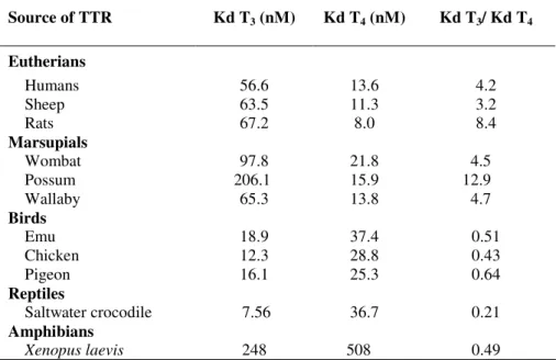
In common with mammals, in teleosts more than 99% of THs do not circulate in the free form (Eales and Shostak, 1985; Weirich et al., 1987) but are covalently bound to plasma proteins (Falkner and Eales, 1973) including lipoproteins (Babin, 1992). In fish thyroid hormone transport by plasma proteins is poorly explored but evidence points to a different general profile. Only two of the three thyroid hormone binding proteins (THBP) present in the blood of larger mammals have been found, albumin (Richardson et al., 1994) and transthyretin (TTR) (Kawakami et al., 2006; Santos and Power, 1999; Yamauchi et al., 1999). The latter protein appears to be the main TH carrier in fish and shows a great ability to bind T3 (Morgado et al., 2006; Yamauchi et al., 1999)unlike
mammalian TTR which preferentially binds T4 (Chang et al., 1999). The biological
significance and adaptive advantage of this transport system is largely unexplored in fish or even in higher vertebrates. As THBP is a major issue of the present work this matter will be further developed in the following section.
1.3.5 Thyroid Hormones Binding Proteins (THBP)
THs exert their action at a cellular level to bring about important physiological processes and therefore the delivery of THs from their site of synthesis to their site of action (target tissues) is a crucial process in vertebrates. Plasma is the immediate major compartment into which the thyroid secretes THs (mostly T4) and the ultimate source of
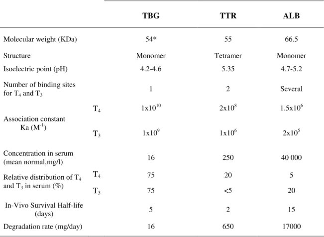
THs for all target tissues. As previously mentioned, in vertebrate’s less than 1% of THs are present in blood as the free form (physiologically active fraction) as the majority is bound to THBP. These proteins are synthesized by the liver and secreted into the bloodstream where they bind and transport THs. In the serum of larger mammals, the major THBPs are thyroxine-binding globulin (TBG), Albumin (ALB) and Transthyretin (prealbumin,TTR) (Larsson et al., 1985). In humans, serum albumin is present at approximately 100-fold the molar concentration of TTR and 2000 fold that of TBG. Although present in much lower concentration, TBG shows the highest affinity for THs (50 fold higher than TTR and 7000 fold higher than ALB) and carries about 75% of all plasma T4 while TTR and ALB transport 20% and 5% respectively. Table 1 shows the
main characteristics of these three THBPs. TBG is a 54KDa acidic glycoprotein (apparent molecular weight 60KDa in SDS-PAGE) which in addition to binding T4 also
binds T3 but with slightly less affinity. It was first recognised as a major THBP in 1952
(Robbins, 1992) and its primary structure deduced in 1989 (Flink et al., 1986). The mature molecule without the 20 amino acids signal peptide is composed of 395 residues (44KDa) and four heterosaccharide groups. It has a single iodothyronine binding site and loses affinity for THs above 55ºC although association with T4 increases its
stability. Alteration of TBG synthesis or degradation processes leads to acquired TBG abnormalities (Refetoff et al., 1976). Human serum albumin (HSA) is a 66.5 KDa protein composed of 585 amino acids with no carbohydrates and forming three main domains. It is the main protein present in serum where it constitutes half of the total protein content. Albumin binds a wide variety of molecules (hormones, drugs, etc.) and thus the association with THs can be considered as non-specific. In spite of containing several iodothyronine binding sites only one has higher affinity for THs but even so it is considerably lower than TH binding to TBG and for the reason the contribution of
albumin to TH transport is relatively minor. TTR is an unglycosylated 55 KDa tetramer which is highly acidic. Despite being present in serum at much higher levels than TBG it plays a lesser role in TH transport and only 0.5 % of circulating TTR is occupied by T4. Its normal concentration in serum (250 mg/l) corresponds to a maximal binding
capacity of approximately 300 µg T4/dl. TTR also binds a great variety of
non-iodothyronine ligands like for example salicylates, penicillin or flavonoid compounds which shown particularly high affinity for the protein. TTR will be characterized in detail in section 1.4 where its features, functions and structural/functional evolution are described.
Although the biological advantage of transporting a high percentage of hormone in a bound, non-active form has not been extensively explored, the existing evidence suggests it has several important functions. THBPs are probably responsible for the maintenance of a large extrathyroidal thyroid hormone pool counteracting permeation of strongly lipophilic hormones into cells (Ekins et al., 1982; Mendel, 1989; Mendel and Weisiger, 1990; Mendel et al., 1987). The latter effect allows an even distribution of hormones to tissues and safeguards the body from the effects of abrupt fluctuations in hormone secretion. THBP can also serve as an additional protection against iodine wastage by imparting macromolecular properties to small iodothyronine molecules limiting their urinary loss (Chan et al., 1972). Furthermore, THBP may promote hormone cellular uptake by interaction with cell membranes and prevent the undue loss of hormones through metabolism and excretion (they prevent glomerular filtration due to the large size of the hormone-protein complex).
The relative abundance and hormone binding properties of THBPs are not conserved in vertebrates. In a series of studies, reviewed by Richardson (2002), serum samples from individuals representative of different vertebrate classes (mammals, birds, reptiles,
amphibians and also fish (Richardson et al., 2005)) were analysed for the presence of THBPs. Albumin was the only THBP present in all species studied, suggesting it may be the evolutionarily oldest TH carrier protein. In addition to albumin, TTR was also present in the blood of several homeotherms (birds and eutherian mammals), a condition controlled by THs. The simultaneous presence of the three THBPs seems to occur only in some eutherian species where TBG was also found in addition to TTR and albumin. The same studies suggest an apparent absence of TTR in the plasma of adult amphibians, reptiles and fishes, however its presence is well established for the early stages of development (Funkenstein et al., 1999; Kawakami et al., 2006; Richardson et al., 2005; Santos and Power, 1999; Yamauchi et al., 1993; Yamauchi et al., 1999; Yamauchi et al., 1998) which are characterised by surging TH blood levels (Hulbert, 2000). The previous findings strongly suggest a straight relationship may exist between the increase in THBP distribution capacity and TH requirements. Such capacity, which obviously increased during the evolution of vertebrates, could be related with the developmentally regulated expression of a gene coding for a new THBP with higher affinity than those already present in blood (Richardson et al., 2005). The presence of three THBP with different T3 and T4 binding capacities may result in a very robust
buffering system enabling a highly effective homeostatic control of free hormone levels. This could also offer some explanation to observations that THBP are not essential for normal thyroid function and questions about their adaptive value. Clinical studies in humans show that wide fluctuations in THBP concentrations or its complete absence do not alter the hormonal economy or metabolic status of the subject (Lissitzky, 1990; Refetoff, 1989). Mutations that lead to the absence of albumin in humans (Kallee and Ott, 1992) and rats (Mendel et al., 1989) or TTR in mice (Palha et al., 1994) are associated with normal phenotypes and the organisms remain eutheroid (with normal
TH levels). These findings are highly suggestive that the THBP system may involve several functionally redundant components. However, the adaptive advantage acquired throughout evolution could rely on such redundancy. One deficient component of the system could be compensated by the others: under conditions of TH stress a buffering system with three carrier proteins would provide a strong selection advantage over a single protein system (Richardson, 2002; Schreiber, 2002a).
The efficiency of the hormone transport and delivery system is certainly crucial for thyroid hormone-dependent processes to take place. Nevertheless, many aspects of the activity of such physiologically important TH transporters and mechanisms by which they regulate TH bioavailability remain unclear and call for further investigation.
Table 1. Properties and metabolic parameters of the three main THBPs in human serum.
From http://www.thyroidmanager.org/Chapter3/3a-frame.htm.
TBG TTR ALB
Molecular weight (KDa) 54* 55 66.5
Structure Monomer Tetramer Monomer
Isoelectric point (pH) 4.2-4.6 5.35 4.7-5.2
Number of binding sites for T4 and T3 1 2 Several T4 1x10 10 2x10⁸ 1.5x10⁶ Association constant Ka (M-1) T3 1x10 9 1x10⁶ 2x10⁵ Concentration in serum (mean normal,mg/l) 16 250 40 000 T4 75 20 5 Relative distribution of T4 and T3 in serum (%) T3 75 <5 20
In-Vivo Survival Half-life
(days) 5 2 15
Degradation rate (mg/day) 16 650 17000
1.4 Transthyretin (TTR)
Transthyretin was identified for the first time in 1942 in human serum and cerebrospinal fluid (Kabat and Moore, 1942; Kabat et al., 1942; Seibert and Nelson, 1942) and was originally called prealbumin due to its greater electrophoretic mobility at pH 8.6 than albumin. In 1958, TTR was first recognized to bind thyroxine (Ingbar, 1958) and designated thyroxine-binding prealbumin (TBPA). Determination of the primary structure by Goodman et al in 1974 (Kanda et al., 1974), led to proposal of a new name transthyretin (TTR) which was adopted in 1981 (Robbins, 1976) and reflects its ability to transport thyroid hormones and retinol.
TTR research has developed over several decades and has been reviewed by different authors (Benson and Uemichi, 1996; Schreiber, 2001; Power et al., 2000a; Schreiber, 2002a; Schreiber and Richardson, 1997). In common with most plasma proteins TTR is synthesized and secreted by the liver (Dickson et al., 1982) and released into the blood stream. It is produced as a single polypeptide chain and folds into a globular protein being is present in plasma as a tetramer of four identical monomers (Blake et al., 1971; Gonzalez and Offord, 1971; Rask et al., 1971) that bind and transport THs. TTR’s role as one of the THBP in the plasma is probably its best characterised function and the biological meaning of the transport network to which it belongs has been discussed in section 1.3.5. However, a number of other characteristics for this protein have also been observed and are now considered.
1.4.1 RBP transport
Another major function attributed to TTR is its ability to bind retinol-binding protein (RBP), the 21 KDa specific carrier of all-trans retinol (Blaner, 1989; Goodman, 1984) from liver storage sites to target cells and is important in vision and bone growth. The
presence of retinol bound to RBP is essential for the formation of a stable complex with TTR and crystallographic studies have established that two RBP molecules can bind one TTR tetramer (Monaco et al., 1995) and do not interfere with TH binding (van Jaarsveld et al., 1973). In mammals holo-RBP (retinol-RBP complex unbound to TTR) circulates almost entirely bound to TTR. It has been proposed that this binding serves to prevent filtration of the relatively small RBP molecule through kidney glomeruli (Kanai et al., 1968; Peterson, 1971). In TTR null mice the levels of plasma RBP and retinol were found to decrease (Episkopou et al., 1993). Surprisingly though, the mice remained healthy showing normal retinol levels in tissues, their embryos develop normally and relatively little RBP was detected in their urine (Wei et al., 1995). In light of these findings, the current understanding of retinol transport is not complete and the participation of TTR in it still requires clarification. Previous studies regarding the evolutionary onset of TTR synthesis suggested it may not be absolutely required for retinol transport (Richardson et al., 1994; Schreiber and Richardson, 1997). Moreover, RBP is known to be present in the blood of fish but it could only be isolated in the uncomplexed form (Berni et al., 1992; Shidoji and Muto, 1977). Although piscine holo-RBP shows binding ability to human TTR it displays an extremely low or inexistent affinity to piscine TTR (Folli et al., 2003). Structural studies show a high degree of conservation in TTR’s TH binding sites but in contrast a few amino acid differences may be responsible for an apparent lack of TTR-RBP binding in fish. This suggests conservation of TTR-TH binding in all vertebrates whereas TTR-RBP binding ability may have been acquired later in evolution.
1.4.2 TTR as a stress and nutritional marker
In mammals TTR is produced by the liver and is a typical negative acute phase protein as its levels strongly decrease in response to injury. The liver plays a crucial role in the adaptive alterations accompanying any stressful condition and is extremely sensitive to nutritional insults. In malnutrition TTR and also ALB and TBG are significantly reduced due to depressed liver synthesis (Ingenbleek, 1985). Moreover, TTR and RBP are the first plasma proteins to decline in parallel as a result of early nutritional deprivation (Ingenbleek, 1985; Ingenbleek et al., 1975). According to the free hormone theory ((Mendel, 1989; Robbins and Rall, 1960), see section 1.3.3) the suppression of TTR and RBP synthesis releases increased amounts of free ligands readily available to target cells. This negative acute phase feature of TTR is commonly used by clinicians to monitor the nutritional status of their patients. In fact TTR has been extensively characterised as a marker of malnutrition or stress (Brugler et al., 2002; Ingenbleek and Bernstein, 1999; Ingenbleek et al., 1972; Ingenbleek and Young, 1994; Ingenbleek and Young, 2002). Food restriction in fish has also been found to cause a dramatic reduction in TTR transcripts (Power et al., 2000b) as previously observed in rats (de Jong and Schreiber, 1987; Le Moullac et al., 1992). However, it is still not resolved if down-regulation of TTR in liver is directly caused by malnutrition or is also a consequence of reduced TH levels in plasma. The factors behind regulation of TTR production are still unclear but it seems likely that in common with other hormonal homeostatic processes the concentration of THs have a role, although studies are still required to clarify this matter.
1.4.3 TTR synthesis in the choroid plexus
In addition to being produced in the liver, high levels of TTR expression have also been reported in the choroid plexus (Dickson et al., 1985; Soprano et al., 1985), where cerebrospinal fluid (CSF) is produced. In fact, evidence shows that abundance of both chicken and rat TTR transcripts in the choroids plexus are far higher than that in the liver (Duan et al., 1991; Schreiber et al., 1990). TTR is the most abundant of all proteins (Dickson et al., 1986) synthesized by the choroids plexus and it is secreted exclusively towards the brain (Schreiber et al., 1990) and is the only THBP produced in the brain (Schreiber and Richardson, 1997; Schreiber et al., 2001) .
Most of the cerebrospinal fluid surrounding the brain is produced in the choroid plexus (Cserr, 1971) and its epithelial cells, where TTR is synthesized, constitute the blood-cerebrospinal fluid barrier. The functional significance of TTR in the brain is probably related to its TH binding capacity and it is proposed to be the brain THBP responsible for hormone distribution (Dickson et al., 1987a; Schreiber et al., 1990). A model for TTR and T4 distribution within the blood-brain barrier was proposed by Southwell et al
(1993). In contrast to the situation in the liver, TTR synthesis in the choroid plexus is constitutive and does not change during the acute phase response which also suggests different regulatory and functional requirements for TTR produced in the brain and in the rest of the body (Dickson et al., 1986).
1.4.4 Molecular Structure
Human TTR is a 55KDa homotetramer composed of four identical subunits, each containing 127 amino acids. The overall shape of the tetramer forms a narrow central channel which buries two identical thyroid hormone binding sites (Blake et al., 1974).
However, under physiological conditions only one of the binding sites is filled at one time due to negative cooperative effect of hormone binding (Neumann et al., 2001). The three-dimensional structure of human TTR (fig. 5) was determined by X-ray crystallography (Blake, et al., 1978; 1974) at 2.5 Å resolution and has subsequently been refined by other authors (Hamilton et al., 1993; Hornberg et al., 2000; Wojtczak et al., 1992). In the first models the N-terminal region of the monomers could not be resolved but in 1993, in the structural model conceived by Hamilton et al. the first 10 N-terminal amino acids were observed as curve structures placed near the opening of the central thyroid hormone binding channel.
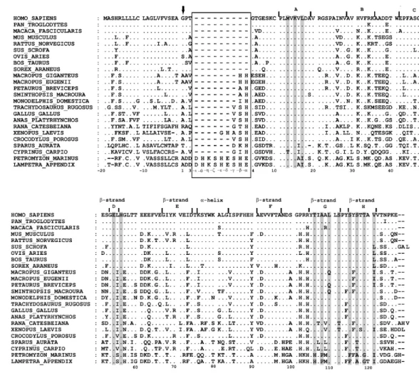
Presently, TTR X-ray structures from several mutant variants of human TTR (for review see Hornberg et al., 2000) and also from chicken, rat and fish are also available (Eneqvist et al., 2004; Folli et al., 2003; Sunde et al., 1996; Wojtczak, 1997). The models show little variation between structures pointing to an overall conservation of TTR three-dimensional structure during vertebrate evolution. X-ray diffraction indicates that the dominant secondary structure is the β-sheet. Each monomer (A, B, C and D) has two 4-stranded β-sheets and a short α-helix. Anti-parallel β-sheet interactions link monomers into dimers and a short loop from each monomer forms the main dimer-dimer interaction resulting in the formation of the central hydrophobic channel (fig. 5). The two TH binding sites situated between monomers A, C and B, D can be divided into three symmetry-related pairs of halogen binding pockets (HBP) comprised of an inner and outer cavity: HBP1 (HBP1’), HBP2 (HBP2’), HBP3 (HBP3’). The main difference between species is the lack of α-helix structures in the chicken TTR subunits. The TH binding site seems to be highly conserved with only one amino acid substitution in the sea bream sequence (Ser117 in the human sequence is substituted by a Thr). However, studies carried out by Eneqvist, et al (2004) point to differences in the
shape of the binding channel between sea bream and human TTR. In sea bream TTR the channel is wider at its entrance and has a narrower inner and outer cavity. Homology models based on the known X-ray crystal structure coordinates have been constructed for Lizard (Achen et al., 1993) and bullfrog (Yamauchi et al., 1998) TTRs and also for fish TTR (Power et al., 2000a) and subsequently X-ray crystallography was carried out. These models showed little variation from the known TTR structures. Despite the high conservation of the structures, comparisons of the electrostatic properties between the piscine homology model and X-ray structures from chicken, human and rat revealed that the surface potential, particularly in the TH binding site is noticeable more negative in chicken and even more-so in the sea bream structure than in human or rat (Power et al., 2000a).
Figure. 5. Diagram of human tetrameric TTR crystal structure with each monomer colored differently
(subunit A, yellow; subunit B, red; subunit C, green; subunit D, blue). TTR structure is predominantly β-sheet; each monomer has a two 4-stranded β-sheets and a short α-helix. A short loop from each monomer forms the main dimer-dimer interaction resulting in the formation of the tetramer. Left panel represents a view (perpendicular to the binding site, Z-axis) of the co-crystal structure of TTR in complex with T4
(shown in stick representation) showing the two TH binding sites. Right panel presents a view (down the Z-axis) rotated by 90° about the Y-axis (relative to the view on left) to display the central channel where TH and other ligands bind. Original diagrams taken from Johnson et al. (2005).
1.4.5 Genomic structure and transcriptional regulation
The genomic structures of TTR have been described for human (Sakaki et al., 1989) rat (Fung et al., 1988) and mouse (Costa et al., 1986). Human TTR is encoded by a single copy gene located on human chromosome region 18q11.2-q12.1 (Sparkes et al., 1987). The gene spans 6.9 Kb with 4 exons, 3 introns and a TATA box like sequence and binding sites for HNF-1, 3 and 4 are located within 150bp from the transcription start site (Sasaki et al., 1985; Tsuzuki et al., 1985). The first exon codes for the 5’ untranslated region, the 18 amino acid signal peptide and the first 3 residues of the mature protein. The second exon encodes residues 4-47 whilst exons 3 and 4 code for residues 47-92 and 93-127, respectively. A similar TTR gene organisation is found in both rat and mouse genomes.
TTR gene expression in the liver is under typical negative acute phase regulation. An extremely well conserved DNA-segment (CTGGGAA) in the 5’-flanking region (Fung et al., 1988) of the gene is thought to be important for such response during inflammation and trauma which causes TTR mRNA in liver to decrease dramatically. Further regulatory elements can be found at least 2000 bp upstream the gene that may be involved in regulation of expression via cis and trans-acting factors (Costa et al., 1989; Yan et al., 1990).
In contrast to the liver, in choroids plexus TTR gene expression does not respond to inflammation or trauma (Dickson et al., 1986; Dickson et al., 1987b). Thyroid hormone status also doesn’t seem to influence TTR expression levels in the choroid plexus (Blay et al., 1993). Very little is known about the regulatory mechanisms of TTR gene expression in that region of the brain. In fish and amphibians where TTR expression seems to be mainly restricted to the liver, the genomic organisation has not yet been

![Figure 2. Analysis of [ 125 I]-T 3 binding to sbrTTR by native glycine-acetate gel electrophoresis](https://thumb-eu.123doks.com/thumbv2/123dok_br/18945369.940088/90.892.212.582.186.407/figure-analysis-binding-sbrttr-native-glycine-acetate-electrophoresis.webp)