Departamento de Ciências Biomédicas e Medicina
Mestrado em Ciências Biomédicas
Dissertação de Candidatura ao Grau de Mestre em Ciências Biomédicas.
The effect of chaperone compounds on
glucocerebrosidase stability and activity in
fibroblasts from Gaucher Disease patients
with different genotypes.
Raquel da Fonseca Gonçalves
Trabalho efectuado sob a orientação de:
Prof. Doutor Gustavo Tiscórnia
"The whole of science is nothing more than a refinement of everyday thinking" Mr Albert Einstein
The effect of chaperone compounds on glucocerebrosidase
stability and activity in fibroblasts from Gaucher Disease patients with
different genotypes.
Declaração de autoria de trabalho:Declaro ser o (a) autor (a) deste trabalho, que é original e inédito. Autores e trabalhos consultados estão devidamente citados no texto e constam da listagem de referências incluída.
Copyright
Raquel Miriam da Fonseca Gonçalves ©, a Universidade do Algarve tem o direito, perpétuo e sem limites geográficos, de arquivar e publicitar este trabalho através de exemplares impressos reproduzidos em papel ou de forma digital, ou por qualquer outro meio conhecido ou que venha a ser inventado, de o divulgar através de repositórios científicos e de admitir a sua cópia e distribuição com objetivos educacionais ou de investigação, não comerciais, desde que seja dado crédito ao autor e editor.
Assinatura
Table of Contents
Abstract ... ix
Resumo ... x
Resumo alargado ... xi
Index of Abbreviations: ... xiii
Chapter 1 ... 1 Introduction ... 1 1.1 Disease ... 1 1.2 Historical Background ... 1 1.3 Clinical Presentation ... 2 1.3.1 Type 1 ... 2 1.3.2 Type 2 ... 3 1.3.3 Type 3 ... 4
1.3.4 Phenotypic continuum (an intermediate phenotype between type 2 and type 3) ... 4
1.4 Genetics ... 5
1.4.1 GBA gene ... 5
1.4.2 GBA Pseudo-‐gene ... 6
1.4.3 Genotype – Phenotype ... 6
1.4.3.2 Gaucher’s Disease in specific ethnic groups ... 9
1.4.3.2.1 Ashkenazi Jews population ... 9
1.4.3.2. Norrbottinian population ... 9
1.4.3.3. Genotype analysis ... 9
1.4.3.4. Inheritance patterns of the disease ... 10
1.5 Molecular mechanisms of GD ... 10
1.5.1 Sphingolipid metabolism ... 10
1.5.2 Molecular mechanisms ... 11
1.6 Therapeutic Approaches ... 13
1.6.1 Enzyme replacement therapy ... 14
1.6.2 Substrate reduction therapy ... 14
1.6.3 Bone marrow transplantation (stem cell transplantation) ... 15
1.6.4 Surgical intervention ... 15
1.7 Pharmacological Chaperones (PCs) ... 15
1.7.1 Chaperone therapy ... 15
1.8 Objective ... 17
Chapter 2 ... 18
Materials and Methods ... 18
2.1 Cell harvest and protein extraction ... 18
2.2 Protein Concentration Assay ... 18
2.3 Glucocerebrosidase Enzymatic Activity Assay ... 19
2.4 Protein Expression ... 20
2.4.1 Western Blot: ... 20
2.4.2 Antibodies used in this study: ... 21
2.5 Chaperone treatment ... 21
Chapter 3 ... 23
Results and Discussion ... 23
3.2 Characterization of Glucocerebrosidase activity in the cell lines used in this
study. ... 23
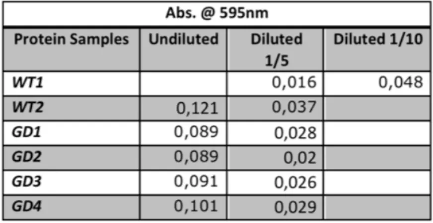
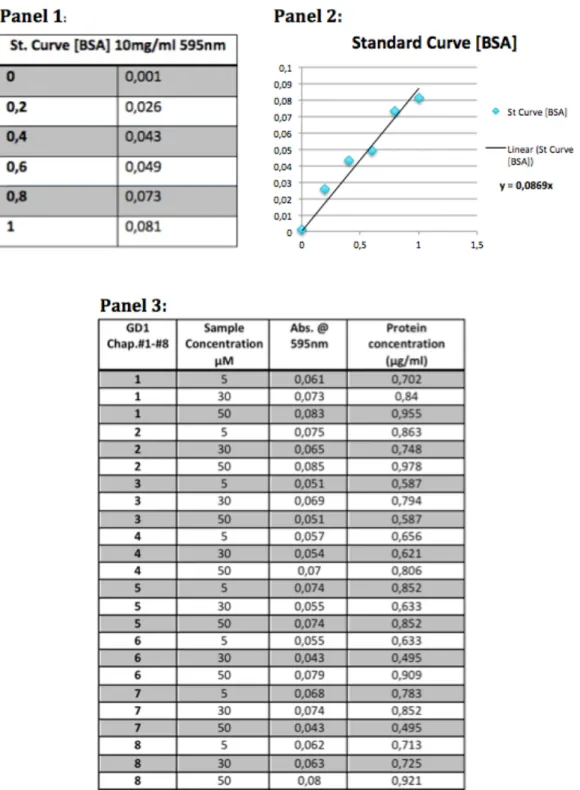
3.2.1 Bradford Assay: ... 23
3.2.2 Glucocerebrosidase Assay: ... 26
3.2.3 Western Blot Analysis ... 30
3.3 Effect of chaperone compounds on glucocerebrosidse activity levels in fibroblasts from 4 GD patients with different phenotypes: ... 31
3.3.1 Effect of 8 chaperone compounds on GD1 fibroblasts (GDT2 genotype N188S/G193W): ... 33
3.3.2 Effect of 8 chaperone compounds on GD2 fibroblasts (GDT2 genotype F213I/RecNaI): ... 41
3.3.3 Effect of 8 chaperone compounds on GD3 fibroblasts (GDT2 genotype L444P/L444P): ... 42
3.3.4 Effect of 8 chaperone compounds on GD4 fibroblasts (GDT2 genotype L444P/R120W): ... 43 Chapter 4 ... 45 Conclusion ... 45 Chapter 5 ... 46 Future Perspectives ... 46 Bibliography ... 47 Annexes ... 50
Figures Index
Figure 1 -‐
Main symptoms observed in Gaucher Disease.__________________2Figure 2 -‐
GBA gene location on the long (q) arm of chromosome 1 at position 21.__________________________________________________________________________ 5Figure 3 -‐
Classification of Gaucher Disease on genotype-‐phenotype.____8Figure 4 -‐
Glucocerebrosidase function._______________________________________11
Figure 5 -‐
Gaucher cells from a bone marrow aspirate._____________________12Figure 6 -‐
BSA absorbance standard curve determined byspectrophotometry._________________________________________________________________24
Figure 7 -‐
Absorbance of protein extracts of WT and GD protein extractssamples._______________________________________________________________________________25
Figure 8 -‐
Protein extract concentration from WT and GD patients.______25
Figure 9 -‐
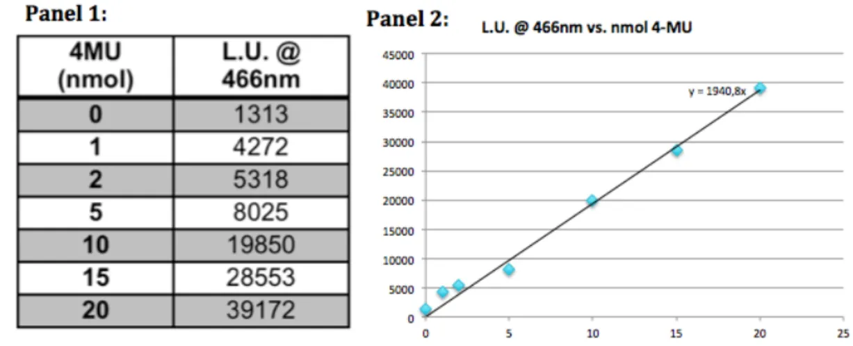
GBA enzymatic activity assay scheme._____________________________26Figure 10 -‐
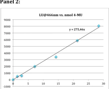
4MU (4-‐methylumbelliferone) fluorescence standard curvedetermined by fluorometry._______________________________________________________27
Figure 11 -‐
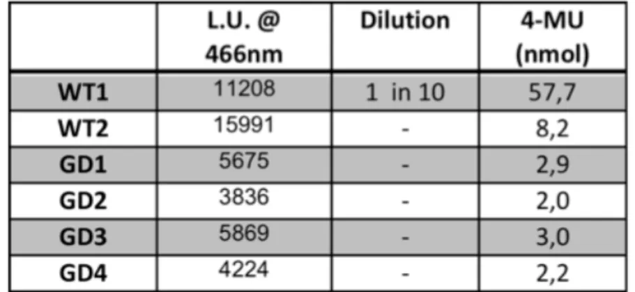
Glucocerebrosidase assay on protein extracts of WT and GDpatient samples._____________________________________________________________________28
Figure 12 -‐
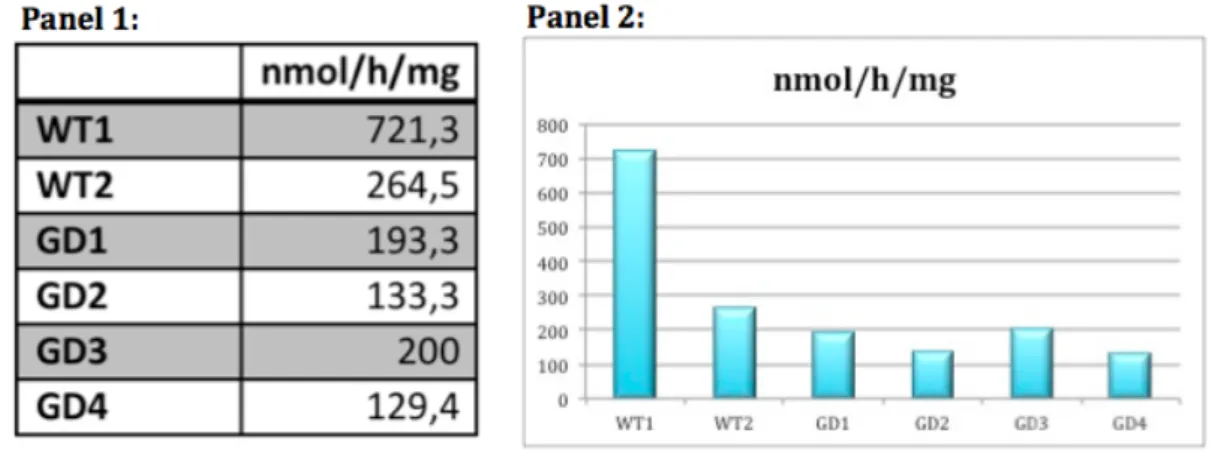
Glucocerebrosidase activity expressed as nmol 4-‐MU/hr/mgtotal protein._________________________________________________________________________29
Figure 13 -‐
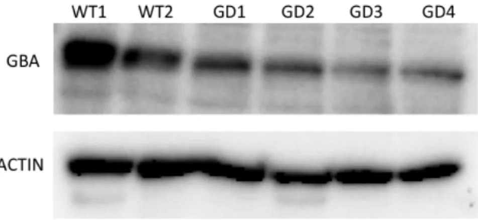
Western Blot of WT and GD protein samples.__________________30
Figure 14 -‐
Bradford Assay on GD1 fibroblasts treated with differentchaperones.___________________________________________________________________________34
Figure 15 -‐
GBA fluorometric assay of GD1 samples and GBA standard curve.__________________________________________________________________________________35Figure 16 -‐
Effect of chaperones 1-‐8 on glucocerebrosidase activity of fibroblasts (GD1) derived from a patient with GDT2 with genotype N188S/G193W.______________________________________________________________________36Figure 17 -‐
Western Blot analysis of GD1 fibroblasts treated withincreasing concentrations (0, 5, 30 and 50µM) of chaperones 1-‐8._____38,39
Figure 18 -‐
Effect of chaperones 1-‐8 on glucocerebrosidase activity of fibroblasts (GD2) derived from a patient with GDT2 with genotype F213I/RecNaI.________________________________________________________________________41Figure 19 -‐
Effect of chaperones 1-‐8 on glucocerebrosidase activity of fibroblasts (GD3) derived from a patient with GDT2 with genotype L444P/L444P.________________________________________________________________________42Figure 20 -‐
Effect of chaperones 1-‐8 on glucocerebrosidase activity of fibroblasts (GD4) derived from a patient with GDT2 with genotype L444P/R120W._______________________________________________________________________43
Agradecimentos
A presente tese de mestrado é o resultado final de um longo percurso académico de aprendizagem e desenvolvimento de qualidades e habilidades adquiridas na licenciatura e no presente mestrado em Ciências Biomédicas. Durante este percurso houve pessoas que de alguma forma contribuíram para tornar possível este mestrado. Essas pessoas foram importantes não só porque me ajudaram, mas também pelo acompanhamento nesta fase da minha vida. Assim, por estas razões nomeio os meus agradecimentos a:
Em primeiro lugar ao Prof. Doutor Gustavo Tiscórnia pela oportunidade de fazer parte do seu laboratório, no qual integrei o seu projecto, onde se insere a presente tese. Agradeço pelo ensinamento, pela paciência e por todos os conselhos na realização desta tese.
Fundamentalmente agradecer à minha família, pois sem eles tudo isto não seria possível. Aos meus pais Luís Gonçalves e Teresa Fonseca e aos meus avós Humbertina Patrício, Rodolfo Cevadinha e Conceição Martins. Agradeço todo o apoio que me concederam ao longo destes anos. Tal como aos meus irmãos e primos que sempre me apoiaram, Rafael Baguinho, Rebeca Baguinho, Bernardo Alves, João Fonseca e Ana Fonseca.
A todos os meus amigos que me deram força, apoiaram e sempre estiveram presentes em todos os momentos, como que uma família. Agradecendo particularmente ao Tiago Justo, Carla Melo, Isa Cruz, Soraia Lagoas, Daniel Barroso, Sónia Vieira, Elsa Gomes, João Castilho, Priscila Sousa, Celina Pereira, Tatiana Ferreira, Elodie Oliveira, Lúcia Mateus, Marco Sousa, Rita Pimenta, Augusto Pimenta, Alda Glória , Vitor Glória, José Martinho e Clara Viegas.
Aos meus colegas da licenciatura e particularmente do mestrado, Bruno Conchinha, Lúcia Santos e Ana Rita Martins.
Sem deixar de agradecer ao laboratório do Prof. Doutor Wolfgang Link, particularmente ao Dr. Richard Hill por me ter ensinado todas as técnicas envolvidas na realização de Western Blot e por me ter facultado todo o material
necessário para o efetuar. Tal como aos seus estudantes Laura Colaço e Ricardo Brandão pela ajuda que me cederam no decorrer deste procedimento.
Às Técnicas que me ensinaram todas as bases para trabalhar no CBME, mais precisamente na sala de cultura, especialmente a Ana Luísa Santos e a Claúdia Florindo. Assim como a todos os colegas do CBME que de algum modo me ajudaram ou cederam material, principalmente na sala de cultura. Particularmente ao Fernando Cristo e à Stéphanie Castaldo pelos conselhos cedidos na elaboração desta tese.
Agradeço também a ajuda prestada pelos colegas de laboratório Dino Matias e Adélia Ova, tal como às colegas do laboratório da Drª Inês Araújo, que me ajudaram e ensinaram durante o ano em que estive a trabalhar no laboratório, principalmente nos primeiros tempos de contacto com o mesmo.
A todos estes, o meu sincero agradecimento.
Abstract
Gaucher disease is the most common lysosomal storage disorder and may present systemic and neurological symptoms. It is caused by mutations in the GBA gene that encodes for glucocerebrosidase (GBA), an enzyme involved in the degradation of sphingolipids in the lysosome of cells. The enzymatic deficiency leads to accumulation of GBA substrate in cell lysosomes. The disease can present three different clinical types: type 1 presents mainly non-‐neuropathic symptoms in multiple organs, whereas type 2 and type 3 are characterized by neurological complications. The only treatments currently available (enzyme replacement therapy and substrate reduction therapy) treat the visceral symptoms, but not the neuropathic manifestations. An alternative approach uses pharmacological chaperones to improve the affected enzyme activity and restore normal activity. This therapy has the potential to treat both neurological and visceral symptoms.
The main purpose of this thesis is to test the effect of 8 different chaperone compounds on glucocerebrosidase stability and activity. These chaperones were tested in fibroblasts from Gaucher disease patients with 4 different genotypes. The results on this experimental work suggest that the effect of a given chaperone depends on the mutation affecting the GBA protein. The techniques established in this work will eventually be used to test the effect of chaperone compounds on neurons derived from induced pluripotent stem cells (IPSc) of Gaucher Disease patients, as potential therapeutic agents.
Key words: Gaucher Disease; Glucocerebrosidase; Chaperones; Gaucher Disease therapies; Lysosomal storage disorders.
Resumo
A doença de Gaucher é a doença de armazenamento lisossomal mais comum, podendo apresentar sintomas sistémicos e neurológicos. Esta é causada por mutações no gene GBA que codifica para a glucocerebrosidase (GBA), uma enzima envolvida na degradação de esfingolípidos nas células do lisossoma. A deficiência enzimática leva à acumulação do substrato de GBA nos lisossomas das células. A doença pode apresentar três tipos clínicos diferentes: o tipo 1 apresenta principalmente sintomas não-‐neuropáticos em múltiplos órgãos, enquanto que o tipo 2 e o tipo 3 são caracterizados por complicações neurológicas. Uma abordagem alternativa usa chaperonas farmacológicas para melhorar a atividade da enzima mutada e recuperar a sua atividade normal. Esta terapia tem o potencial de tratar os sintomas neurológicos, assim como os sintomas viscerais. Atualmente os únicos tratamentos disponíveis (terapia de substituição da enzima e terapia de redução do substrato) tratam os sintomas viscerais, mas não os sintomas neuropáticos
O principal objectivo desta tese é testar o efeito de oito compostos de chaperonas diferentes, na atividade e estabilidade da glucocerebrosidase. Estas chaperonas foram testadas em fibroblastos de pacientes com quatro génotipos diferentes da doença de Gaucher. Os resultados deste trabalho experimental sugerem que o efeito de uma dada chaperona depende da mutação que afecta a proteína GBA (glucocerebrosidase). As técnicas estabelecidas neste trabalho serão usadas, eventualmente, para testar o efeito dos compostos de chaperona em neurónios derivados de IPSc de pacientes com a doença de Gaucher, como potenciais agentes terapêuticos.
Palavras chave: Doença de Gaucher; Glucocerebrosidase; Chaperonas; Terapias para a doença de Gaucher; Doenças de acumulação lisosomal.
Resumo alargado
A doença de Gaucher é considerada uma patologia rara, no entanto tem vindo a atingir várias populações e famílias com uma taxa crescente, sendo uma doença que afecta adultos, crianças e bebés.
Esta patologia divide-‐se principalmente em três tipos: o tipo 1 que se caracteriza por afectar tanto crianças como adultos, podendo estes indivíduos viver apenas durante a sua infância ou eventualmente atingir a idade adulta. Uma das principais características que identificam o tipo 1 e diferem-‐no dos outros tipos da doença de Gaucher é o facto de o sistema nervoso central não ser afectado. O tipo 2 caracteriza-‐se por afectar maioritariamente crianças, possuindo uma esperança média de vida curta, tendo em conta que os indivíduos vivem em média cerca de nove meses. Ocorre no tipo 2, entre outras complicações, o envolvimento severo do sistema nervoso central. O tipo 3 afecta tanto crianças como adultos, sendo a esperança média de vida dos indivíduos o período da infância e, em alguns casos, a idade jovem adulta. No tipo 3 o sistema nervoso central está afectado, assim como no tipo 2. Existe ainda o tipo intermediário, entre o tipo 2 e o tipo 3, que se inicia com idade avançada, porém com rápida progressão de complicações neurológicas (Grabowski, 2008).
A presente doença é classificada como doença de depósito lisossómico, devido ao armazenamento que existe nos lisossomas das células intituladas células de Gaucher, onde decorre uma acumulação do substrato da enzima que sofre mutações. O gene GBA dá origem à enzima glucocerebrosidase, porém quando surgem mutações neste gene sucedem alterações no funcionamento normal da enzima. Atualmente estão referenciadas mais de 250 mutações neste gene, as quais contribuem para a atividade reduzida da enzima glucocerebrosidase. A anomalia na clivagem normal da enzima possibilita ao substrato desta acumular-‐se nos macrófagos, originando as denominadas células de Gaucher. Este armazenamento sucede principalmente no baço, fígado e medula óssea. Como resultado dessa acumulação, alguns órgãos são afectados provocando a doença de Gaucher (Grabowski, 2008).
A doença de Gaucher tem sido foco de investigação para métodos terapêuticos da mesma, uma vez que as terapias que existem atualmente não
possuem um alcance geral por parte de todos os pacientes, visto que as atuais formas de tratamento possuem um elevado custo e algumas delas não tratam completamente todos os tipos da doença. Assim, surge a necessidade de implementar novas terapias que possam combater as atuais limitações dos tratamentos convencionais para doença de Gaucher (Tropak et al., 2008).
Deste modo, sucede uma nova estratégia terapêutica para esta doença: um tratamento que utiliza chaperonas, proteínas que aumentam a atividade da glucocerebrosidase, a enzima mutada na doença de Gaucher. Este método inovador pretende aumentar a eficácia da terapia, melhorando os seus efeitos nos pacientes e alcançando todos os tipos existentes da doença. As chaperonas ajudam a estabilizar o folding da enzima mutada, aumentando e corrigindo o seu tráfego no retículo endoplasmático de forma a que exerça corretamente a sua função. Este processo tem sido investigado com o objectivo de melhorar as atuais condições dos pacientes da doença de Gaucher (Tropak et al., 2008).
É no âmbito da presente investigação na doença de Gaucher que se insere este trabalho experimental, o qual tem como objectivo testar os efeitos das chaperonas na atividade da glucocerebrosidase em fibroblastos de pacientes com diferentes genótipos.
Os fibroblastos de diferentes genótipos foram submetidos a um tratamento com as chaperonas medindo a concentração e a respectiva atividade da enzima glucocerebrosidase. Os resultados obtidos confirmam que em certos casos, as chaperonas utilizadas aumentam de facto a atividade da enzima mutada, revelando ser um método promissor para o tratamento da doença de Gaucher. O presente estudo demonstra ainda que o aumento sucede consoante a chaperona utilizada e a respectiva concentração da mesma. Existem também alterações devido aos diferentes genótipos das linhas celulares utilizadas neste estudo.
No futuro, as técnicas estabelecidas para medição da concentração e da atividade da glucocerebrosidade poderão ser utilizadas para medir os efeitos das chaperonas na enzima mutada em neurónios diferenciados de IPSc de pacientes com a doença de Gaucher. A fim de estabelecer o potencial desta opção terapêutica para a doença de Gaucher, particularmente para as suas formas neuropáticas.
Index of Abbreviations:
-‐ 4MU 4-‐methyl-‐umbelliferone;
-‐ aa Amino acids; -‐ Ala Alanine; -‐ Asn Asparagine; -‐ bp Base pairs;
-‐ BSA Bovine serum albumin;
-‐ DMEM Dulbecco’s modified eagle’s medium;
-‐ ECL Enhanced chemiluminescence reagent;
-‐ EET Enzyme enhancement therapy;
-‐ ER Endoplasmic reticulum;
-‐ ERT Enzyme replacement therapy;
-‐ FBS Fetal bovine serum;
-‐ FS++ Immortal human fibroblast cell line used;
-‐ GBA Glucocerebrosidase;
-‐ GBA Gene GBA;
-‐ GBAP Pseudo gene GBA;
-‐ GD Gaucher Disease;
-‐ GD1 1st Fibroblast cell line from GD patients (with type 2 GD);
-‐ GD2 2nd Fibroblast cell line from GD patients (with type 2 GD);
-‐ GD3 3rd Fibroblast cell line from GD patients (with type 2 GD);
-‐ GD4 4th Fibroblast cell line from GD patients (with type 2 GD);
-‐ GDT1 Gaucher disease type 1;
-‐ GDT2 Gaucher disease type 2;
-‐ GDT3 Gaucher disease type 3;
-‐ Glu Glutamic acid;
-‐ GSL Glycosphingolipids;
-‐ HFF Primary cell line from a human foreskin dissection used;
-‐ hr Hour(s);
-‐ IPSc Induced pluripotent stem cells;
-‐ kb Kilo basepair;
-‐ Leu Leucine;
-‐ LU Light units;
-‐ min Minutes;
-‐ mRNA Messenger RNA;
-‐ nm Nanometers;
-‐nmol Nanomoles;
-‐ PBS Phosphate buffered saline;
-‐ PC Phosphate Citrate;
-‐ PCR Polymerase chain reaction;
-‐ PCs Pharmacological Chaperones;
-‐ PE Protein extract;
-‐ Pro Proline;
-‐ PVDF Polyvinylidene difluoride membrane;
-‐ rpm Rotation per minute;
-‐ RT Room temperature;
-‐ SRT Substrate reduction therapy;
-‐ TBST Tris-‐buffered saline and tween 20;
-‐ WB Western blot;
-‐ WT1 First wild-‐type cell line used;
-‐ WT2 Second wild-‐type cell line used; -‐ µg Micrograms; -‐ µl Microliters; -‐ µM Micromolar.
Chapter 1
Introduction
1.1 Disease
Lysosomal storage disorders (LSD) are a group of rare diseases caused by accumulation of a diverse set of metabolic intermediates in cell lysosomes. Gaucher disease (GD) is a lysosomal storage disorder that can present both systemic and neurological symptoms and is inherited in an autosomal recessive way. It is the most common LSD, with a prevalence of approximately 1/75000 live births worldwide. However the disease presents higher prevalence in specific ethnic groups. GD is caused by mutations in the gene coding for glucocerebrosidase (GBA), enzyme that catalyses the breakdown of sphingolipids in the cell lysosome. As the result of this enzymatic deficiency, GBA substrates accumulate in the lysosome (Beck, 2007; Bohra & Nair, 2011; Butters, 2007; Vairo et al., 2013; Zhao, Keddache, Bailey, Gl, & Gaucher, 2003).
1.2 Historical Background
Gaucher disease was first described by the french physician Philippe Gaucher in 1882. In his doctoral thesis, Gaucher described a patient presenting progressive enlargement of the spleen (splenomegaly), nosebleeds, bruising, bleeding gums, weakness and weight loss. The spleen tissue presented an infiltration by large irregular shaped cells (now called Gaucher cells) that he noted under microscopic examination. Gaucher proposed that the woman suffered a rare form of spleen cancer (Brady, Kanfer, & Shapiro, 1965; Mehta, 2006). By 1904, it had been recognized that this was a distinct disorder that could run in families and affect both children and adults. Brill et al. described the familiar nature of GD (Brady et al., 1965; Mehta, 2006).
Neurologic symptoms (in neuronopathic subtypes) Pulmonary involvement Skeletal involvement Thrombocytopenia and anaemia Hepatosplenomegaly 1.3 Clinical Presentation
Gaucher disease is a rare disorder in which patients can show a multisystemic clinical presentation, with a range of symptoms affecting a broad range of tissues and organs. Its symptoms can be broadly divided into systemic, affecting liver, spleen, bone marrow, lungs and skeletal system, and neurological, affecting the central nervous system (Figure 1)(Cassinerio, Graziadei, & Poggiali, 2013; G. A. Grabowski, Barnes, & Burrow, 2011; Gupta, Oppenheim, Kauvar, Tayebi, & Sidransky, 2011). Depending on the clinical presentation, Gaucher disease has been classically divided in three main types.
Figure 1 -‐ Main symptoms observed in Gaucher Disease. Adapted from: “Hepatosplenomegaly: MedlinePlus Medical Encyclopedia Image,” 2012.
1.3.1 Type 1
Gaucher disease type 1 (GDT1) is the most common form of the disease (~90% of Gaucher disease patients). Its onset is variable and can occur anywhere from 20 years of age and older. The typical major symptoms are hepatosplenomegaly, pancytopenia, leucopenia, bone loss, vertebrae colapse, pulmonar disease, cholelithiasis, cardiac and renal complications and secondary neurological complications related to bone disease (Figure 1). The most common
sign in type 1 variants is asymptomatic splenomegaly. Spleen volume can become 50 times bigger than the normal size, inducing early satiety and causing mechanical interference with respiratory function. Over 50% of the patients present hepatomegaly. In certain patients the liver can become massive, overflowing much of the abdomen. A majority of patients (75%-‐90%) have bone injuries, the most frequent being the Erlenmeyer flask deformity of the distal femur. Furthermore, destruction of the marrow cavity, lytic changes in bone cortex and osteoporosis/osteopenia may be observed. Osteopenia is a progressive process that is more suitable to be noticed in adulthood. There is a consistent absence of primary central nervous system disease in GDT1. Life expectancy for GDT1 patients is decreased (68 years vs. 77 years), regardless of gender (Cassinerio et al., 2013; G. A. Grabowski et al., 2011; Gupta et al., 2011).
1.3.2 Type 2
Gaucher disease type 2 (GDT2), also called ‘neuronopathic’ is the most severe form of the disease. Onset occurs shortly after birth with first symptoms occurring between 2-‐6 months of age. Typically, patients present growth arrest, bulbar and pyramidal signs, oculomotor abnormalities and severe neurological manifestations, including central nervous system symptoms such as spasticity, and eye paralysis. The cognitive involvement is general and usually starts as developmental stagnation, but later progresses to neurological degeneration.
Mechanisms underlying neurodegeneration remain poorly understood. Patients
gradually evolve microcephaly, stridor, trismus and epilepsy. Most children with Gaucher disease also develop hepatosplenomegaly, severe cachexia and joint contractures. Muscle-‐skeletal disease is normally not as evident in GDT2 as in other GD types. Patients eventually die of pneumonia or other infections, usually around 2 years of age. Severe cases show prenatal or perinatal symptoms. Prenatal symptoms include poor fetal movement, cardiomegaly, intrauterine growth retardation, non-‐immune hydrops fetalis, splenomegaly and hepatomegaly, and joint contractures or arthrogryposis (as a consequence of fetal akinesia). Approximately 35% of babies diagnosed perinatally with GDT2
are dysmorphic, with low-‐set ears, a small nose with a flat nasal bridge and hypertelorism (Cassinerio et al., 2013; G. A. Grabowski et al., 2011; Gupta et al., 2011). Fortunately, GDT2 is relatively rare (~10% of cases of Gaucher disease – 1:100 000 in the general population) (Cassinerio et al., 2013; G. A. Grabowski et al., 2011; Gupta et al., 2011).
1.3.3 Type 3
Gaucher disease type 3 presents both systemic and neurological symptoms. It is the chronic form of the disease, having an earlier onset than type 1, usually during childhood or early adulthood. The most common symptoms are hepatosplenomegaly, anaemia, thrombocytopenia, bone involvement, pulmonary infiltrates and esophageal varices associated with liver cirrhosis. The lifespan is severely reduced, with most patients dying in their 4th decade of life. There are three subgroups of type 3: type 3a – is characterized by progressive myoclonic epilepsy and dementia; type 3b -‐ presents systemic disease and neurologic manifestations largely limited to horizontal supranuclear gaze palsy; and type 3c – presents neuropathic involvement with severe valvular and aortic arch calcifications (Cassinerio et al., 2013; G. A. Grabowski et al., 2011; Gupta et al., 2011).
1.3.4 Phenotypic continuum (an intermediate phenotype between type 2 and type 3)
There is an intermediate phenotype with onset later than usually identified in type 2, however with much severe neurological involvement than commonly observed in type 3. Signs and symptoms as horizontal supranuclear gaze palsy and refractory myoclonis seizures were described, as also as rapid decline due to these seizures in neuronal function with related ataxia, dementia and opisthotonus. Death occurs at 3 years old, caused by progressive brainstem involvement and aspiration pneumonia (Cassinerio et al., 2013; Goker-‐Alpan et al., 2003; G. A. Grabowski et al., 2011; Gupta et al., 2011).
1.4 Genetics 1.4.1 GBA gene
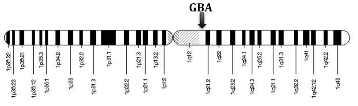
The GBA gene is located at chromosome 1q21, contains 11 exons and 10 introns and is approximately 7kb in length. The GBA gene location is represented in Figure 2. Alternative splicing results in multiple transcript variants. The region surrounding human GBA has been found to be particularly rich in genes, including seven genes and two pseudo-‐genes within a 85kb region of chromosome 1q. The putative TATA and CAAT like boxes of glucocerebrosidase promoter have been recognized about 260 bp upstream from the upstream ATG start codon (G. a Grabowski, 2012).
Figure 2 -‐ GBA gene location on the long (q) arm of chromosome 1 at
position 21. Adapted from: “GBA -‐ glucosidase, beta, acid,” 2014; NCBI, 2014.
Several different mRNA species have been detected on Northern blots, which can be attributed to splice variants, alternate polyadenylation sites, or transcription of the pseudo-‐gene (G. a Grabowski, 2012).
The mRNA has 2324bp that translates into a 536-‐aminoacid protein Glucocerebrosidase. There are 3 isoforms of the protein. In normal conditions it is trafficked to the lysosome, where it catalyses the breakdown of glycolipid gluosylceramide to ceramide and glucose. The protein has four glycosylation sites at Asn58, Asn98, Asn185, and Asn309 and its catalytic residues are located at Glu274 and Glu379.
The tissue levels of GBA mRNA vary among different cell types, with high, moderate, low and negligible levels reported in epithelial, fibroblast, macrophage and B-‐cell lines, respectively (Hruska, LaMarca, Scott, & Sidransky, 2008).
1.4.2 GBA Pseudo-‐gene
There is a 5kb GBA pseudo-‐gene (GBAP) that is located approximately 16kb downstream from the GBA gene, on chromosome 1. GBAP is transcribed, an unusual characteristic for a pseudo-‐gene, but the open-‐reading frame is short due to several stop codons. Nonetheless, it has maintained a high degree of homology with the functional gene, sharing approximately 96% of the sequence in coding regions and the same organization of exons and introns. The different length of GBA and GBAP is explained by the presence of several Alu insertions in introns of GBA. The existence of this pseudo-‐gene is significant, as recombination events between GBA and its pseudo-‐gene can result in mutated GBA genes. Furthermore, the high degree of homology can complicate the identification of GBA mutations. The pseudo-‐gene carries a 55-‐bp deletion in exon 9, which may be of use for molecular diagnostic applications (Hruska et al., 2008).
1.4.3 Genotype – Phenotype
GBA carries out its function in the lysosome, the main site of cellular digestion. The complex molecules being digested can, ultimately, be external material phagocytized by the cell or can be constituted by cellular material destined for turn over. A complex catabolic network resides in the lysosome. Catalytic insufficiency on any one of the many enzymes involved in these catabolic pathways will result in accumulation of that enzyme’s substrate, interruption of the metabolic pathway and indirect effects on other pathways. Glucocerebrosidase catalyses the breakdown of glucocerebroside into glucose and ceramide, and mutations in the GBA gene lead to Gaucher disease, characterized by glucocerebroside accumulation. The phenotype is essentially determined by the combination of mutations on both alleles, but environmental
factors may also play a role in the severity of the clinical presentation. GBA mutations are identified based on the amino acid position in the mature enzyme, with Ala40 designated as the first residue (G. A. Grabowski, 2008; Hruska et al., 2008). More than 250 different mutations in the GBA gene have been described, including point mutations, insertions and deletions (G. A. Grabowski et al., 2011; G. a Grabowski, 2012; Gupta et al., 2011; Lieberman, 2011). Recombination events between GBA and its highly homologous downstream pseudo-‐gene have produced a significant number of complex mutations; these can be the result of gene conversion, fusion or duplication. The 250 mutations described to date include 203 missense mutations, 18 nonsense mutations, 36 small insertions or deletions that lead to either frameshifts or in-‐frame alterations, 14 splice junction mutations, and 13 complex alleles resulting of recombination with GBA pseudo-‐gene. Some complex alleles can carry two or more mutations in cis. (Hruska et al., 2008).
A main point of interest in characterizing mutations for any genetic disease is the ability to predict clinical prognosis. In GD, we find a broad range of mutations causing a wide range of clinical presentations and a poor genotype-‐ phenotype correlation. This makes it difficult to summarize our current knowledge on the subject; however, a number of observations can be made:
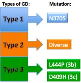
1. The most prevalent mutations are L444P (c1448T>C) and N370S (c.1226A>G).
2. The most common mutations in GD are: N370S, 84GG, L444P and IVS2+1.
3. The presence of the N370S mutation in one or both alleles is often associated to GDT1. The mutation is considered neuroprotective as individuals carrying this mutation rarely present neurological symptoms (Figure 3).
4. The presence of the mutations L444P or D409H is frequently associated to the development of neurological symptoms and frequently found in
GDT2 and GDT3 patients (Guggenbuhl, Grosbois, & Chalès, 2008; Tsuji et al., 1988) (Figure 3).
5. Many point mutations have been identified and homozygosity for those mutations has been associated with all types of GD.
Figure 3 -‐ Classification of Gaucher Disease on genotype-‐phenotype. Mutation N370S is one of the few recognized genotype-‐phenotype correlations related in patients with type 1 GD. In type 2 GD the L444P mutation is frequently found, however the recombinant and rare alleles are particularly predominant on this type. In type 3 GD patients commonly carry the L444P mutation and, as well, an atypical phenotype is related with mutation D409H on this type. Adapted from: Bohra & Nair, 2011; Hruska et al., 2008.
While it is possible to identify individual mutant alleles in GD patients, this capability has limited advantages, as it is difficult to predict phenotype from genotype. The majority GBA mutations can be found in all three types of GD patients.
Patients who share the same genotype may present different symptoms, clinical progress and responses to therapy. Conversely, patients with the same genotypes can show marked differences in clinical presentation. There are even distinct differences between siblings and twin brothers with the same genotype (Hruska et al., 2008; Park et al., 2003).
1.4.3.2 Gaucher Disease in specific ethnic groups 1.4.3.2.1 Ashkenazi Jews population
Ashkenazi Jews are the group with the highest prevalence of GD (1:855 live births). Usually, the carrier frequency for GBA mutations amongst this group is set between 1 in 14 and 1 in 18. The most frequent mutation is the A-‐>G transition at 1226G, the mutation that causes an Asn-‐>Ser substitution at amino acid 370 of the mature protein. Mutation N370S accounts for approximately 70% of mutant alleles (Sidransky, 2013). This mutation is present in approximately 6% of the Ashkenazi Jews population and is the main cause of the high incidence of Gaucher disease in this ethnic group. The second most frequent mutation is an insertion of a guanine at nt.84 of the cDNA and has an incidence of about 0.6%. Among this population, the combined frequency of the N370S and c.84dupG mutations is 0.0343, resulting in a combined incidence of 1:855. This population generally presents type 1 disease (Sidransky, 2013).
1.4.3.2. Norrbottinian population
In the Norrbottinian population, in Northern Sweeden, there is an AT à C transition at nt.1448, which produces a Leu à Pro substitution at amino acid 444 of the mature protein. It has a moderately high frequency amongst this population even though it is revealed in other populations at lower frequencies. This population commonly presents type 3 GD (Sidransky, 2013).
1.4.3.3. Genotype analysis
A number of PCR based genotyping assays are available for the most frequent GD mutations, but the identification of mutant GBA alleles can be problematic, as primers must be designed to discriminate between the functional gene and the pseudo-‐gene (Hruska et al., 2008). Furthermore, detection of
recombinant alleles can pose particular difficulties. Southern blot can help resolve ambiguities, but ultimately, direct sequencing is being increasingly used to characterize mutant alleles, as this technique can detect structure as well as frequent, rare or novel mutations (Hruska et al., 2008).
1.4.3.4. Inheritance patterns of the disease
GD inheritance is autosomal recessive. Heterozygotes are generally asymptomatic. Assuming both parents are heterozygotes, each child of the couple has a 25% probability of being affected, a 50% chance of being an asymptomatic carrier and a 25% possibility of not carrying a GD allele (G. a Grabowski, 2012; Pastores & Hughes, 2014). Prenatal diagnosis may be offered to high-‐risk families and the diagnosis consists on glucocerebrosidase assays in amniotic fluid cells or chorionic villi. Based on this information and the identification of GBA or GBAP mutations in family members, an expected phenotype can be deduced (Guggenbuhl et al., 2008).
1.5 Molecular mechanisms of GD 1.5.1 Sphingolipid metabolism
Sphingolipids are synthesized in the endoplasmic reticulum from nonsphingolipid precursors. Sphingolipids perform important functions in membrane biology and therefore have important effects on cell metabolism. Although there is a great diversity on both structure and function of sphingolipids, all of them share a common anabolic and catabolic pathway (Gault, Obeid, & Hannun, 2011). The metabolism of sphingolipids can be viewed as a set of interconnected networks that diverge from a single common entry point and flock to a single common breakdown pathway. Their simplest forms – sphingosine, phytosphingosine or dihydrosphingosine – serve as the backbone of more complex molecules. For example, the acylation of sphingosine, phytosphingosine or dihydrosphingosine with a variety of acetyl-‐CoA molecules by the action of various ceramide synthase gives rise to long molecules such as
ceramide or phytoceramida dihydriceramida (Gault et al., 2011). A particular class of sphingolipids are the glycosphingolipids (GSL) that are characterized by the kind of sugar residues attached to their ceramide headgroup. GSL are broadly divided into two categories: glycosphingolipids and galactosphingolipids (Gault et al., 2011).
Figure 4 -‐ Glucocerebrosidase function. Glucosylceramide is cleaved by Glucocerebrosidase into Ceramide and Glucose, in the normal function of the enzyme, as represented in the red square. Whereas Glucosylsphingosine is cleaved by Glucocerebrosidase into Glucose and Sphingosine. Glucocerebrosidase is mutated in GD resulting in the accumulation of Glucosylceramide mainly in the lysosomes. Adapted from: Sidransky, 2013. 1.5.2 Molecular mechanisms
The enzyme glucocerebrosidase (also known as GBA; acid beta-‐ glucosidase; beta-‐glucocerebrosidase; glucosylhydrolase; glucosylceramidase) is synthesized in the ER and trafficked to the lysosome. While on route, it suffers post-‐translational modifications, mainly in the form of addition of mannose
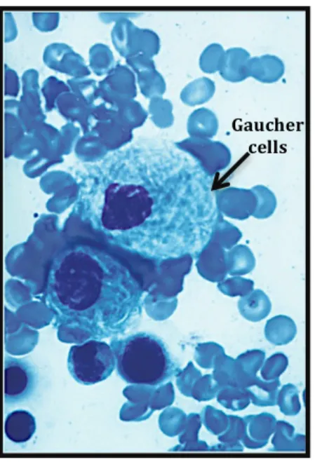
carbohydrates at selected Asn residues. Once in the lysosome, it hydrolyses the glucocerebroside (also called glucosylceramide) to glucose and ceramide, as shown in Figure 4. As a direct result of mutation, enzyme structure is altered with several possible consequences, depending on the nature of the mutation. A mutation in the active site of the enzyme will affect its catalytic function. A mutation elsewhere in the enzyme can also affect catalytic activity indirectly. Another consequence of a mutation can be a modification of the stability of the protein, resulting in increased protein degradation, which ultimately diminishes the amount of active enzyme reaching the lysosome. As a result, the enzyme’s substrate, glucosylceramide is accumulated. In monocyte-‐lineage cells, these lipid-‐laden lysosomes result into the characteristic “Gaucher cells”. They present a large reticuloendothelial system, a small and eccentrically placed nuclei and a striated cytoplasm as shown in Figure 5 (G. A. Grabowski, 2008; Vairo et al., 2013).
Figure 5 -‐ Gaucher cells from a bone marrow aspirate. These cells derive from macrophages with stored glycolipids. They measure around 20 – 100µm in diameter. Gaucher cells are also swollen and have a cytoplasm that looks lacy. Adapted from: Elstein, Abrahamov, Hadas-‐ halpern, & Zimran, 2001; Vairo et al., 2013.
Gaucher cells