1. Pediatric Rheumatology Unit, Faculdade de Medicina da Universidade São Paulo, São Paulo, Brazil
2. Division of Rheumatology, Faculdade de Medicina da Universidade São Paulo, São Paulo, Brazil
3. Obstetrics and Ginecology Department, Faculdade de Medicina da Universidade São Paulo, São Paulo, Brazil
4. Adolescent Unit, Faculdade de Medicina da Universidade São Paulo, São Paulo, Brazil
INTRODUCTION
Infections are an important cause of morbidity and mortality in our childhood-systemic lupus erythe-matosus (C-SLE) patients,1including genital infections, such as human papillomavirus (HPV)2.
Adult patients with SLE, particularly those on im-munosuppressive therapy, have cervical dysplasia with detection of HPV infection varying from 4.7 to 50%3-10. In C-SLE, we previously reported suggestive le-sions of HPV infection in 2% of these patients2.
In addition, this genital abnormality is induced by the proliferation of squamous epithelial cells secondary to this virus infection and was rarely described in adult SLE11. To our knowledge, none case was reported and the prevalence of condyloma acuminatum in children and adolescents C-SLE patients was not performed.
Therefore, from January 1983 to May 2012, 5,682 patients were followed at the Pediatric Rheumatology Unit from Instituto da Criança da Faculdade de Medi -cina da Universidade de São Paulo and 289 (5%) of them met the American College of Rheumatology (ACR)12classification criteria for SLE. Four (1.4%) of our female C-SLE patients had condyloma acumina-tum with confirmation of HPV DNA testing by Hybrid Capture 2 (HC2 high-risk; Digene Corporation, cur-rently QIAGEN, Gaithersburg, MD, USA), using DNA of oncogenic group (16, 18, 31, 33, 35, 39, 45, 51, 52, 56, 58, 59 e 68). None of our male C-SLE patients had condyloma acuminatum.
Pap smears were evaluated by the same cytopatholo -gist blinded to gynecology examination in our Univer-sity Hospital. They were performed according to the 2001 Bethesda Classification System13in 5 patterns. Guided biopsies were performed on all identifiable le-sions at colposcopy.
This study was approved by the Local Ethics Com-mittee of our University Hospital. Demographic data, clinical and laboratory findings, disease activity14and
Condyloma acuminatum in childhood-systemic
lupus erythematosus patients
Gabriella E. Lube1, Nadia E. Aikawa1,2, Maricy Tacla3, Marta M. Leal4, Benito Lourenço4, Luiz E.V. Silva4, Lígia B. Queiroz4, Edmund C. Baracat3, Clovis A. Silva1,2
ABSTRACT
Introduction: Infections are frequent in
childhood-sys-temic lupus erythematosus (C-SLE) patients, inclu ding human papillomavirus (HPV). HPV infection may cause genital and anal warts named condyloma acumi-natum (CA). To our knowledge, none case was report-ed and the prevalence of CA in C-SLE population was not performed.
Case Reports: From January 1983 to May 2012, 5,682
patients were followed at the Pediatric Rheumatology Unit from of our University Hospital and 289 (5%) of them met the American College of Rheumatology clas-sification criteria for C- SLE. Four (1.4%) of our female patients had CA. The median age at diagnosis was 13 years. Three of them were sexually active and all of them had active disease and had high risk HPV anoge -nital warts. Pap smears showed low-grade squamous intraepithelial lesion, guided biopsies identified chro -nic cervicitis, vulvar, vaginal, anal and/or cervix intraepi thelial neoplasia. All of them were under corti-costeroids and immunosuppressive drugs. The visible genital warts lesions were eradicated.
Discussion: Our patients requires rigorous gynecologic
follow-up due to the severe anogenital dysplasia. HPV vaccine should be indicated in all CSLE prior to se -xual activity.
Keywords: Adolescent; Childhood; Systemic Lupus
Erythematosus; Human Papillomavirus; Infection; Condyloma acuminatum.
CASE REpORT
CASE 1
A 12 year-old female was admitted to our University TABLE I. DEmOgRAphIC DATA, CLINICAL AND LABORATORy fINDINgS, pAp SmEARS, DISEASE
ACTIvITy/DAmAgE, TREATmENTS AND OUTCOmE IN ChILDhOOD-SySTEmIC LUpUS ERyThEmATOSUS (C-SLE) pATIENTS AT CONDyLOmATA ACUmINATUm (CA) DIAgNOSIS
Cases
Variables 1 2 3 4
Demographic data
Age at JSLE diagnosis, years 12 8 14 15
Period between JSLE and CA, months 22 108 1 48
Age at menarche, years 10 11 10 12
Age at first sexual intercourse, years 14 16 - 16
Clinical features at CA Vulvar warts Vulvar, vaginal, Vulvar and Vulvar warts and anal warts anal warts
Pap Smears at CA LSIL/ASCH LSIL - LSIL
Histological findings CC, metaplasia CIN 3, VIN 2, AIN 1 Papilomatosis CC
Laboratory findings at CA Haemoglobin, g/dL 11.2 11.8 12.2 10.8 Leukocytes/mm3 5,600 5,800 5,500 9,100 Lymphocytes/mm3 1,792 1,102 440 600 Platelets count/mm3 276,000 254,000 140,000 338,000 Urinalysis Leukocytes/mL 670,000 16,000 140,000 11,000 Erythrocytes/mL 1,800 19,000 3,000 79,000 Urea, mg/dL 20 33 33 25 Creatinine, mg/dL 0.2 0.9 0.96 1.0 Proteinuria, g/24h 0.35 0.8 0.23 0.8 CRP, mg/dL 9.85 2.41 6.6 0.62 ESR, mm/1sth 42 27 26 48
Disease activity and damage at CA
SLEDAI-2K 6 14 20 19
SLICC-ACR/DI 0 0 NA 0
HPV isolation HPV DNA of HPV DNA of HPV DNA of HPV DNA of
oncogenic group oncogenic group, oncogenic group, oncogenic group and cervix biopsy cervix biopsy, HPV 16
HPV 16
Treatments at CA
JSLE therapy CT, AZA, AM CT, AZA, AM CT, IVCYC, AM CT, AM
HPV LEEP CO2-laser Surgical removal LEEP
vaporization
Recurrence of HPV/CA Yes – – –
ASCH = Atypical squamous cells of undetermined significance that cannot exclude high-grade squamous intraepithelial lesion, LSIL = Low-grade squamous intraepithelial lesions, CC = chronic cervicitis, CIN = cervical intraepithelial neoplasia, VIN = vulvar intraepithelial neoplasia, AIN 1 = anal intraepithelial neoplasia, CRP = C-reactive protein, ESR = erythrocyte sedimentation rate, CT = corticosteroid, AZA = azathioprine, AM = antimalarials, IVCYC = intravenous cyclophosphamide, LEEP = loop electrosurgical excisional procedure, SLEDAI-2K - Systemic Lupus Erythematosus Disease Activity Index 2000, SLICC/ACR-DI - Systemic Lupus International Collaborating Clinics/ACR - Damage Index, NA – not applicable.
damage15 scores, treatment regimens and outcome of our four female C-SLE patients at IA diagnosis are shown in Table I, and their cases were reported he rein.
CASE 2
The 8-year-old female was admitted to the pediatric unit of our university hospital with fever, arthralgia (knees, ankles and wrist), malar rash, oral ulcers, pleu-ritis, pericarditis and arterial hypertension. Her laboratory exams identified hemoglobin 8.4 g/dL, hema -tocrit 26%, leucocytes 25,000/mm³ (61% neutrophils 36% lymphocytes, 1% eosinophils and monocytes 2%), platelets 70,000/mm³, creatinine 1.2 mg/dL, urea 89 mg/dL, C3 42 mg/dL (normal 79-152) and C4 3.3 mg/dL (normal 15-38). The proteinuria was 0.15 g/24h and abnormal urinalysis (155,000 erythrocytes/mL and 302,000 leukocytes/mL). Immunological tests showed ANA 1/200 (homogeneous pattern) and positive anti--dsDNA, and negative anti-Ro, anti-La, anti-Sm and anticardiolipin antibodies. ESR was 66 mm/1st hour and renal biopsy showed diffuse proliferative nephritis (class IV of World Health Organization). The dia -gnosis of C-SLE was confirmed according to the ACR criteria and the SLEDAI2K score was 21. She was trea -ted with prednisone (2mg/kg/day) with progressive dose decrease, chloroquine 250mg/day and intra-venous cyclophosphamide (500-1000 mg/m²) for three consecutive years. The age at menarche was 11 years with regular menstrual cycles after 13 years. At 17-years--old, the gynecologic clinical examination of the geni-talia showed diffuse warts on vagina, vulva and peri neal regions suggesting condyloma acuminatum.
Chlamy-dia trachomatis was not isolated by HC2 CT-ID and
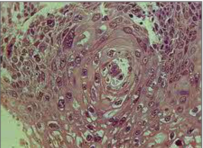
follow ing serologic tests were negative: hepatitis virus A, hepatitis virus B, hepatitis virus C, EBV, CMV, and HIV. At that moment, she was under prednisone 20 mg/day, chloroquine 250mg/day and azathioprine 150mg/day, and her laboratory findings, SLEDAI-2K 14 and SLICC/ACR-DI 0 are shown in Table I. Pap smears showed LSIL and Digene HPV test by HC2 show ed DNA of oncogenic group. The age onset of se -xual activity was 16 years and only one se-xual partner. Colposcopically guided biopsies were performed in three locations and demonstrated: condyloma with koilocytosis, eosinophilic border and nuclear atypia (Figure 1), CIN 3 in the cervix (Figure 2), vulvar in-traepithelial neoplasia (VIN 2) and anal inin-traepithelial neoplasia (AIN 1). HPV-16 was identified in vulva and endocervix by immunohistoquimic assay. She was trea -ted with CO2-laser vaporization in the genitalia wi thout eradication of all visible lesions and LEEP in the cervix.
CASE 3
A 14-year-old female was admitted in our University Hospital with malaise, fever, malar rash, and arthritis
in ankles and knees. Her laboratory exams identified hemoglobin 8.4g/L, white blood cell count (WBC) 5.000/mm³ (56% neutrophils, 38% lymphocytes, 1% eosinophils and monocytes 5%), platelets 262,000/ /mm³ and negative Coombs test. Immunological tests showed antinuclear antibodies ANA 1:320 (fine speck-led pattern), and positive anti-double-stranded DNA (anti-dsDNA), anti-Sm, anti-RNP and IgG anticardi-olipin antibodies (14.0 GPL). C3 was 0.33 mg/dL (nor-mal 0.5-1.8), C4 0.051mg/dL (nor(nor-mal 0.1-0.4), urea 25 mg/dL (normal 10-42), creatinine 0.5 mg/dL mal 0.6-0.9), C-reactive protein (CRP) 3.2 mg/L (nor-mal < 5) and erythrocyte sedimentation rate (ESR) 56 mm/1sthour. The proteinuria was 0.6 g/24h and uri-nalysis showed granular casts, hematuria 208,000/ml, leukocyturia 162,000/ml, and the diagnosis of C-SLE was confirmed according to the ACR criteria. The Sys-temic Lupus Erythematosus Disease Activity Index 2000 (SLEDAI-2K) score was 27, and she was treated with chloroquine 5mg/kg/day and prednisone 60mg/day. The menarche was 10 years and at 13 years and 10 months, her menstrual cycles were irregular. The age onset of sexual activity was 14 years, with three sexual partners. At that moment, the gynecologic cli -nical examination of the genitalia showed vulvar warts suggesting condyloma acuminatum. Chlamydia
tra-chomatis was also identified by Hybrid Capture 2 (HC2
CT-ID; Digene Corporation, currently QIAGEN, Gaithersburg, MD, USA). The following serologic tests were negative: hepatitis virus A, hepatitis virus B, hepa -titis virus C, Epstein-Barr virus (EBV), cytomegalovirus (CMV), human T-lymphotropic virus (HTLV) and hu-man immunodeficiency virus (HIV). The Pap smear showed LSIL according the 2001 Bethesda System13. Laboratory findings, SLEDAI-2K and Systemic Lupus International Collaborating Clinics/ACR-Damage Index (SLICC/ACRDI) were 6 and 0, respectively (Ta -ble I). She was under prednisone 20mg/day, azathio-prine 150mg/day and chloroquine 250mg/day. Exter-nal genital warts were treated with loop electrosurgery and all visible lesions were eradicated. At 16 years old, she had new vulvar warts, Pap smears showed ASCH and Digene HPV test by HC2 showed DNA of onco-genic group. At that moment, colposcopically guided biopsies demonstrated chronic cervicitis with meta-plasia and HPV identification. External genital warts were treated with loop electrosurgery and genital warts in the cervix with loop electrosurgical excisional pro-cedure (LEEP). All visible lesions were eradicated.
Hospital with fever, malar rash, petechiae, psychosis and severe alveolar hemorrhage. Her examinations identified hemoglobin 9.1 g/dL, hematocrit 26.9%, WBC 4,930/mm³ (78% neutrophils 16% lymphocytes, 1% eosinophils and 5% monocytes), platelets 37,000/mm³, urea 22 mg/dL and creatinine 0.74 mg/dL. Immunological tests showed ANA 1/1280 (ho-mogeneous pattern), positive rheumatoid factor and anti-dsDNA, and negative anti-Sm, anti-RNP, anti-Ro, anti-La and IgG and IgM anticardiolipin antibodies. C3 was 26 mg/dL (normal 79-152), C4 2 mg/dL (normal 15-38), ESR 62 mm/1sthour and CRP 15.8 mg/L. Uri-nalysis showed 1000 leukocytes/mL and 1000 ery-throcytes/mL, and proteinuria was 1.023 g/24h. The diagnosis of C-SLE was confirmed according to the ACR criteria and the SLEDAI-2K score was 20. She was hospitalized in the intensive care unit and received methylprednisolone 1 g/day for three days, intravenous cyclophosphamide (750 mg/m²), hydroxychloroquine 300mg/day and prednisone (2.0 mg/kg/day) with im-provement of psychosis and alveolar hemorrhage. After one month of hospitalization, the gynecologic clinical examination of the genitalia showed vulvar and anal warts suggesting condyloma acuminatum with in-tact hymen. The following serologic tests were nega-tive: hepatitis virus A, hepatitis virus B, hepatitis virus C, CMV and HIV. At that moment, her menstrual cy-cles were irregular and the age at menarche was 10 years. No history of sexual intercourse and sexual abuse was reported. Laboratory findings, SLEDAI-2K and SLICC/ACR-DI are shown in Table I. Perineum and anal biopsies demonstrated papilomatosis, with iden-tification of HPV-16 by chromogenic in situ
hybridiza-tion. The Digene HPV test by HC2 showed DNA of oncogenic group in vaginal ostium undertaken by
Cy-tobrush ®. She was treated with warts surgical removal
procedure in the perineum and anus with total im-provement.
CASE 4
An 8-year-old female was admitted in our University Hospital with chronic idiopathic thrombocytopenic purpura with petechiae and bruising on the lower limbs. She had autoimmunity family history of SLE (her brother, maternal aunt and maternal grandmoth-er) and Sjögren syndrome (her mothgrandmoth-er). Her examina-tions identified hemoglobin 10.2 g/dL, hematocrit 32.3%, WBC 6,300mm³ (48% neutrophils, 41% lym-phocytes, 6% eosinophils and 5% monocytes) and platelets 15,000/mm³. Immunological tests showed ANA 1:200 (speckled pattern) and positive lupus an-ticoagulant, and negative anti-dsDNA, anti-Ro and anti-La antibodies, and rheumatoid factor. C3 was 91 mg/dL (normal 79-152), C4 9 mg/dL (normal 15-38) and ESR 37mm/1sthour. She was treated with predni-sone (2.0 mg/kg/day) with gradual dose reduction. At 15 years old, she had malar rash, arthritis in knees, upper limbs vasculitis, arterial hyuppertension and her exa -minations showed proteinuria was 0.6 g/24h and uri-nalysis showed granular casts, hematuria 15,000/ml, leukocyturia 31,000/ml. The diagnosis of C-SLE was confirmed according to the ACR criteria and the SLEDAI2K score was 24. She was treated with predni -sone (1.0 mg/kg/da) and chloroquine (250mg/day). At 19 years, the gynecologic clinical examination of the genitalia showed vulvar warts suggesting condyloma fIgURE 1. Condyloma with koilocytosis, eosinophilic border
and nuclear atypia
fIgURE 2. Cervical intraepithelial neoplasia (CIN 3) in the cervix
acuminatum. The following serologic tests were nega-tive: hepatitis virus B, hepatitis virus C, CMV, Chlamy-dia trachomatis and HIV. Her menstrual cycles were
regu lar after menarche at 12 years. The age onset of sexual activity was 16 years and only one sexual part-ner. Laboratory findings, SLEDAI-2K 19 and SLICC/ /ACR-DI 0 are shown in Table I. She reported condom in all sexual intercourses without hormonal contra-ceptive use. At 19 years, she received prednisone 5 mg/day and chloroquine 250mg/day, the Pap smears showed LSIL and Digene HPV test by HC2 showed DNA of oncogenic group and colposcopically guided biopsies demonstrated chronic cervicitis. She was treat-ed with LEEP with total improvement.
DISCUSSION
This was the first report that evaluated the prevalence of condyloma acuminatum in a large population of C--SLE patients from a university pediatric hospital, and clearly showed that this infection occurred in female sexually active and virgin patients with disease activi-ty and under immunosuppressive agents.
Of note, female C-SLE patients are becoming ado-lescents and became sexually active with a consequent higher risk of sexually transmitted disease (STD), such HPV.2HPV is known as the most common STD in Uni -ted States females and the major risk factor is younger age at coitarche (first sexual intercourse)16, as observed in three of our C-SLE patients.
In addition, the incidence of anogenital warts was approximately 1% in sexually active adults and may be up to 3% in sexually active adolescents17. The most fre-quently types associated with these lesions are HPV 6 and 11, contrasting with our four cases and other re-ports18that presented the oncogenic group with a marked risk to dysplasia and cervical cancer.
On the other hand, condyloma acuminatum was also observed in our lupus patients without coitarche and sexual abuse history. Indeed, HPV infection may be related to autoinoculation or heteroinoculation, through direct contact or fomites in these patients, as described in one of our cases. A recent study reported anogenital HPV infections in girls before sexual activi-ty, diagnosed by the same routine Hybrid Capture II HPV DNA test, as used in the present cases18.
Interestingly, Chlamydia trachomatis is the most com-mon bacterial cause of STD and the prevalence of this endocervical infection was 3% in a recent adult SLE
Brazilian study5. The same study did not observe a higher frequency of HPV associated with this genital infection5. Chlamydia trachomatis infection was obser -ved in one of our sexually active lupus patient, and to our knowledge this genital infection was not reported in juvenile systemic lupus erythematosus population. The most important risk factors associated to general infections in C-SLE population are related to disease itself (activity1, 2and lymphopenia) and treatments (cor-ticosteroids and immunosuppressive drugs)1,10. Of note, in adult SLE immunosuppressant, especially aza-thioprine and cyclophosphamide use, was the main risk factor for HPV infection3,4,6-11.
The treatment of condyloma acuminatum includes physical destruction in the anogenital area, such as sur-gical removal with loop electrosursur-gical excisional and CO2-laser vaporization,17as indicated in our cases.
Therefore, we would recommend routinely gyneco-logic evaluation and HPV infection testing before start immunosuppressive drugs in adolescent SLE patients. Importantly, HPV vaccine should be indicated in all C--SLE patients, particularly before start sexual inter-course. Recently, the quadrivalent HPV vaccine was safe and effective in adult lupus patients older than 18 years and did not induce disease activity or flares19. Immu-nogenicity and safety of this immunization should be also known in the C-SLE population.
In conclusion, condyloma acuminatum was rarely observed in C-SLE population, mainly after coitarche and in virgin patients with disease activity and/or un-der immunosuppressive agents. Our patients requires rigorous gynecologic follow-up due to the severe anogenital dysplasia.
ACKNOWLEDgmENTS
This study was supported by Fundação de Amparo à Pesquisa do Es-tado de São Paulo – FAPESP (grant # 2011/12471-2 to CAS), by CNPQ (302724/2011-7 to CAS), Federico Foundation to CAS and by Núcleo de Apoio à Pesquisa “Saúde da Criança e do Adolescen-te” da USP (NAP-CriAd) to CAS. We thank Dr. Elsa Gay Pereyra and Dr Katia Pincerato for your suggestions and collaborations and Dr Helio Hehl Caiaffa-Filho for the HPV test evaluation.
CORRESpONDENCE TO Clovis Artur Almeida da Silva Rua Araioses, 152/81 – Vila Madalena São Paulo – SP – Brazil, CEP 05442-010 E-mail: clovis.silva@icr.usp.br
REfERENCES
1. Faco MM, Leone C, Campos LM, Febronio MV, Marques HH, Silva CA. Risk factors associated with the death of patients hos-pitalized for juvenile systemic lupus erythematosus. Braz J Med
Biol Res 2007; 40: 993-1002.
2. Febronio MV, Pereira RM, Bonfa E, Takiuti AD, Pereyra EA, Sil-va CA. Inflammatory cervicoSil-vaginal cytology is associated with disease activity in juvenile systemic lupus erythematosus. Lu-pus 2007; 16: 430-435.
3. Santana IU, Gomes Ado N, Lyrio LD, Rios Grassi MF, Santiago MB. Systemic lupus erythematosus, human papillomavirus in-fection, cervical pre-malignant and malignant lesions: a syste-matic review. Clin Rheumatol 2011; 30: 665-672.
4. Rojo-Contreras W, Olivas-Flores EM, Gamez-Nava JI, et al. Cer-vical human papillomavirus infection in Mexican women with systemic lupus erythematosus or rheumatoid arthritis. Lupus 2012; 21: 365-372.
5. Costapinto L, Olavarria VG, Grassi MF, et al. Prevalence of Chla-mydia trachomatis endocervical infection in systemic lupus erythematosus patients and evaluation of the risk for HPV-in-duced lesions. Rheumatol Int 2012;33:631-636
6. Klumb EM, Araujo ML, Jr., Jesus GR, et al. Is higher prevalen-ce of prevalen-cervical intraepithelial neoplasia in women with lupus due to immunosuppression? J Clin Rheumatol 2010; 16: 153-157. 7. Lee YH, Choe JY, Park SH, et al. Prevalence of human papillo-ma virus infections and cervical cytological abnorpapillo-malities among Korean women with systemic lupus erythematosus. J Korean Med Sci 2010; 25: 1431-1437.
8. Nath R, Mant C, Luxton J, et al. High risk of human papillo-mavirus type 16 infections and of development of cervical squa-mous intraepithelial lesions in systemic lupus erythematosus patients. Arthritis Rheum 2007; 57: 619-625.
9. Tam LS, Chan PK, Ho SC, et al. Natural history of cervical pa-pilloma virus infection in systemic lupus erythematosus - a pros-pective cohort study. J Rheumatol 2010; 37: 330-340. 10. Tam LS, Chan PK, Ho SC, et al. Risk factors for squamous
in-traepithelial lesions in systemic lupus erythematosus: a pros-pective cohort study. Arthritis Care Res (Hoboken) 2011; 63: 269-276.
11. CostaPinto L, Grassi MF, Serravalle K, Travessa AC, Olavarria VN, Santiago MB. Giant disseminated condylomatosis in SLE. Lupus 2012; 21: 332-334.
12. Hochberg MC. Updating the American College of Rheumato-logy revised criteria for the classification of systemic lupus eryt-hematosus. Arthritis Rheum 1997; 40: 1725.
13. Solomon D, Davey D, Kurman R, et al. The 2001 Bethesda Sys-tem: terminology for reporting results of cervical cytology. JAMA 2002; 287: 2114-2119.
14. Gladman DD, Ibanez D, Urowitz MB. Systemic lupus erythe-matosus disease activity index 2000. J Rheumatol 2002; 29: 288-291.
15. Gladman D, Ginzler E, Goldsmith C, et al. The development and initial validation of the Systemic Lupus International Col-laborating Clinics/American College of Rheumatology damage index for systemic lupus erythematosus. Arthritis Rheum 1996; 39: 363-369.
16. Hariri S, Unger ER, Sternberg M, et al. Prevalence of genital hu-man papillomavirus among females in the United States, the National Health And Nutrition Examination Survey, 2003--2006. J Infect Dis 2011; 204: 566-573.
17. Thornsberry L, English JC, 3rd. Evidence-based treatment and prevention of external genital warts in female pediatric and ado-lescent patients. J Pediatr Adolesc Gynecol 2012; 25: 150-154. 18. Doerfler D, Bernhaus A, Kottmel A, Sam C, Koelle D, Joura EA. Human papilloma virus infection prior to coitarche. Am J Obs-tet Gynecol 2009; 200: 487 e481-485.
19. Mok CC, Ho LY, Fong LS, To CH. Immunogenicity and safety of a quadrivalent human papillomavirus vaccine in patients with systemic lupus erythematosus: a case-control study. Ann Rheum Dis 2013;72:659-664.