Article
Printed in Brazil - ©2019 Sociedade Brasileira de Química
*e-mail: [email protected], [email protected]
Antimicrobial,
Anti-Inflammatory
and
Antioxidant
Activities
of
Polyoxygenated
Chalcones
YessenyA.Vásquez-Martínez,aMauricioE.Osorio, *,bDiegoA.SanMartín,b MarcelaA.Carvajal,cAlejandraP.Vergara,cElizabethSanchez,cMarcelaRaimondi,d
SusanaA.Zacchino,dCarolinaMascayano,eClaudiaTorrent,eFranciscoCabezas,e SophiaMejias,fMargaritaMontoyafandMarceloCortez-SanMartínf
aPrograma Centro de Investigaciones Biomédicas Aplicadas, Facultad de Ciencias Médicas,
Universidad de Santiago de Chile, 9170022 Santiago, Chile
bLaboratorio de Productos Naturales, Departamento de Química,
Universidad Técnica Federico Santa María, 2390123 Valparaíso, Chile
cCentro de Biotecnología CB-DAL, Universidad Técnica Federico Santa María,
2390136 Valparaíso, Chile
dFarmacognosia, Facultad de Ciencias Bioquímicas y Farmacéuticas,
Universidad Nacional de Rosario, Suipacha 531, 2000 Rosario, Argentina
eDepartamento de Ciencias del Ambiente, Facultad de Química y Biología,
Universidad de Santiago de Chile, 9170022 Santiago, Chile
fDepartamento de Biología, Facultad de Química y Biología,
Universidad de Santiago de Chile, 9170022 Santiago, Chile
It was synthesized nine polyoxygenated chalcones with a potential and safe use as antioxidant, antimicrobial and anti-inflammatory therapies. Chalcones obtained by Claisen-Schmidt condensation were studied as antioxidant, inhibitors of human 5-lipoxygenase, antifungal, antibacterial and antibiotic resistance modifiers. Two chalcones with catecholic moieties were able to strongly decrease the minimum inhibitory concentration (MIC) of methicillin against methicillin-resistant Staphylococcus aureus, increase the antiradical activity and significantly
inhibit the human 5-lipoxygenase. Only one of these chalcones was active synergistically with methicillin. Chalcones with methoxyl substituents at different positions displayed the best activities against Cryptococcus neoformans. Only one chalcone showed good activity against the plant
pathogenic bacteria Pseudomonas syringae whose half maximal inhibitory concentration (IC50) value (2.5 µg mL-1) was similar to that observed with the antibiotic streptomycin (2.9 µg mL-1). These simple chalcones have safe potential uses in antioxidant, antimicrobial and anti-inflammatory therapies.
Keywords: antioxidant activity, polyoxygenated chalcones, human 5-lipoxygenase inhibitors,
antibacterial, antifungal
Introduction
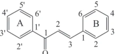
Chalcones (1,3-diphenyl-prop-2-en-1-ones) essentially differ from other flavonoids by its open C-ring, thus possessing a linear chain of three carbon atoms binding
A with B ring (Figure 1).1 Chalcones have been used
as intermediates for the preparation of compounds with therapeutic value. A number of synthetic routes have been reported for the synthesis of chalcones, the Claisen-Schmidt
condensation under homogeneous conditions in the presence of acid or base being the most used. Traditionally, strong alkaline media2-8 and the use of several Lewis acids9-15
have been employed for their synthesis. Some improved strategies include solvent-free conditions,16 microwave17
and ultrasound irradiation18 and the grinding technique.19
The synthesis of chalcones in acid condition using SOCl2
in ethanol to generate HCl in situ have been used to prepare
chalcones with one free hydroxyl group,20 however, the
synthesis of chalcones with additional oxygenated groups have not been proven using this method. Chalcones display
Antimicrobial,
Anti-Inflammatory
and
Antioxidant
Activities
of
Polyoxygenated
Chalcones
YessenyA.Vásquez-Martínez,aMauricioE.Osorio, *,bDiegoA.SanMartín,b MarcelaA.Carvajal,cAlejandraP.Vergara,cElizabethSanchez,cMarcelaRaimondi,d
a large number of different biological activities, such as antibacterial, antifungal, anti-cancer, anti-inflammatory, analgesic, antiviral, anti-malarial, antipyretic and cytotoxic activities.20,21
Multi-drug resistance is one of the major causes of the alarming level of infectious diseases worldwide. The discovery of new drugs with potent antimicrobial activity, particularly against the resistant strains, is, therefore, highly needed. In addition, since many of the currently available antifungal drugs have undesirable side effects, are ineffective against new or re-emerging fungi, or develop a rapid resistance in the pathogen,22 there is an urgent
need of a next generation of new antifungal agents that can overcome the above disadvantages. Several reports highlighting the significance of chalcones as antimicrobial and antiparasitic agents have been documented in the
literature.21 Chalcone derivatives with –OH and –OCH
3
in the rings A and B have demonstrated potential anti-leishmanial and anti-trypanosomal activities by acting on a number of molecular targets.23
Fungal plant pathogens such as Botrytis cinerea
(B. cinerea),24Gibberella fujikuroi (G. fujikuroi)25 and Phytophthora cinnamomi (P. cinnamomi)26,27 attack a
wide range of agriculturally and ornamentally important plants28 and significantly affect the economy of developing
countries causing large economic losses in agriculture.29
Numerous fungicides such as carboxin, captan, thiram, tebuconazole and metalaxyl have been used to control fungal phytopathogens, but all of them possess secondary effects in other non-target microorganisms and living beings.30 Natural and synthetic chalcones with –OH and
–OCH3 groups linked to different positions of the A and
B rings have shown bactericidal, antifungal, anthelmintic, insecticidal, insect antifeedant, antiviral and phytotoxic activities.31
Plant pathogenic bacteria cause serious problems for industrial agriculture because they affect crops of nutritional
and commercial interest. Agrobacterium tumefaciens
(A. tumefaciens), Pseudomonas syringae (P. syringae) and Erwinia carotovora (E. carotovora) are Gram-negative
bacteria that are responsible for a number of economically important diseases. These bacteria usually infect a wide variety of fruits, vegetables, and ornamental plants,32-34
and different types of compounds have been used for their control. However, these control efforts have not always been successful and less toxic new products to replace the existing ones are highly needed. To achieve this goal, the worldwide trend is to explore new compounds against these phytopathogens in order to minimize the risks associated with the development of pathogen populations insensitive to these chemical compounds.35 Also, many countries have
limited the use of some commercial antimicrobials based on recommendations from the Codex Alimentarius36 according
to the maximum residue limits (MRLs) for residues of pesticides or veterinary drugs in foods.
Endogenous free-radical species like reactive oxygen (ROS) and nitrogen species (RNS) and other reactive small molecules have emerged as important regulators of many physiological and pathological processes. ROS and RNS are essential to maintain homeostasis and health of human beings, but uncontrolled and excess ROS/RNS have been implicated in the pathogenesis of various diseases including cancer, cardiovascular and neurodegenerative diseases as well as ageing. The human organism has developed defense systems to neutralize excessive levels of ROS and RNS and compensate for the oxidative stress.37 When
endogenous antioxidant protection is not able to maintain the proper balance of free radicals due to their unlimited or uncontrolled production, several health problems appear. In such cases, additional external antioxidants are required to restore the proper balance between free radicals and antioxidants in the body.38 Chalcone derivatives with an
–OH substituent on ring A, and –SCH3 and –OCH3 groups
at the para position of ring B showed good anti-oxidant
activity.21
Different natural or synthetic chalcones has been studied for their anticancer activities because they exert cytotoxicity through multiple mechanisms, which include cell cycle disruption as well as inhibition of angiogenesis, tubulin polymerization, apoptosis and cell signaling.39 On
the other hand, high expression of 5-lipoxygenase (5-LOX) has been described in cancer cells and its inhibition has been widely characterized to control tumor cell growth.40,41
Moreover, ROS also have to be closely linked with tumor initiation, development and progression, by promoting cell survival, cell proliferation and migration.42 Thus,
inhibition of 5-LOX or the decrease in ROS levels can also have impact on cancer cell survival and/or proliferation. 5-LOX belongs to a group of closely related non-heme iron containing dioxygenases. These enzymes catalyze the addition of molecular oxygen into poly-unsaturated fatty acids (PUFAs) containing cis, cis-1,4-pentadiene
structures to give their hydroperoxy derivatives. LOXs are further classified into 5-, 8-, 9-, 11-, 12-, and 15-LOXs
according to the positional specificity of arachidonate oxygenation. In addition, the 5-LOX is the source of potent pro-inflammatory mediators present in a variety of inflammatory and allergic diseases.43 As 5-LOX have been
shown to have a major role in the pathogenesis of various inflammatory disorders, including cancer, inhibitors of 5-LOX will have a wide range of therapeutic applications.44
Di-O-prenylated chalcone derivatives have shown to
possess good inhibitory potency of 5-LOX (half maximal
inhibitory concentration (IC50) = 4 µM), potent
anti-proliferative effects (50% growth inhibition (GI50) = 9 µM)
on the MCF-7 breast cancer cell line, and no appreciable effects up to 100 µM on the immortalized nontumorigenic human epidermal (HaCaT) cell line.43
In this paper, we report a facile synthesis of nine chalcones to evaluate their effects against phytopathogens and human pathogenic bacteria and fungi as possible alternatives to antimicrobials currently in commercial use. Additionally, as endogenous free radicals and human 5-lipoxygenase (5-hLOX) have been shown to have major roles in the pathogenesis of various health problems including cancer, cardiovascular and neurodegenerative diseases and inflammatory disorders, the chalcones were tested as 5-hLOX inhibitors and DPPH (2,2-diphenyl-1-picrylhydrazyl) radical scavengers.
Results and Discussion
Synthesis
Chalcones 11-19 were obtained in poor to good
yields (11-90%) via Claisen-Schmidt condensation of
acetophenones 1-6 with benzaldehydes 7-10 in acidic or
alkaline media (Scheme 1). Acidic medium was mostly
preferred when one or more free hydroxyl groups are bound to the aromatic system of the chalcones. When Claisen-Schmidt condensations using other 2’-hydroxy or 2’-methoxy acetophenones were conducted in acidic conditions, chalcones were not produced (12 and 19). This
was probably due to either the hydrogen-bond formed by the OH or OMe groups on C-2’ with the carbonyl group (that could hinder the formation of the enol intermediate) or by problems of steric hindrance with the 2’-methoxyl group.
The yields of all the synthesized chalcones were similar or better than those reported in the literature and all were obtained in only one step. Chalcone 12 is the only new
molecule in the series. The solid-state reactions (without solvent) were not attempted.
The synthesis of chalcones with a hydroxyl group in 4-position was incompatible with alkaline medium,
except for 12. Chalcones with 2’-OH group can be
synthesized in alkaline medium, but with low yield (11% for 19). Protecting groups resistant to alkaline medium
(i.e., methoxymethyl (MOM), tetrahydropyranyl (THP)) should be considered to obtain chalcones in good yields with two or more free hydroxyl groups. Chalcones with 3,4-diOH substituents (catechol group) on the B ring can be synthesized in acid condition with moderate yields (47% for 11 and 57% for 18).
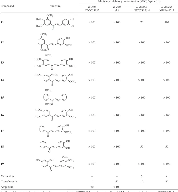
In vitro antibacterial effects of chalcones 11-19 on bacteria
pathogenic to humans
Determination of the minimum inhibitory concentration (MIC) of the chalcones against bacteria
The result of the bacterial growth inhibition test (Table 1) showed that chalcones 11 and 18 were active
against sensitive S. aureus strains with MICs of 50 and
70 µg mL-1, respectively. These compounds also inhibited
the growth of MRSA 97-7 strain (MICs of 50 and
90 µg mL-1, respectively).
When comparing the structural characteristics of 11 and 18, which are weakly active against S. aureus with the rest
of the chalcones, it can be observed that both compounds possess a catechol moiety in their structures, which could play a key role in the activity. Then, our results indicated that the presence of catechol group at positions 3,4 on the B ring is necessary to exhibit the anti-staphylococcal activity, decreasing this activity when the hydroxyl at position 3
Table1. Results of the in vitro antibacterial assay
Compound Structure
Minimum inhibitory concentration (MIC) / (µg mL-1)
E. coli ATCC25922
E. coli 33.1
S. aureus NTCC8325-4
S. aureus MRSA 97-7
11 > 100 > 100 70 100
12 > 100 > 100 > 100 > 100
13 > 100 > 100 > 100 > 100
14 > 100 > 100 > 100 > 100
15 > 100 > 100 > 100 > 100
16 > 100 > 100 > 100 > 100
17 > 100 > 100 > 100 > 100
18 > 100 > 100 50 50
19 > 100 > 100 > 100 > 100
Methicillin – – 5 50
Ciprofloxacin 1 50 10 80
Ampicillin 60 > 100 – –
is methylated (chalcones 11, 13, 14, 16 and 17). These
results are similar to those obtained by Batovska et al.,45
who reported that the presence of hydroxyl group on ring B alone is not enough for the chalcones to exhibit anti-staphylococcal activity and the introduction of a methoxy group next to o- and p-hydroxyl groups led to more inactive
chalcones, while the activity was kept when the methoxy group was inserted next to the m-hydroxyl group. They also
showed that the lipophilicity of ring A is important for the antibacterial activity against S. aureus.
Chalcones with hydroxyl and methylthio groups on B ring at position 4 are being considered for a next work.
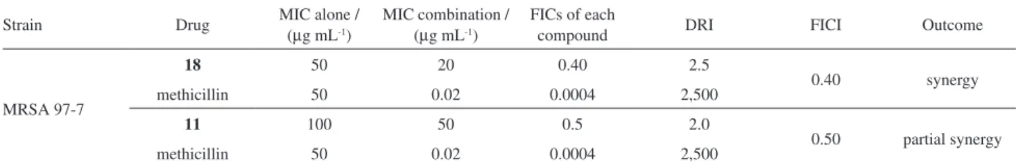
Evaluation of the ability of chalcones 11 and 18 to potentiate the antibiotic activity of methicillin
The detection of what are known as resistance-modifying agents (RMAs) represents an attractive strategy to mitigate the spread of bacterial drug resistance, since it could facilitate the recycling of well-established antibiotics.46 Drug repositioning can accelerate the drug
development process as the already established compounds are characterized by known safety profiles, pharmacology and administration routes.47,48
Chalcones 11 and 18 showed an important capacity
to reduce the MIC of methicillin against MRSA 97-7, re-sensitizing these resistant bacteria 2,500 times methicillin
and 2.5 times the synthetic chalcone 18. Compound 18
was found to act synergistically in conjunction with the
commercial antibiotic (Table 2), while compound 11
showed only partial synergy, although the dose was reduced by the same amount as 18.
These findings add new data to the previously reported49
enhancing capacity of chalcones to antibiotics activity showing that five chalcones possessing either 2-OH or 4-OH on the B ring enhanced the antibacterial activity of the non β-lactam antibiotics doxycycline, ciprofloxacin and/or gentamicin. In the present paper, the 3,4-diOH chalcones either unsubstituted (18) or 3’,4’,5’-trimethoxylated (11)
on the A ring, showed potentiated activity of the β-lactam antibiotic methicillin.
In vitro antifungal effects on fungi pathogenic to humans
In the last decades, fungi have emerged as major causes of human morbidity and mortality, mainly among the immunocompromised and seriously-ill hospitalized
patients.50 Most of these mycoses-related deaths were
associated with Candida albicans (C. albicans) and
Cryptococcusneoformans (C.neoformans) and, although
there are several antifungal drugs in clinical use to treat both opportunistic fungal species, fungal infections remain very difficult to eradicate.
Since chalcones with different substituents than the ones synthesized here have shown antifungal activities in several previous papers,51-58 the antifungal properties
of these compounds could add new interesting data on the antifungal behavior of chalcones. Thus, the nine chalcones 11-19 were tested for antifungal properties
against the clinically important yeasts C. albicans
(ATCC 10231) and C. neoformans (ATCC 32264). To
assess antifungal activities, the broth microdilution method M27-A3 for yeasts of the Clinical and Laboratory Standards Institute (CLSI)59 was used.
The percentage of inhibition of each fungus was determined for each compound at two-fold dilutions in the range 250-3.9 µg mL-1 (Table 3). In the right column, IC
50
represents the minimum concentration that inhibits fungal growth by 50%. This value is accepted in the literature as representative of the in vitro activity of target compounds.60
Although Table 3 shows that all compounds inhibit both C. albicans and C. neoformans at different tested
concentrations, it is interesting to note that C. albicans
is more sensitive than C. neoformans. In Table 3, it
can be observed that IC50 of all compounds against
C. albicans ranged from 125 to > 250 µg mL-1, with
five compounds (11, 12, 14, 16 and 19) possessing an
IC50≥ 250 µg mL-1, one (13) with IC50 = 250 µg mL-1, two
(15 and 18) displaying IC50 = 125 µg mL-1 and one (17)
with IC50 = 50 µg mL-1. In the case of C. neoformans the
IC50 ranged from 7.8 to > 250 µg mL-1 with two compounds
(15 and 17) displaying an IC50 between 7.8-15.6 µg mL-1,
Table2. Synergistic effects of chalcones 11 and 18 with methicillin in MRSA 97-7
Strain Drug MIC alone / (
µg mL-1)
MIC combination / (µg mL-1)
FICs of each
compound DRI FICI Outcome
MRSA 97-7
18 50 20 0.40 2.5
0.40 synergy
methicillin 50 0.02 0.0004 2,500
11 100 50 0.5 2.0
0.50 partial synergy
methicillin 50 0.02 0.0004 2,500
one compound (16) with an IC50 = 31.25 µg mL-1, three
compounds with IC50≥ 125 µg mL-1 (12, 13 and 14) and
only two compounds (11 and 18) with IC50 > 250 µg mL-1.
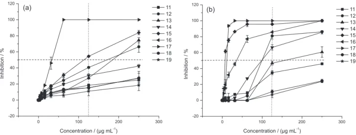
The comparative behavior of all compounds against
C. albicans and C. neoformans can be clearly corroborated
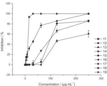
by comparing the dose-responses curves in Figure 2. Against C. albicans (Figure 2a), four (13, 15, 17 and 18)
out of the nine compounds produced an inhibition ≥ 50% at 250 µg mL-1, one (15) produces ≥ 50% inhibition at
125 µg mL-1 and one (17) at 50 µg mL-1. While, against C. neoformans, six compounds (12-17) produced an
inhibition ≥ 50% at 250 µg mL-1 and five of them (12,
14-17) produced the same inhibition at 125 µg mL-1
(Figure 2b).
To see the influence of the methylation of the 3-OH group (on the B ring) on the growth of C. neoformans, the
anticryptococcal activity of 18 (with 3,4-diOH on the B ring)
was compared with 17 (with 3-OMe, 4-OH on the B ring), Table3. Percentages of inhibition of chalcones 11-19 against C. albicans ATCC 10231 (Ca) and C. neoformans ATCC 32264 (Cn) at different concentrations. The compounds were ordered by structural similarity in order to facilitate the structure-activity analysis and not in numerical order
Compound Structure Fungi Concentration / (µg mL
-1)
250 125 62.5 31.2 15.6 7.8 3.9 IC50a
18 Ca
Cn
66.3 ± 1.9 24.4 ± 2.1
40.6 ± 0.0 10.9 ± 2.0
24.4 ± 0.1 0
17.8 ± 0.2 0
11.1 ± 1.9 0
8.1 ± 0.7 0
3.6 ± 0.2 0
125
> 250
17 Ca Cn 100 100 100 100 100 100
45.9 ± 1.5 100
18.2 ± 0.6 93.5 ± 0.1
8.8 ± 0.0 15.4 ± 0.1
5.2 ± 0.5 8.6 ± 0.4
50 15.6
11 Ca
Cn
25.0 ± 2.8 45.9 ± 0.2
18.4 ± 1.6 34.7 ± 2.7
12.6 ± 0.8 9.1 ± 0.2
8.4 ± 0.4 7.6 ± 0.5
5.1 ± 1.0 3.3 ± 0.5
3.4 ± 0.2 2.8 ± 0.1
2.3 ± 0.9 1.7 ± 0.5
250 250
13 Ca
Cn
74.6 ± 3.3 60.6 ± 1.6
27.51 ± 1.35 46.6 ± 0.4
10.4 ± 1.0 0
6.7 ± 0.3 0
3.8 ± 0.7 0
2.4 ± 1.2 0
1.8 ± 0.1 0
250 125
15 Ca
Cn
84.0 ± 2.7 100
54.5 ± 0.0 95.6 ± 0.8
31.0 ± 2.3 95.5 ± 2.9
17.0 ± 2.0 86.3 ± 0.5
8.0 ± 0.2 75.3 ± 1.5
3.7 ± 0.0 61.5 ± 1.0
0 0 125 7.8 12 Ca Cn
27.4 ± 8.3 86.0 ± 0.0
15.1 ± 2.1 80.9 ± 1.9
13.5 ± 0.3 5.5 ± 0.6
9.4 ± 1.1 0.00
4.9 ± 0.9 0.00
3.4 ± 0.3 0.00
2.3 ± 0.3 0.00
> 250 125
14 Ca
Cn
42.3 ± 2.1 85.6 ± 2.1
30.7 ± 1.9 66.5 ± 1.3
19.6 ± 1.5 27.4 ± 0.0
13.5 ± 1.2 3.2 ± 0.1
8.5 ± 1.2 0
6.0 ± 0.9 0
3.6 ± 0.0 0
> 250 125
16 Ca
Cn
27.1 ± 1.3 100
15.3 ± 0.9 85.8 ± 0.0
12.1 ± 0.6 77.1 ± 1.3
8.2 ± 0.7 45.5 ± 0.1
6.2 ± 0.1 32.0 ± 0.5
2.5 ± 0.8 19.9 ± 0.0
0.2 ± 0.5 14.8 ± 0.2
> 250 31.25
19 Ca
Cn
18.4 ± 1.6 23.7 ± 0.5
11.0 ± 0.8 7.1 ± 3.7
5.9 ± 0.2 0
5.1 ± 0.2 0
2.6 ± 0.0 0
1.8 ± 0.3 0
1.6 ± 0.1 0
> 250
> 250
Amphotericin B Ca
Cn 100 100 100 100 100 100 100 100 100 100 100 100 100 100 1.5 1.0 aIC
both with an un-substituted ring A (Figure 3a). With the same purpose, the anticryptococcal activity of 11 (with 3,4-diOH
on the B ring) was compared with 13 (with 3-OMe, 4-OH
on the B ring), both with three methoxyl groups on the A ring (Figure 3b). Results clearly show that the methylation of the 3-OH increased the activity in different degrees on the A ring unsubstituted and substituted chalcones. However, in the compounds with an unsubstituted A ring the difference in inhibition between 17 and 18 (Figure 3a) was much greater
than that observed between 11 and 13 (Figure 3b), showing
that the substitution of ring A could play an important role in the activity.
To see the influence of different substituents on ring A on the anticryptococcal activity, all compounds possessing 3-OMe, 4-OH substituents on the B ring (12-14, 16, 17)
were compared in Figure 4.
From Figure 4, it is clear that the chalcone with 3-OMe, 4-OH substituents and unsubstituted-ring A chalcone (17) displayed the best activity reaching the
highest inhibition (100%) at 7.8 µg mL-1. The activity
decreases in the following order: 17 (unsubstituted-ring
A) > 16 (3’,4’-(MeO)2 A ring) > 12 (2’5’-(MeO)2 A ring)
> 14 (2’,4’-(MeO)2 A ring) > 13 (3’,4’,5’-(MeO)3 A ring).
In addition, the (MeO)2 derivatives possessing one of
the methoxyl groups at position 2’ (12 and 14) displayed
lower activities than 16 (3’,4’-(MeO)2 derivative), probably
due to steric hindrance of the OMe at position 2’.
Our results showedthat these chalcones were more active against C. neoformans than against C. albicans. Regarding
the relation between structure and anticryptococcal activity, the methylation of the 3-OH of the B-ring led to an improved activity. In turn, the pattern of substitution of
Figure2. Comparative antifungal activities of chalcones 11-19 against (a) C. albicans; (b) C. neoformans. Thehorizontaldotted line was drawn to clearly see the number of compounds that displayed ≥ 50% inhibition of fungal growth. The vertical dotted lines were drawn to clearly see the effects of the different compounds at 125 µg mL-1.
Figure3. Influence of the methylation of the 3-OH group on ring B on growth of C. neoformans, when ring A is (a) unsubstituted (chalcones 17 and 18)
the A ring appears to play some role in the anticryptococcal activity.
In vitro antibacterial effects on bacteria pathogenic to plants
Results of the antibacterial assays (Table 4) indicate an outstanding antibacterial activity of compound 12 on
the growth of P. syringae with a low IC50 value (8 µM
or 2.5 µg mL-1). This effect is similar to that observed
with streptomycin whose IC50 value is close to 5 µM
(2.9 µg mL-1). Although the best inhibitory effect was
observed in the previous case, the remaining compounds of the list do not show activity or show a moderate effect (i.e.,
19 shows an IC50 =191 µM and 17, IC50 = 343 µM) only
against P. syringae. The other two phytopathogenic bacteria
were less sensitive to the compounds. However, some sensitivity was observed in the presence of compound 16
for A. tumefaciens, and in the presence of the compound 19
against E. carotovora and P. syringae.
Chalcones 19 and 12 showed an interesting effect on P. syringae that is a Gram-negative bacterium, which is
classified as pathovars according to its host specificity. Infection with this pathogen produces important economic losses that may affect seriously the net production as well as the quality of the product obtained.
In vitro fungicidal effects on fungi pathogenic to plants
The results of the antifungal activity assay using phytopathogens (Table 5) indicate a high sensitivity to the chalcones, with growth inhibition percentages higher than 51%, at all concentrations tested. Although the positive controls, based on commercial antifungal agents (pure active ingredients), showed strong inhibition (100%) at all concentrations tested in the assays, some chalcones showed inhibition values similar or slightly lower on the
growth of the oomycete P. cinnamomi (for the case of
compound 15) and G. fujikuroi (for the case of compounds 14, 15 and 17). This difference was statistically significant
with respect to the negative control. The foregoing indicates that this type of compound may have great potential for
the treatment of these pathogens. Compound 15 was the
most active of this series inhibiting the growth of the two fungi (B. cinerea and G. fujikuroi) and the oomycete
(P. cinnamomi). P. cinnamomi was the pathogen most
sensitive to 15, registering 100% growth inhibition with
the lowest concentration tested (250 µM), achieving effects similar to those observed with metalaxyl.
Additionally, an interesting sensitivity of the fungus
G. fujikuroi was observed compared to the complete series
of chalcones, with percentages of inhibition higher than 75.8%.
In vitro 5-hLOX enzyme assay
Three chalcones displayed an interesting activity as human 5-hLOX inhibitors (Table 6). The analysis of the relationship between structure and activity showed that the most potent inhibitors 11, 18, and 19 possess free hydroxyl
groups on the A ring (19) or catechol groups on the B ring
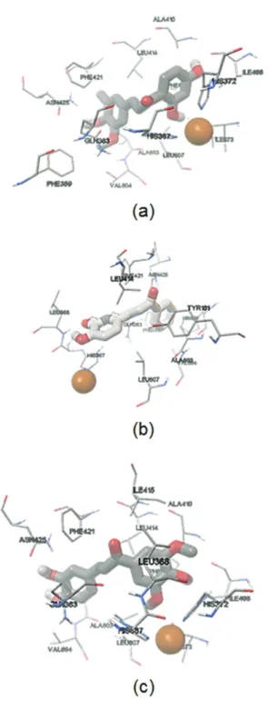
(11 and 18) in the structure. The docking studies (Figure 5)
revealed that the catechol group present in chalcone 18 was
oriented towards the metal, which agrees with a competitive mechanism found in a kinetics experiment (Figure 6). Compound 11, which was the best inhibitor found, presented
a mixed mechanism and was unable to orient the catechol
Figure4. Comparative anticryptococcal activity of 3-OMe, 4-OH chalcones with different substituents on ring A [unsubstituted (17); 3’,4’,5’-(OMe)3 (13); 2’,4’-(OMe)2 (14), 2’,5’-(OMe)2 (12), 3’,4’-(OMe)2 (16)].
Table4. IC50 values of chalcones towards phytopathogenic bacteria
Compound IC50 / µM
A. tumefaciens E. carotovora P. syringae
11 > 500 500 457
12 > 500 > 500 8
13 > 500 > 500 > 500
14 > 500 > 500 > 500
15 > 500 > 500 > 500
16 392 > 500 494
17 > 500 > 500 500
18 > 500 > 500 343
19 500 442 191
group towards the iron, generating an interaction between Asn425 and this group. Compound 19, that showed the worst
IC50 value and had a competitive mechanism, only oriented
a hydroxyl group towards the metal.
It is known that flavonoids containing ortho-diOH
groups have inhibitory activities against LOXs,61,62 although
it is not the main requirement. We show that there are differences in the type and potency of the inhibition between 11 and 18, so the presence of a catechol would
not be an absolute structural requirement for this activity.
Moreover, the most active compound (11) has three
additional methoxy groups in the 3’,4’,5’ positions.
DPPH radical scavenging activity
The results obtained from the antioxidant activity assays are shown in Table 7. All the compounds were
compared with Trolox. Compounds 11 and 18 showed
more potent antioxidant activity than Trolox. The presence of the catechol group resulted in a significant increase of the antioxidant activity with respect to the compounds
without this group. Hydroxy groups in meta positions
(i.e., 19) and methoxy groups in meta and ortho positions
of the A ring (i.e., 14 and 16) and methoxy groups in
position 3 of the B ring (i.e., 13 and 17) decrease the
antioxidant activity. Hydroxy and methoxy groups on the B and A rings, respectively, increase the antioxidant activity.
In vitro cytotoxic activity of 18
One of the most effective synthesized chalcone as antioxidant and 5-hLOX inhibitory capacity was 18. Given
that both biological activities are related to proliferation
Table5. Percentage of growth inhibition of phytopathogenic fungi
Compound
Mycelial growth inhibition of fungal plant pathogens / %
P. cinnamomi B. cinerea G. fujikuroi
250 µM 500 µM 1000 µM 250 µM 500 µM 1000 µM 250 µM 500 µM 1000 µM
11 60.14 ± 1.26 66.67 ± 1.26 68.84 ± 1.26 56.65 ± 0.97 66.28 ± 0.63 55.96 ± 0.0 77.11 ± 1.45 83.28 ± 1.21 85.30 ± 2.35
12 64.13 ± 1.09 67.75 ± 1.66 73.55 ± 0.63 51.83 ± 1.95 69.27 ± 0.79 50.46 ± 0.0 79.54 ± 1.37 81.09 ± 0.65 83.29 ± 0.61
13 61.23 ± 0.63 64.13 ± 1.09 69.20 ± 2.26 60.09 ± 1.95 67.66 ± 0.97 71.56 ± 0.79 78.36 ± 0.69 81.39 ± 2.91 83.89 ± 3.23
14 69.57 ± 1.09 82.97 ± 0.77 100 ± 0.0 50.92 ± 0.79 72.94 ± 0.79 100 ± 0.0 84.64 ± 2.04 87.58 ± 0.91 85.96 ± 2.19
15 100 ± 0.0 100 ± 0.0 100 ± 0.0 69.27 ± 0.79 72.02 ± 0.79 76.15 ± 0.79 85.02 ± 0.57 86.97 ± 0.54 88.20 ± 0.52
16 65.58 ± 1.66 74.64 ± 0.63 81.16 ± 1.26 60.09 ± 1.95 53.26 ± 0.07 73.39 ± 1.59 77.11 ± 1.45 82.21 ± 0.63 84.34 ± 0.59
17 63.77 ± 1.26 73.55 ± 0.63 75.36 ± 1.26 57.34 ± 1.95 74.77 ± 0.79 74.77 ± 5.55 89.06 ± 0.98 91.38 ± 0.54 82.44 ± 3.65
18 59.06 ± 1.66 67.03 ± 1.66 74.28 ± 1.66 51.38 ± 1.59 59.63 ± 1.56 67.89 ± 1.59 79.92 ± 1.79 80.71 ± 0.65 81.46 ± 1.22
19 60.51 ± 0.63 67.39 ± 2.17 76.09 ± 2.17 54.13 ± 1.59 57.80 ± 1.56 74,31 ± 1.59 75.83 ± 1.94 81.46 ± 1.12 85.02 ± 0,57
C+ 100 ± 0.0a 100 ± 0.0a 100 ± 0.0a 100 ± 0.0b 100 ± 0.0b 100 ± 0.0b 100 ± 0.0b 100 ± 0.0b 100 ± 0.0b
C– 0 0 0 0 0 0 0 0 0
The percentage of mycelial growth inhibition is based on mycelium area measurements after 72 h of incubation. Each point represents the mean of three independent experiments. C+: positive control (ametalaxyl; bcaptan); C–: negative control (stock solution 1% ethanol/water).
Table6. Results of 5-hLOX activity assays in presence of chalcones 11-19
Compound Inhibitiona / % IC
50 / µM Ki / µM Type
16 W.A N.D N.D N.D
17 W.A N.D N.D N.D
18 88.36 ± 2.18 0.023 ± 0.004 0.01592 competitive
19 58.60 ± 5.42 0.707 ± 0.037 1.962 competitive
11 96.30 ± 1.47 0.011 ± 0.004 0.006326 mixed
12 W.A N.D N.D N.D
13 W.A N.D N.D N.D
14 41.35 ± 4.33 N.D N.D N.D
15 11.67 ± 13.13 N.D N.D N.D
NDGA 93.42 ± 5.87 N.D N.D N.D
aChalcone at 10 µM; IC
and viability of cancer cells, we measured 18-induced
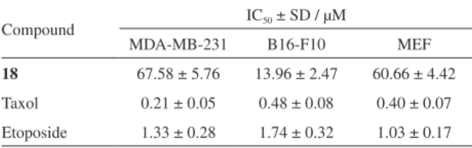
cytotoxic activity against MDA-MB-231 (human breast adenocarcinoma cell line), B16-F10 (mouse melanoma cells) and MEF (primary mouse embryonic fibroblast). Our results in the Table 8 show that 18 exhibits a 4-fold
higher selectivity upon B16-F10 compared to the effect on non-cancerous cells (MEF). 18 also exhibited
Figure6. The Lineweaver-Burk plot of the inhibition of 5-hLOX by three
chalcones (11, 18 and 19).
Figure5. Docking results for chalcones with 5-hLOX. (a) 19, with a
∆Gb = –3.68 kcal mol-1 and an experimental ki = 1.96 µM; (b) 18, with a ∆Gb = –4.9 kcal mol-1 and an experimental ki = 0.016 µM; (c) 11, with a ∆Gb = –4.25 kcal mol-1 and an experimental ki = 0.06 µM. The orange ball represents the iron atom, located in the active site.
Table 7. Screening results of DPPH radical scavenging activity of
chalcones 11-19
Compound IC50 ± SD / µM
11 11.75 ± 0.21
12 33.50 ± 1.52
13 49.04 ± 1.25
14 54.40 ± 5.82
15 NA
16 73.03 ± 7.53
17 104.26 ± 5.44
18 12.45 ± 0.39
19 NA
Trolox 22.54 ± 0.61
cytotoxic activity against MDA-MB-231, but with no
selectivity over MEF. When the cytotoxic effect of 18
against B16-F10 was compared with commonly used
chemotherapeutic drugs, it could be verified that 18
showed 29- and 8-fold lesser activity than taxol and etoposide, respectively. Furthermore, 18 exhibited 300-
and 50-fold lesser cytotoxic activity against MDA-MB231 cell line. It is noteworthy that 18 exhibited antioxidant
activity and 5-LOX inhibition at lower concentrations than the needed to exert cytotoxic activity.
Conclusions
Apparently a 3,4-diOH substitution on the chalcone skeleton plays a key role in the potentiation capacity of the β-lactam antibiotic methicillin. For this reason, the chalcone
18 was subjected to cytotoxicity studies reinforcing a
possible use in the control of resistant pathogens in vivo.
These studies showed a low cytotoxicity against MEF cell line. This clearly suggests that 18 could be useful for the
treatment against resistant bacteria, although further studies are needed to confirm this possibility. Although chalcones
with 3,4-O-di-hydroxy groups are sometimes considered
interferers of all assays,63 it appears not to be applied here
because the most active chalcones against phytopathogens and human pathogenic fungi (12, 15 and 17) do not have
this substitution pattern.
In both cases, bacteria and fungi, the sensitivity for each molecule can vary, not only by cellular structural characteristics such as wall and plasma membrane, but also by differences in their mechanisms of tolerance to stress and detoxification.64
Due to the versatility of biological properties showed here to this kind of compounds, simple and safe polyoxygenated chalcones not only have potential uses in different biological areas, whose specificity or action target depends largely on their functional groups, but also they would help us to fight resistant pathogens to commercial antibiotics through the specific re-sensitization.
Experimental
Chemistry
General data
All purchased chemical reagents (Merck or Sigma-Aldrich) were of the highest available purity and were used without previous purification. Melting points (mp in °C) were measured on a Stuart-Scientific SMP3 melting point apparatus (Stone, United Kingdom) and are uncorrected.
Infrared (IR) spectra were recorded as a thin film (14
and 15) or in KBr disks (11, 12, 13, 16, 17, 18 and 19)
in a Thermo Scientific Nicolet 6700 FT-IR spectrometer (Massachusetts, USA), and wave numbers are reported in cm-1. Low resolution mass spectra (MS) were recorded on a
Thermo Scientific, Trace GC Ultra, ISQ mass spectrometer (Austin, USA) at 70 eV ionizing voltage and are given as
m/z (rel. int., in percentage). High-resolution mass spectra
(HRMS) were recorded on an ExactiveTM Plus Orbitrap
spectrometer (Thermo Scientific, Bremen, Germany) by infusion, applying a voltage of 5 kV in the positive ionization mode. The spectra were recorded using full scan mode, covering a mass range from m/z 140 to 430. The resolution
was set to 140000, and maximum loading time for the ion cyclotron resonance (ICR) cell was set to 200 ms. 1H and
13C nuclear magnetic resonance (NMR), 2D heteronuclear
single quantum correlation (HSQC) and 2D heteronuclear multiple bond correlation (HMBC) spectra were recorded in CDCl3 or DMSO-d6 solutions and referenced to the residual
solvent peaks at d 7.26 and 2.50 ppm for 1H and d 77.0
and 39.5 ppm for 13C, respectively, on a Bruker Avance
400 NMR spectrometer (Rheinstetten, Germany) operating
at 400.1 MHz for 1H and 100.6 MHz for 13C. Chemical
shifts are reported as d (ppm), and coupling constants (J) are
given in Hz. Silica gel (200-400 mesh, Merck, Darmstadt, Germany) was used for column chromatography and HF-254 silica gel plates for thin layer chromatography (TLC). TLC spots were detected both under a UV lamp and by heating after drenching in 10% H2SO4 in H2O. Antioxidant effects
were determined in a Thermo Scientific Multiskan GO 96-well plate photometer (Vantaa, Finland).
General experimental procedure
General procedure in alkaline condition
To a solution of acetophenone (1 mol equiv.) and appropriate benzaldehyde (1 mol equiv.) in methanol (5 mL), KOH (4 mol equiv.) was added in one portion. The mixture was warmed to 50 °C and stirred until completion of reaction (monitored by TLC). Then the solvent was removed under reduced pressure, poured into ice water and
Table8. Cytotoxic activities of 18 against cancer and non-cancerous cells
Compound IC50 ± SD / µM
MDA-MB-231 B16-F10 MEF
18 67.58 ± 5.76 13.96 ± 2.47 60.66 ± 4.42
neutralized with aqueous 10% HCl solution. The residue was extracted with ethyl acetate (15 mL × 3 times). The combined organic layer was washed with brine, dried over anhydrous sodium sulfate and filtered; the solvent was evaporated under reduced pressure. The mixture was subjected to silica gel flash column chromatography (ethyl acetate/hexanes as the mobile phase) to yield the products. The fractions of the product were dried and recrystallized from ethanol to obtain pure chalcones.
General procedure in acid condition
To a stirred mixture of acetophenone (1 mol equiv.) and benzaldehyde (1 mol equiv) in absolute ethanol (5 mL), thionyl chloride (0.5 mL, 3 mol equiv.) was added dropwise and the reaction was monitored by TLC. After standing for 16 h, ice water (30 g approximately) was added to the reaction mixture in order to decompose the excess of SOCl2. Then, ethyl acetate (30 mL) was added
and the organic layer was separated, and a new extraction with ethyl acetate was performed. The pooled organic solutions were dried over anhydrous sodium sulfate and filtered; the solvent was evaporated under reduced pressure. The mixture was subjected to silica gel flash column chromatography (ethyl acetate/hexanes as the mobile phase) to yield the products. Solid chalcones were recrystallized from ethanol.
All structures were confirmed by IR and NMR spectra as discussed below.
Physical data of chalcones
1-(3,4,5-Trimethoxyphenyl)-3-(3,4-dihydroxyphenyl)-prop-2-en-1-one (11)
It was obtained from 1-(3’,4’,5’-trimethoxyphenyl) ethanone (1, 0.5 g, 2.4 mmol) and 3,4-dihydroxybenzaldehyde
(7, 0.327 g, 2.4 mmol) in 5 mL of ethanol and 0.5 mL of
SOCl2 (0.82 g, 6.9 mmol), stirring for 16 h as described above.
mp 151-153 °C (lit.65 158-159 °C); IR (KBr) ν / cm-1 3331,
2942, 1645, 1586, 1558; HRMS m/z, observed: 331.1178;
C18H19O6 [M + H]+ requires: 331.1182; 1H NMR (400 MHz,
CDCl3) d 7.75 (d, 1H, J 15.5 Hz, 2-CH=3-CH), 7.32 (d,
1H, J 15.5 Hz, 2-CH=3-CH), 7.27 (s, 1H, 3-ArH-2), 7.25
(s, 2H, 1-ArH-2,6), 7.14 (dd, 1H, J 8.2, 1.8 Hz, 3-ArH-6),
6.94 (d, 1H, J 8.2 Hz, 3-ArH-5), 6.81 (s, 1H, ArOH-3),
6.59 (s, 1H, ArOH-4), 3.96 (s, 3H, 1-ArOCH3-4), 3.94 (s,
6H, 1-ArOCH3-3,5); 13C NMR (100 MHz, CDCl
3) d 56.4
(1-ArOCH3-3,5), 61.0 (1-ArOCH3-4), 106.1 (1-ArCH-2,6),
115.0 (3-ArC-2), 115.6 (3-ArCH-5), 119.3 (2-CH=3-CH),
122.8 (3-ArCH-6), 127.7 (3-ArC-1), 133.5 (1-ArC-1), 142.5
(1-ArC-4), 144.2 (3-ArC-3), 145.9 (2-CH=3-CH), 147.1
(3-ArC-4), 153.1 (1-ArC-3,5), 190.3 (1-C=O).
1-(2,5-Dimethoxyphenyl)-3-(4-hydroxy-3-methoxyphenyl)-prop-2-en-1-one (12)
It was obtained from 1-(2,5-dimethoxyphenyl)
ethanone (2, 0.5 g, 2.78 mmol) and
4-hydroxy-3-methoxybenzaldehyde (8, 0.426 g, 2.8 mmol) in 10 mL
of methanol and KOH (0.83 g, 14.8 mmol), stirring for 2 h as described above. mp 96-98 °C; IR (KBr) ν / cm-1
3391, 3067, 3002, 2941, 2836, 1650, 1577, 1513; MS
m/z (%): 314 (100), 299 (25), 283 (19), 177 (95), 137
(83); HRMS m/z, observed: 315.1227; C18H19O5 [M + H]+
requires: 315.1232; 1H NMR (400 MHz, CDCl
3) d 7.55
(d, 1H, J 15.8 Hz, 2-CH=3-CH), 7.24 (d, 1H, J 15.8 Hz,
2-CH=3-CH), 7.18-7.16 (m, 2H, 1-ArH-6’, 3-ArH-6), 7.10
(d, 1H, J 1.6 Hz, 3-ArH-2), 7.04 (dd, 1H, J 8.9, 3.1 Hz,
1-ArH-4), 6.97-6.94 (m, 2H, 1-ArH-3, 3-ArH-5), 5.96
(s, 1H, 3-ArOH-4), 3.95 (s, 3H, ArOCH3), 3.87 (s, 3H,
ArOCH3), 3.83 (s, 3H, ArOCH3); 13C NMR (100 MHz,
CDCl3) d 55.8 (ArOCH3), 55.9 (ArOCH3), 56.6 (ArOCH3),
110.0 (3-ArCH-2), 113.4 (1-ArCH-3’), 114.4 (3-ArCH-6),
114.8 (3-ArCH-5), 118.6 (1-ArCH-4), 123.2 (1-ArCH-6),
124.7 (2-CH=3-CH), 127.6 (1-ArC-1), 130.0 (3-ArC-1),
144.1 (2-CH=3-CH), 146.7 (3-ArC-3), 148.1 (3-ArC-4),
152.3 (1-ArC-2), 153.6 (1-ArC-5), 192.8 (1-C=O).
1-(3,4,5-Trimethoxyphenyl)-3-(4-hydroxy-3-methoxy-phenyl)-prop-2-en-1-one (13)
It was obtained from 1-(3’,4’,5’-trimethoxyphenyl)
ethanone (1, 0.5 g, 2.4 mmol) and
4-hydroxy-3-methoxybenzaldehyde (8, 0.361 g, 2.4 mmol) in 5 mL
of ethanol and 0.5 mL of SOCl2 (0.82 g, 6.9 mmol), stirring
for 16 h as described above. mp 88-91 °C (lit.45 75-76 °C);
IR (KBr) ν / cm-1 3512, 3429, 3086, 2994, 2963, 2941,
2839, 1647, 1586, 1561, 1516; MS m/z (%): 344 (100), 329
(65), 313 (29), 145 (28); 1H NMR (400 MHz, CDCl 3) d 7.75
(d, 1H, J 15.6 Hz, 2-CH=3-CH), 7.32 (d, 1H, J 15.6 Hz,
2-CH=3-CH), 7.30 (s, 2H, 1-ArH-2,6), 7.25 (dd, 1H,
J 9.6, 1.5 Hz, 3-ArH-6), 7.12 (d, 1H, J 1.5 Hz, 3-ArH-2),
6.97 (d, 1H, J 8.2 Hz, 3-ArH-5), 5.97 (s, 1H, 3-Ar-OH-4),
3.96 (s, 3H, 3-ArOCH3-3), 3.95 (s, 6H, 1-ArOCH3-3,5),
3.94 (s, 3H, 1-ArOCH3-4); 13C NMR (100 MHz, CDCl
3)
d 56.0 (ArOCH3), 56.4 (1-ArOCH3-3,5), 61.0 (ArOCH3),
106.1 (1-ArCH-2,6), 110.4 (3-ArCH-2), 114.9 (3-ArCH-5),
119.6 (2-CH=3-CH), 123.0 (3-ArCH-6), 127.5 (3-ArC-1),
133.8 (1-ArC-1), 142.3 (1-ArC-4), 145.2 (2-CH=3-CH),
146.8 (3-ArC-3), 148.3 (3-ArC-4), 153.1 (1-ArC-3,5),
189.5 (1-C=O).
1-(2,4-Dimethoxyphenyl)-3-(4-hydroxy-3-methoxyphenyl)-prop-2-en-1-one (14)
(8, 0.93 g, 6.1 mmol) in 5 mL of ethanol and 0.81 mL of
SOCl2 (1.33 g, 11.1 mmol), stirring for 1 h as described
above. Pale yellow oil (lit.66 sticky-solid); IR (film) ν / cm-1
3370, 2940, 2840, 1648, 1601, 1512; MS m/z (%): 314
(100), 299 (49), 178 (90), 165 (98), 137 (70); 1H NMR
(400 MHz, CDCl3) d 7.71 (d, 1H, J 8.7 Hz, 1-ArH-6), 7.60
(d, 1H, J 15.7 Hz, 2-CH=3-CH), 7.33 (d, 1H, J 15.7 Hz,
2-CH=3-CH), 7.15 (dd, 1H, J 8.2, 1.6 Hz, 3-ArH-6),
7.06 (d, 1H, J 1.6 Hz, 3-ArH-2), 6.92 (d, 1H, J 8.2 Hz,
3-ArH-5), 6.54 (dd, 1H, J 8.6, 2.1 Hz, 1-ArH-5), 6.48 (d,
1H, J 2.1 Hz, 1-ArH-3), 6.22 (s, 1H, 3-ArOH-4), 3.90 (s,
3H, 3-ArOCH3-3), 3.88 (s, 3H, 1-ArOCH3), 3.85 (s, 3H,
1-ArOCH3); 13C NMR (100 MHz, CDCl3) d 55.5 (ArOCH3),
55.7 (ArOCH3), 55.8 (ArOCH3), 98.6 (1-ArCH-3), 105.0
(1-ArCH-5), 110.2 (3-ArCH-2), 114.8 (3-ArCH-5), 122.2
(1-ArC-1), 122.7 (3-ArCH-6), 124.8 (2-CH=3-CH), 127.8
(3-ArC-1), 132.6 (1-ArCH-6), 142.8 (2-CH=3-CH), 146.7
(3-ArC-3), 147.8 (3-ArC-4), 160.1 (1-ArC-OCH3), 163.9
(1-ArC-OCH3), 190.9 (1-C=O).
1-(2,5-Dimethoxyphenyl)-3-phenyl-prop-2-en-1-one (15)
It was obtained from 1-(2’,5’-dimethoxyphenyl)
ethanone (2, 2.0 g, 11.1 mmol) and benzaldehyde (9,
1.3 g, 12.2 mmol) in 10 mL of methanol and KOH (2.5 g, 44.4 mmol), stirring for 2 h as described above. Yellow oil (lit.67 yellow oil); IR (film) ν / cm-1 3060, 3001, 2942, 2911,
2835, 1658, 1594, 1575; MS m/z (%): 268 (100), 253 (15),
237 (25), 177 (60), 165 (70), 151 (61), 103 (65); 1H NMR
(400 MHz, CDCl3) d 7.65 (d, 1H, J 15.8 Hz, 2-CH=3-CH),
7.61-7.59 (m, 2H, 3-ArH-2,6), 7.42 (d, 1H, J 15.8 Hz,
2-CH=3-CH), 7.40-7.38 (m, 3H, 3-ArH-3,4,5), 7.19 (d, 1H, J 3.1 Hz, 1-ArH-6), 7.03 (dd, 1H, J 9.0, 3.1 Hz, 1-ArH-4),
6.94 (d, 1H, J 9.0 Hz, 1-ArH-3), 3.86 (s, 3H, ArOCH3),
3.81 (s, 3H, ArOCH3); 13C NMR (100 MHz, CDCl
3) d 55.8
(ArOCH3), 56.5 (ArOCH3), 113.4 (1-ArCH-6), 114.4
(1-ArC-3), 119.2 (2-CH=3-CH), 126.9 (1-ArCH-4), 128.4
(3-ArCH-2,6), 128.8 (3-ArCH-3,4,5), 129.6 (3-ArC-1),
130.2 (1-ArC-OCH3), 135.1 (1-ArC-OCH3), 143.2
(2-CH=3-CH), 152.6 (1-ArC-2), 153.6 (1-ArC-5), 192.4
(1-C=O).
1-(3,4-Dimethoxyphenyl)-3-(4-hydroxy-3-methoxyphenyl)-prop-2-en-1-one (16)
It was obtained from 1-(3’,4’-dimethoxyphenyl)
ethanone (4, 0.5 g, 2.8 mmol) and
4-hydroxy-3-methoxybenzaldehyde (8, 0.42 g, 2.8 mmol) in 5 mL of
ethanol and 0.41 mL of SOCl2 (0.67 g, 5.6 mmol), stirring for
2 h as described above. mp 127-128 °C (lit.68 128-129 °C);
IR (KBr) ν / cm-1 3341, 3078, 2992, 2930, 2835, 1643,
1585, 1557, 1510, 1438, 1418, 1267; MS m/z (%): 314
(100), 283 (65), 207 (90), 165 (58), 145 (39); 1H NMR
(400 MHz, CDCl3) d 7.73 (d, 1H, J 15.5 Hz, 2-CH=3-CH),
7.68 (dd, 1H, J 8.4, 1.8 Hz, 1-ArH-6), 7.62 (d, 1H,
J 1.7 Hz, 3-ArH-6), 7.40 (d, 1H, J 15.5 Hz, 2-CH=3-CH),
7.23 (dd, 1H, J 8.2, 1.6 Hz, 3-ArH-6), 7.12 (d, 1H, J 1.5 Hz,
3-ArH-2), 6.95-6.92 (m, 2H, 1-ArH-5, 3-ArH-5), 5.95
(s, 1H, 3-ArOH-4), 3.97 (m, 9H, ArOCH3); 13C NMR
(100 MHz, CDCl3) d 56.0-56.1 (ArOCH3), 109.9
(1-ArCH-5), 110.2 (3-ArCH-2), 110.8 (1-ArCH-2), 114.8
(3-ArCH-5), 119.4 (2-CH=3-CH), 122.8 (1-ArC-6), 123.0
(3-ArC-6), 127.6 (1-ArC-1), 131.5 (3-ArC-1), 144.3
(2-CH=3-CH), 146.8 (3-ArC-4), 148.1 (3-ArC-3), 149.2
(1-ArC-3), 153.1 (1-ArC-4), 188.7 (1-C=O).
1-Phenyl-3-(4-hydroxy-3-methoxyphenyl)-prop-2-en-1-one (17)
It was obtained from acetophenone (5, 0.48 g, 4 mmol)
and 4-hydroxy-3-methoxybenzaldehyde (8, 0.502 g,
3.3 mmol) in 8 mL of ethanol and 0.24 mL of SOCl2 (0.39 g,
3.3 mmol), stirring for 6 h as described above. mp 91-92 °C (lit.69 93 °C); IR (KBr) ν / cm-1 3322, 3001, 2938, 2835,
1655, 1583, 1564, 1525, 1454, 1420, 1369, 1248; MS m/z
(%): 254 (100), 237 (22), 223 (20), 165 (35), 145 (48), 124
(60); 1H NMR (400 MHz, CDCl
3) d 8.01 (d, 2H, J 7.3 Hz,
1-ArH-2,6), 7.75 (d, 1H, J 15.6 Hz, 2-CH=3-CH), 7.60-7.56
(m, 1H, 1-ArCH-4), 7.52-7.48 (m, 2H, 1-ArH-3,5), 7.38
(d, 1H, J 15.6 Hz, 2-CH=3-CH), 7.22 (dd, 1H, J 8.2,
1.4 Hz, 3-ArH-6), 7.13 (d, 1H, J 1.4 Hz, 3-ArH-2), 6.96
(d, 1H, J 8.2 Hz, 3-ArH-5), 5.97 (s, 1H, 3-ArOH-4), 3.96
(s, 3H, 3-ArOCH3-3); 13C NMR (100 MHz, CDCl3) d 56.0
(3-ArOCH3-3), 110.0 (3-ArCH-2), 114.9 (3-ArCH-5), 119.8
(2-CH=3-CH), 123.4 (3-ArCH-6), 127.5 (3-ArC-1), 128.4
(1-ArCH-3,5), 128.6 (1-ArCH-2,6), 132.6 (1-ArCH-4),
138.5 (1-ArC-1), 145.2 (2-CH=3-CH), 146.8 (3-ArC-3),
148.3 (3-ArC-4), 190.7 (1-C=O).
1-Phenyl-3-(3,4-dihydroxyphenyl)-prop-2-en-1-one (18)
It was obtained from acetophenone (5, 0.5 g, 4.2 mmol)
and 3,4-dihydroxybenzaldehyde (7, 0.58 g, 4.2 mmol) in
5 mL of ethanol and 0.31 mL of SOCl2 (0.5 g, 4.2 mmol),
stirring for 16 h as described above. mp 198-200 °C dec. (lit.70 200-201 °C); IR (KBr) ν / cm-1 3482, 3304, 1647,
1582, 1563, 1441, 1365, 1365, 1284; MS m/z (%): 240
(100), 223 (25), 163 (14), 110 (21), 77 (23); HRMS
m/z, observed: 241.0861; C15H13O3 [M + H]+ requires:
241.0865; 1H NMR (400 MHz, DMSO-d
6) d 9.42 (br
s, 2H, 3-ArOH-3,4), 8.09 (d, 2H, J 7.5 Hz, 1-ArH-3,5),
7.65-7.62 (m, 1H, 1-ArH-4), 7.60 (s, 2H, 2-CH=3-CH),
7.56-7.52 (m, 2H, 1-ArH-2,6), 7.25 (s, 1H, 3-ArH-2), 7.18
(d, 1H, J 8.1Hz, 3-ArH-6), 6.80 (d, 1H, J 8.1 Hz, 3-ArH-5);
13C NMR (100 MHz, DMSO-d
6) d 115.5 (3-ArCH-2), 115.7
(3-ArC-1), 128.3 (1-ArCH-3,5), 128.7 (1-ArCH-2,6), 132.7
(1-ArCH-4), 138.0 (1-ArC-1), 145.0 (2-CH=3-CH), 145.6
(3-ArC-3), 148.8 (3-ArC-4), 189.0 (1-C=O).
1-(2,4-Dihydroxyphenyl)-3-(3,4,5-trimethoxyphenyl)-prop-2-en-1-one (19)
It was obtained from 1-(2’,4’-dimethoxyphenyl)ethanone
(3, 0.5 g, 3.3 mmol) and 3,4,5-trimethoxybenzaldehyde
(10, 0.65 g, 3.3 mmol) in 6 mL of methanol and KOH
(2 g, 35.6 mmol), stirring for 2 h as described above. mp 181-183 °C (lit.71 188-190 °C); IR (KBr) ν / cm-1
3329, 3016, 2978, 2943, 2833, 1623, 1583, 1506, 1434,
1375, 1333, 1303; MS m/z (%): 330 (48), 194 (37), 181
(100); 1H NMR (400 MHz, DMSO-d
6) d 13.51 (s, 1H,
1-ArOH-2), 10.75 (s, 1H, 1-ArOH-4), 8.23 (d, 1H, J 9.0 Hz,
1-ArH-6), 7.91 (d, 1H, J 15.4 Hz, 2-CH=3-CH), 7.75 (d,
1H, J 15.3 Hz, 2-CH=3-CH), 7.23 (s, 2H, 3-ArCH-2,6),
6.44 (dd, 1H, J 8.8, 2.3 Hz, 1-ArH-5), 6.29 (d, 1H,
J 2.3 Hz, 1-ArH-3), 3.86 (s, 6H, 3-ArOCH3-3,5), 3.71 (s,
3H, 3-ArOCH3-4); 13C NMR (100 MHz, DMSO-d
6) d 56.1
(3-ArOCH3-3,5), 60.1 (3-ArOCH3-4), 102.6 (1-ArCH-3),
106.7 (3-ArCH-2,6), 108.2 (1-ArCH-5), 113.0 (1-ArC-1),
120.2 (2-CH=3-CH), 130.1 (3-ArC-1), 133.2 (1-ArCH-6),
139.8 (3-ArC-4), 144.2 (2-CH=3-CH), 153.1 (3-ArC-3,5),
165.2 (1-ArC-4), 165.9 (1-ArC-2), 191.5 (1-C=O).
Cytotoxicity assays
In vitro antibacterial activity assays, human pathogens
Minimum inhibitory concentration assay
Two clinical isolates of methicillin resistant S. Aureus
(622-4 and 97-7), and one clinical isolate of E. coli 33.1,
kindly donated by Dr Marcela Wilkens from Universidad de Santiago de Chile (Chile) were used, and as control strains S. aureus (NCTC8325-4) and E. coli (ATCC25922)
were used. The antimicrobial activities of chalcones against strains E. coli (ATCC25922), E. coli multi-resistant (33.1), S. aureus (NCTC8325-4) and methicillin-resistant S. aureus
97-7, were determined using the microdilution method established by CLSI.72 Briefly, stock solutions (5 mg mL-1)
of compounds in DMSO were diluted in Mueller-Hinton broth (MHB) to the different two-fold assay concentrations. The final concentration of DMSO was ≤ 2.5% and does not affect the microbial growth. The obtained solution was then added to MHB, and serially two-fold diluted (in a 96-well microplate). 100 µL of inoculum 1.5 × 10⁶ colony forming
unit (CFU) mL-1, prepared in MHB, was then added. The
plates were sealed with a tight-fitting plastic cover, then incubated at 37 ºC for 18 h. The assay was repeated three times. Wells containing MHB, 100 µL of inoculum and
DMSO (final concentration of ≤ 2.5%) served as negative controls. The MIC was defined as the lowest concentration of compounds resulting in the complete inhibition of visible growth.
Checkerboard dilution test
The type of interaction of the combination of chalcones
11 and 18 with methicillin was investigated with the
checkerboard method.73 Briefly, the uppermost row (A)
of a 96-well microtiter plate contained substance X in a concentration of about four times the expected MIC of the microorganism examined. Each following row (B-H) contained half the concentration of the previous one. The same procedure was carried out along the columns (1-12) with substance Y. So, each well contained a unique combination of the two substances (X and Y). At last 100 µL of Mueller-Hinton broth containing about
105 CFU mL-1 were added to the wells and incubated
at 37 °C for 24 h. The concentrations of the first wells without visible growth along the stepwise boundary between inhibition and growth were used to calculate the fractional inhibitory concentration index (FICI) as follows: FICI = fractional inhibitory concentration of A (FICA) +
fractional inhibitory concentration of B (FICB) = (MIC of
drug A in combination / MIC of drug A alone) + (MIC of drug B in combination / MIC of drug B alone). FICI was interpreted as follows: synergy, < 0.5; partial synergy, 0.5-0.75; additive effect, 0.76-1.0; indifference, 1.0-4.0; and antagonism, > 4.0.74
In vitro antifungal activity assays against human pathogens
Microorganisms and media
For the antifungal evaluation, standardized strains from the American Type Culture Collection (ATCC, Rockville,
MD, USA), C. albicans ATCC 10231 and C. neoformans
ATCC 32264, were used. Strains were grown on Sabouraud-chloramphenicol agar slants for 48 h at 30 °C and sub-cultured every 15 days to prevent pleomorphic transformations. Inocula were obtained according to reported procedures59 and adjusted to 1-5 × 10³ CFU mL-1.
Fungal growth inhibition
The fungal growth inhibition percentage was determined with broth microdilution techniques performed in 96-well microplates according to the CLSI reference method for broth dilution antifungal susceptibility testing of yeasts,
approved standard M27-A3.59 For the assay,
medium, to final concentrations range 250-3.9 µg mL-1.
An inoculum suspension (100 µL) was added to each well (final volume in the well: 200 µL). A growth control well (GCW) (containing medium, inoculum, and the same amount of DMSO used in a CTW, but compound-free) and a sterility control well (SCW) (sample, medium, and sterile water instead of inoculum) were included for each fungus tested. Microtiter trays were incubated in a moist, dark chamber at 30 °C for 48 h for both yeasts. Microplates were read in a VERSA Max microplate reader (Molecular Devices, Sunnyvale, CA, USA). Amphotericin B (Sigma-Aldrich, St. Louis, USA) was used as positive control. Tests were performed in triplicate. Reduction of growth for each compound concentration was calculated as follows: inhibition(%) = 100 (OD405 CTW – OD405 SCW) / (OD405
GCW – OD405 SCW), where OD405 is the optical density
at 405 nm. The concentration of each compound that
produces 50% fungal growth reduction (IC50) was taken
as the MIC endpoint.
In vitro antibacterial activity assays, phytopathogens
Liquid-dilution methods were used to evaluate the effects of the compounds on the growth of E. carotovora
(NCPPB 312), A. tumefaciens (strain C58C1) and
P. syringae (NCPPB 281). Bacteria were grown in sterile
tubes with 10 mL of Mueller-Hinton (MH) medium and incubated at 27 °C for 12 h with shaking, to produce an initial culture. The antimicrobial activity was evaluated by observing the growth response of both microorganisms in samples with different concentrations of the compounds.75-77
All assays were performed on sterile 96-well microplates with a final volume of 200 µL containing MH broth inoculated with 1 µL aliquots of bacterial suspension (105-106 CFU mL-1, initial culture) in the presence of
different concentrations of test compounds (3.9, 7.8, 15.6, 31.3, 62.5, 125 and 250 µM). MH was used as the negative control (C–), and MH with streptomycin78 was used as the
positive control (C+). The plates were incubated for 7 h at 27 °C. Bacterial growth was monitored by measuring the optical density at 595 nm every hour with a microplate reader. All tests were performed in ten repetitions for each microorganism evaluated. Bacterial growth was shown as the arithmetic mean expressed in terms of the negative control (100% growth). The lowest concentration of the compound preventing the appearance of turbidity was considered to be the MIC.
The first experiment (first kinetic assay) in which the compound was exposed to the bacterial cultures in MH was carried out over a period of 6 h. Subsequently, to determine the minimal bactericidal concentration (MBC), a second
experiment (second kinetic assay) was conducted; this experiment involved taking inoculum from the first kinetic assay and adding it to MH in a new 96-well microplate, which was then cultured for 7 h. The aim of this second culture (second kinetic assay) was to observe and quantify the ability of bacterial cultures to recover from the cytotoxic effect caused by the compounds, which is evidenced by the presence or absence of bacterial growth. This second experiment allows a more accurate conclusion to be reached: if the bacterial culture’s growth declines with respect to the culture of the first kinetic assay, then it is related to a bactericidal property. If, on the other hand, the growth continues in line with that of the culture of first kinetic assay, then it is related to a bacteriostatic property.
In vitro fungicidal activity assays, phytopathogens
A virulent isolate of B. cinerea obtained from
naturally infected grape berries79 was prepared and
maintained by placing it on potato dextrose agar (PDA)
at 5 °C. P. cinnamomi was kindly provided by the
National Institute of Agricultural Research (INIA, La Platina, Santiago, Chile). The fungus G. fujikuroi was
kindly provided by Dr B. Fraga, Natural Products and Agrobiology Institute, CSIC, Canary Islands, Spain. The agar dilution technique was used to evaluate the effect
of the chalcones on the growth of P. cinnamomi,80,81
G. fujikuroi82 and B. cinerea.83,84 Fungicidal activity assays
of these compounds were performed in microcultures by growing the fungi in sterile Petri dishes (35 mm) at a final volume of 2 mL medium containing different compound concentrations. Clarified V8 (Campbell Soup) medium containing metalaxyl85 for P. cinnamomi or PDA medium
for B. cinerea and G. fujikuroi with captan86,87 was used
as the positive control (C+), and without fungicide as the negative control (C−). The medium in each slot was then inoculated with a small block (4 mm) of clarified V8 or PDA medium containing fungal hyphae excised from the edge of an actively growing culture. The fungal growth was monitored at different times (24-72 h). To determine the growth of mycelium in the agar plate, the surface of the mycelium was measured using Sigma Scan Pro 5 software.88 The final results are expressed as percent growth
inhibition, calculated according to the control without fungicide. Each treatment was independently performed in triplicate.
Statistics
treatment and control groups. Tukey’s honest significant difference test was applied to compare the means of every treatment against the control and simultaneously establish their significance (p < 0.05). All data are presented as
mean ± standard deviation (S.D.).
In vitro 5-LOX enzyme assay
The commercially available enzyme 5-hLOX by Cayman Chemicals Inc. (Ann Arbor, MI, USA) was diluted (1:500) in the assay buffer 50 mM (4-(2-hydroxyethyl)-1-piperazineethanesulfonic acid (HEPES), 2 mM ethylenediamine tetraacetic acid (EDTA), 10 µM adenosine triphosphate (ATP) and 10 µM CaCl2 at pH 7.5) and mixed
with 10 µM of H2DCFDA dye the reaction mixture and
incubated for 15 min in the assay plate. Later, 280 µL
of buffer and 10 µM of inhibitor were added per well,
and finally incubated for 30 min at 37 °C. Finally, the reaction was started by adding a suitable concentration of arachidonic acid (0.5 µM) and the fluorescence was read in a multimode detector Synergy™ HT Multi-Mode Microplate Reader (Biotek, Bad Friedrichshall, Germany) at 480 nm excitation/520 nm emission after an incubation
time of 1 h at room temperature. The IC50 values were
obtained for analysis of oxidation of H2DCFDA dye to
the highly fluorescent 2’,7’-dichloro-fluorescein (DCF) product and obtained by using the non-linear curve-fitting program. The kinetic assays to determine the mechanism of inhibition were done with the best inhibitors, using different concentrations of the inhibitor (0-1 µM) and substrate (0.2-5 µM). Experimental conditions were carried out as described previously. All data were collected in duplicate and the assays were performed on different days to ensure reproducibility of the method. The graphics for IC50 and
kinetics studies were performed by GraphPad Prism Demo V.7.89
Docking of chalcones with human 5-LOX
The chalcone structures were built with the Molecular
Operating Environment software.90 ChelpG charges were
obtained at the B3LYP/6-31G** level theory, employing the Gaussian 09 package.91 Docking of all inhibitors into
the active site of the crystal structure of 5-hLOX (PDB code: 3O8Y, 2.39 Å resolution) was performed with the
AutoDock4 package,92 using a Lamarckian algorithm and
assuming total flexibility of the inhibitors. The grid maps were made up to 60 × 60 × 60 points, with a grid-point spacing of 0.375 Å and the Fe3+ as the center of the grid
map. The AutoTors option was used to define the ligand torsions, and the docking results were then analyzed by a
ranked cluster analysis, resulting in conformations with the highest overall binding energy (most negative Gibbs free energy binding value, –∆G).
General procedure to determine the DPPH radical scavenging activity
The radical scavenging activity of the chalcones towards the DPPH radical was measured as described,93 adapted to
a screen of 96-well plates. Briefly, stock solutions of each compound were prepared in methanol at 1 mM concentration (10 mL). Dilutions (1-200 µM) were prepared from the stock solutions. Methanol (90 µL), each dilution (150 µL), and DPPH (60 µL, Sigma-Aldrich, St. Louis, USA) in methanol (0.5 mM), resulting in a final concentration of 0.1 mM DPPH, were added in a 96-well plate. Methanol was used as the blank sample. The mixtures were left for 30 min at room temperature, and the absorbances were then measured at 517 nm. Trolox was used as the standard antioxidant. The radical scavenging activity was calculated as follows: Inhibition(%) = [(blank absorbance – sample absorbance) / blank absorbance] × 100. The mean of three IC50 (concentration causing 50% inhibition) values for each
compound was determined graphically.
In vitro cytotoxic activity of 18
The cell lines used in this work included MDA-MB-231 human breast adenocarcinoma cells, B16-F10 mouse metastatic melanoma cells and MEF primary mouse embryonic fibroblast. Cells were maintained in DMEM high glucose medium (Mediatech, Manassas, VA, USA) supplemented with 10% (MDA-MB-231 and B16-F10) or 15% (MEF) heat-inactivated fetal bovine serum (HyClone Laboratories, South Logan, USA), 100 IU mL-1 penicillin
and 100 µg mL-1 streptomycin and maintained at 37 °C
in a 5% CO2 humidified atmosphere. Cell viability was
measured using CyQuant Direct Cell Proliferation Assay Kit (Life Technologies, Grand Island, NY, USA) following the manufacturer’s instruction. Briefly, 5000 cells well-1
were seeded onto a flat-bottomed 96-well plate in 200 µL final volume. Six hours after seeding, the culture medium was replaced with the medium containing the tested compounds at concentrations ranging from 0 up to 100 µM dissolved in DMSO (0.1% final concentration) during 72 h. The concentrations used to calculate the IC50 values