Symbiotic Bacteria of Threatened Amphibians –
Implications for Disease Management and Patterns of
Decline
Joshua H. Daskin*¤, Sara C. Bell, Lin Schwarzkopf, Ross A. Alford
School of Marine and Tropical Biology, James Cook University, Townsville, Queensland, Australia
Abstract
Chytridiomycosis, caused by the fungus Batrachochytrium dendrobatidis (Bd), is a widespread disease of amphibians responsible for population declines and extinctions. Some bacteria from amphibians’ skins produce antimicrobial substances active against Bd. Supplementing populations of these cutaneous antifungal bacteria might help manage chytridiomycosis in wild amphibians. However, the activity of protective bacteria may depend upon environmental conditions. Biocontrol ofBdin nature thus requires knowledge of how environmental conditions affect their anti-Bdactivity. For example,Bd-driven amphibian declines have often occurred at temperatures belowBd’s optimum range. It is possible these declines occurred due to reduced anti-Bdactivity of bacterial symbionts at cool temperatures. Better understanding of the effects of temperature on chytridiomycosis development could also improve risk evaluation for amphibian populations yet to encounterBd. We characterized, at a range of temperatures approximating natural seasonal variation, the anti-Bd activity of bacterial symbionts from the skins of three species of rainforest tree frogs (Litoria nannotis, Litoria rheocola,and Litoria serrata). All three species declined during chytridiomycosis outbreaks in the late 1980s and early 1990s and have subsequently recovered to differing extents. We collected anti-Bdbacterial symbionts from frogs and cultured the bacteria at constant temperatures from 8uC to 33uC. Using a spectrophotometric assay, we monitored Bd growth in cell-free supernatants (CFSs) from each temperature treatment. CFSs from 11 of 24 bacteria showed reduced anti-Bdactivityin vitro when they were produced at cool temperatures similar to those encountered by the host species during population declines. Reduced anti-Bd activity of metabolites produced at low temperatures may, therefore, partially explain the association betweenBd-driven declines and cool temperatures. We show that to avoid inconsistent antifungal activity, bacteria evaluated for use in chytridiomycosis biocontrol should be tested over a range of environmental temperatures spanning those likely to be encountered in the field.
Citation:Daskin JH, Bell SC, Schwarzkopf L, Alford RA (2014) Cool Temperatures Reduce Antifungal Activity of Symbiotic Bacteria of Threatened Amphibians – Implications for Disease Management and Patterns of Decline. PLoS ONE 9(6): e100378. doi:10.1371/journal.pone.0100378
Editor:Michael Sears, Clemson University, United States of America
ReceivedNovember 11, 2013;AcceptedMay 27, 2014;PublishedJune 18, 2014
Copyright:ß2014 Daskin et al. This is an open-access article distributed under the terms of the Creative Commons Attribution License, which permits unrestricted use, distribution, and reproduction in any medium, provided the original author and source are credited.
Funding:Research funding was provided by the Australian Research Council (http://www.arc.gov.au/) Discovery Grants Understanding and managing resistance to the amphibian chytrid fungus Batrachochytrium dendrobatidis in Australian tropical rainforest frogs (DP0986537) and Understanding the
Postgraduate Fulbright Scholarship from the Australian-American Fulbright Commission (http://www.fulbright.com.au). The funders had no role in study design, data collection and analysis, decision to publish, or preparation of the manuscript.
Competing Interests:The authors have declared that no competing interests exist.
* Email: [email protected]
¤ Current address: Department of Ecology and Evolutionary Biology, Princeton University, Princeton, New Jersey, United States of America
Introduction
Emerging wildlife diseases can cause species declines and extinction [1], and disease emergence and pathogenicity may depend on environmental context [2]. Therefore, patterns of decline and disease management strategies may be understood by examining the effects of environmental context on disease emergence. One way environmental context might affect disease dynamics is by altering species interactions in the complex assemblage of microbiota inhabiting wildlife [3]. Chytridiomyco-sis, a disease caused by the fungal pathogen Batrachochytrium dendrobatidis(Bd) and responsible for rapid and extensive population declines in over 200 amphibian species since the late 1970s [4,5], serves as a model system for understanding other wildlife diseases [6]. Twenty years of research on Bd and its interaction with
amphibians and their environment can provide insight for anyone interested in the effects of diseases on the conservation of wildlife [5,7].
Currently, no effective treatments or preventative actions are available to manage chytridiomycosis in wild populations of threatened amphibians [8]. However, bioaugmentation, by supplementing populations of anti-Bd bacteria, has proved effective in laboratory trials and may be a viable option for disease management if it also provides increased protection in nature [9–11]. Previous experimental work on anti-Bd bacteria was conducted under constant laboratory conditions [11,12]. However, naturally-occurring symbiotic bacteria, including anti-biotic producers, vary in abundance and physiological activity with environmental context [13–15]. This variation can affect the success of biocontrol programs [16–18], and can have strong
tipping point between epidemic and endemic disease: amphibian chytridiomycosis as a model system" (DP130101635). JHD was supported by a U.S. ''
evolutionary and ecological impacts [3]. Bacteria chosen for probiotic use based on high levels of anti-Bd activity under constant conditions in the laboratory could have lower levels of anti-Bd activity in the more variable conditions occurring in nature. If this occurs, then choosing bacteria for use in the management of chytridiomycosis, will require knowledge of how candidate bacteria are affected by a range of environmental conditions [9].
Environmentally induced variation in the protection afforded to amphibian hosts by bacterial symbionts might also partially explain patterns of past chytridiomycosis-driven declines. In the tropics, where the impacts of chytridiomycosis have been most severe, higher elevations and cooler seasons have been associated with higher prevalences of infection, more intense infections, and more frequent declines [19–24]. Bd’s relatively cool thermal optimum (17–25uC) [25] may partially explain this pattern [21], but many declines have occurred at temperatures well below this window, where one might expect chytridiomycosis to be less severe [26].
There are a number of reasons why declines might occur at very low temperatures. In natural environments, Bd may respond to low temperatures by increasing fecundity [27], and amphibian hosts’ immune defenses may be weaker [28]. Another possible contributor to the high incidence of Bd-driven declines at temperatures belowBd’sin vitrothermal optimum is that symbiotic cutaneous bacteria, which would otherwise reduce the severity of chytridiomycosis, may have reduced activity or population density at cooler temperatures. At present, there is no published information on how the composition or antifungal activity of assemblages of anti-Bd bacteria responds to changes in environ-mental context.
To examine the effect of temperature on the production of
anti-Bdmetabolites by bacteria, we sampled bacterial symbionts from the Australian hylid frogsLitoria serrataandLitoria nannotisin 2010. We also examined bacteria isolated by Bell [29] and Bell et al. [30] from Litoria rheocola, L. serrata, and L. nannotisin 2009. All three species experienced population declines following chytridiomycosis outbreaks, although some populations have since recovered or recolonized [23,31]. We identified the bacteria and characterized their antifungal activity across a range of the temperatures experienced by their amphibian hosts.
Materials and Methods
Ethics Statement
All procedures involving animals received clearance from the James Cook University Animal Ethics Review Committee (approval number A1316) and all field sampling was permitted by the Queensland Department of Environment and Resource Management (permit WITK05922209).
Collection and Isolation of Bacteria
To screen for anti-Bdactivity, we collected bacteria from twelve frogs (six L. serrata, six L. nannotis) caught at Windin Creek in Wooroonooran National Park, Queensland, Australia (,750 m
a.s.l., S 17u219570 E 145u429540) in February 2010. We rinsed each frog with a stream of sterile distilled water to remove non-resident bacteria [32,33], then swabbed its dorsal and ventral surfaces and legs twice, using a sterile rayon swab (MW112, MWandE, Bath UK). We streaked each frog’s swab onto a low nutrient agar plate (R2A, Becton, Dickinson and Company, New Jersey, US). We used new gloves and plastic bags to catch, handle, and hold each animal to prevent disease transmission or contamination of samples. We released frogs at their point of
capture immediately after swabbing. After returning our samples to the laboratory, we isolated each morphologically-distinct bacterium into axenic (pure) culture using standard microbiolog-ical techniques, and checked Gram stains of each isolate using light microscopy to ensure purity [34]. Axenic isolates were stored on ceramic Microbank microbeads (Microbank, Pro-Lab Diagnostics U.K.). The beads are stored dry at280uC. When a live culture is required, a bead can be dropped into liquid media or streaked across solid agar.
Identification of Anti-Bd Bacteria
To determine whether bacterial isolates inhibited growth of
Bd in vitro, we performed challenge assays using a method developed by Bell et al. [30] with slight modifications described here. We inoculated bacteria from Microbank microbeads into one mL TGhL broth (eight g tryptone, one g gelatine, two g lactose per liter of water) in 24-well plates (Costar 3524, Corning, New York, US) and incubated them for 48-hours at 23uC. We then centrifuged each liquid culture for five minutes at 75006g,
and filtered the supernatant through a 0.22mm syringe filter (Millex GV, Millipore, Massachusetts, US). This left a cell-free supernatant (CFS) containing bacterial metabolites.
In 96-well assay plates (Costar 3595, Corning, New York, US), we inoculated 1.06105liveBdzoospores (isolate Gibbo River, L. Les, 06-LB-1 isolated by L. Berger in 2006 from aLitoria lesueriand passaged weekly) suspended in 50mL of fresh TGhL into each of five replicates containing 50mL of each CFS. We included replicates of positive and negative controls consisting of 1.06105 live and heat-killed Bd zoospores, respectively, suspended in 100mL TGhL. We incubated the plates at 23uC and monitored progress ofBdgrowth in each well with daily spectrophotometric readings at 492 nm [35]. We continued monitoring until maximum growth was observed in the positive control and in the majority of wells containing CFS (four days). This is the most conservative point in the assay at which to determine anti-Bd
activity; i.e., this is the point at which the ratio ofBdgrowth in CFSs to Bd growth in the positive control is maximized. We examined 96-well plates using light microscopy and excluded from our analyses any replicates that had become contaminated.
We transformed mean optical densities at 492 nm (OD492) for each isolate-temperature combination on each day, to correct for initial coloration of CFSs (by subtracting the mean initial OD492) and background absorbance of inoculated zoospores on that day (by subtracting the mean negative control OD492) [30]. Using the corrected value on the maximum growth day for both the positive control and for each CFS by temperature combination, we standardized isolate-specific values against the similarly corrected value for the positive control. This produced a measure of Bd
We extracted DNA from each isolate that strongly inhibitedBd, first by three freeze-thaw cycles of 10 minutes each at280uC and 70uC, and then, if freeze-thaw cycles alone did not yield sufficient DNA for successful PCR, using a Qiagen (Hilden, Germany) DNeasy blood and tissue kit with pretreatment for Gram-negative bacteria, as per the manufacturer’s protocol. DNA was amplified using universal bacterial 8F and 1492R primers [36], and sequenced by Macrogen, Inc. (Seoul, South Korea). We aligned forward and reverse sequences in Geneious [37] and matched to sequences in the NCBI GenBank database (http://ncbi.nlm.nih. gov) to identify bacteria. We have submitted genetic sequence data to GenBank.
Experimental Challenge Assays
To test for temperature-induced changes in bacterial anti-Bd
activity we performed additional challenge assays and quantitative analysis using bacteria identified in the initial screening assay as strongly inhibitory. In addition to the bacteria isolated from frogs sampled in February 2010, we included strongly inhibitory bacteria isolated fromLitoria nannotis, L. serrata, and L. rheocola at Windin Creek in the Austral winter of 2009 and tested using the methods described above [29]. In total, we tested 24 isolates in the experimental challenge assays, all of which inhibitedBdgrowth by more than the 63.5% threshold in initial challenge assays (Table 1). We inoculated each bacterium from a Microbank microbead into a 25 cm2flask (TPP, Trasadingen, Switzerland) containing 10 mL TGhL broth and incubated it at 23uC for at least 48 hours until growth was observed. We then added 500mL of each bacterial inoculum to one mL TGhL in each of six, 24-well plates. One plate was placed in each of six incubators set at 8, 13, 18, 23, 28, and 33uC, respectively. We chose temperatures to approxi-mate the range of conditions experienced by Litoria spp. in the Australian Wet Tropics [38].
We grew bacterial cultures for two to five days, based on the time to maximal absorbance at 492 nm (a surrogate for maximal bacterial concentration) and used these cultures to produce the CFSs included in the next round of challenge assays. Growing cultures to maximum absorbance minimized any differences among treatments that might have arisen if metabolite production was triggered by quorum sensing, a mechanism of detection and response in bacterial colonies based on population density [39]. When OD492 stopped increasing (qualitatively determined from the plateauing of absorbance values) we produced bacterial CFSs as described for the initial screening assay. We harvested each individual bacterium as it reached maximum growth; i.e, we did not wait for absorbance of all bacteria in a plate to plateau before harvesting. This method does not account for possible differences in the maximum achievable concentration of any given bacterium at different temperatures. CFSs were held at220uC until use in the challenge assays. We completed challenge assays at 23uC with five replicates inoculated from each growth treatment temperature for each bacterium’s CFS, and with 2.96104zoospores initially inoculated in each 100mL assay well. Bd was therefore not cultured at a range of temperatures; only the bacteria used to produce CFSs were. This allowed us to evaluate the effects of temperature on the bacteria independent of the well-known effects of temperature on the growth ofBd[25]. We removed from the analysis any replicates that appeared to be contaminated when observed by light microscopy. As in the initial assays, the challenge assays continued until maximum growth was achieved in the positive controls (here six days).
Statistical Analysis
Corrected and standardized absorbance data were averaged across replicates of each CFS-temperature combination. The resulting averages were not normally distributed and could not be normalized by standard transformations. Therefore, we used a Kruskal-Wallis test to examine the effects of temperature on the anti-Bd activity of CFSs from tested bacteria. We performed analyses in S-PLUS (version 8.0, Insightful Corporation, Seattle) and R 3.0 (R core team, 2013).
Results
Identification of Anti-Bd Bacteria
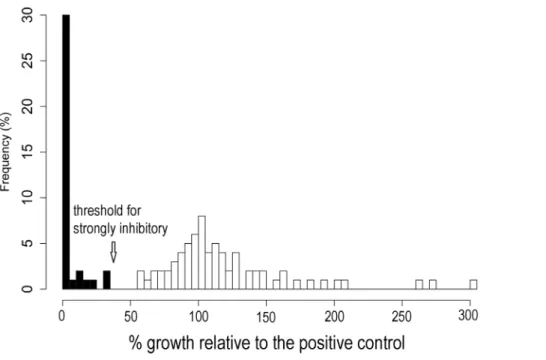
Four of 720 replicates (0.6%) were removed from the initial challenge assay due to contamination. Bd growth expressed relative to the positive control was not normally distributed among CFSs in the initial screening assay (Figure 1). Thirty-seven percent of CFSs tested at this stage inhibitedBdgrowth strongly.
Bdgrew poorly or not at all when exposed to these CFSs (Figure 2). We also observed a small number of CFSs (e.g.,Pseudomonassp. SBR3-slima, Figure 3) that apparently enhanced Bd growth, a phenomenon also reported by Bell et al. [30].
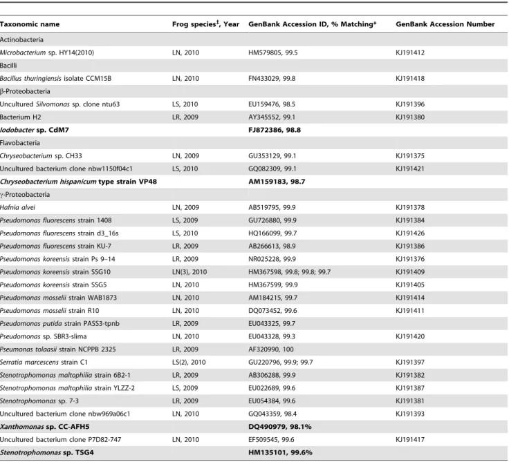
After removing duplicate isolates that came from the same individual frog and matched identical GenBank entries, 16 distinct, strongly inhibitory bacterial isolates remained from samples collected in 2010. These 16 matched the 13 operational taxonomic units (OTUs) listed for 2010 in Table 1; one OTU was found on two frogs, and one was found on three. Pseudomonads constituted six of the 13 OTUs, making them the most common group among inhibitory bacteria. The strongly inhibitory isolates collected in 2009 and included in experimental assays are also listed in Table 1.
Experimental Challenge Assay
The experimental challenge assay continued for six days, at which pointBdin the positive control and in 136 of 157 CFSs had reached its maximum growth. All the 21 remaining CFSs hadBd
growth and absorbances already well above the point at which they would have been considered inhibitory. Forty-one of 785 replicates (5.2%) became contaminated and were subsequently removed from analysis of the initial challenge assay. Bacterial responses to temperature included all of increased, decreased, and non-directional changes to CFS anti-Bdactivity (Figure 3).
The temperature at which bacteria were cultured significantly affected the antifungal activity of their CFSs (Kruskal-Wallis test, x2= 15.35, df = 5, p,0.01). Antifungal activity tended to be lowest in CFSs produced at 8uC (Figure 4). Using the cutoff from the initial screening assay of 63.5% or greater inhibition ofBdgrowth relative to the positive control, 46% of tested CFSs had no strong antifungal activity when produced at 8uC, whereas no more than 28% produced at any of the warmer temperatures lost strong antifungal activity.
Discussion
Variation in Antifungal Activity
The metabolites of many of the defensive bacterial symbionts of amphibians we tested had reduced antifungal activity when produced at 8uC. Such reductions in anti-Bdactivity were likely caused by changes in the quantity, identity, or both of bacterially-produced substances, and could have contributed to chytridiomy-cosis-driven declines that occurred in high elevation populations of the three Litoria species sampled here [23,31,40]. Winter air temperatures in the high elevation habitats of these species can often be lower than 8uC [26,41]. Therefore, frogs may experience decreased bacterial protection from Bd in winter, which in
Australia and elsewhere is when chytridiomycosis causes greater morbidity and mortality [19,23,42]. WhileBdphysiology may also be altered under variable temperature regimes and it will be important to test bacteria andBdexposed together to a range of temperatures, our design and the in vitroassay we used did not allow for testing such effects. Nonetheless, our experiment is an incremental step towards understanding context-dependency in amphibian-Bd-bacteria interactions.
Anti-Bdbacteria occur on a wide geographic and phylogenetic range of amphibians [12,43,44], and the pattern that chytridio-mycosis is more virulent at cool temperatures is also widespread [19–21,23]. Many bacteria can alter their rates of antibiotic production in response to environmental temperatures [17,45,46]. Table 1.Bacteria identified as strongly inhibitory ofBatrachochytrium dendrobatidisand used in the experimental challenge assay.
Taxonomic name Frog species`
, Year GenBank Accession ID, % Matching* GenBank Accession Number
Actinobacteria
Microbacteriumsp. HY14(2010) LN, 2010 HM579805, 99.5 KJ191412
Bacilli
Bacillus thuringiensisisolate CCM15B LN, 2010 FN433029, 99.8 KJ191418
b-Proteobacteria
UnculturedSilvomonassp. clone ntu63 LS, 2010 EU159476, 98.5 KJ191396
Bacterium H2 LR, 2009 AY345552, 99.1 KJ191380
Iodobactersp. CdM7 FJ872386, 98.8
Flavobacteria
Chryseobacteriumsp. CH33 LN, 2009 GU353129, 99.1 KJ191375
Uncultured bacterium clone nbw1150f04c1 LS, 2010 GQ082309, 99.1 KJ191421
Chryseobacterium hispanicumtype strain VP48 AM159183, 98.7
c-Proteobacteria
Hafnia alvei LN, 2009 AB519795, 99.9 KJ191378
Pseudomonas fluorescensstrain 1408 LS, 2009 GU726880, 99.9 KJ191384
Pseudomonas fluorescensstrain d3_16s LS, 2010 HQ166099, 99.7 KJ191426
Pseudomonas fluorescensstrain KU-7 LR, 2009 AB266613, 98.9 KJ191386
Pseudomonas koreensisstrain Ps 9–14 LR, 2009 NR025228, 99.9 KJ191376
Pseudomonas koreensisstrain SSG10 LN(3), 2010 HM367598, 99.8; 99.8; 99.7 KJ191409
Pseudomonas koreensisstrain SSG5 LN, 2010 HM367599, 99.9 KJ191405
Pseudomonas mosseliistrain WAB1873 LN, 2010 AM184215, 99.7 KJ191414
Pseudomonas mosseliistrain R10 LN, 2010 DQ073452, 99.6 KJ191411
Pseudomonas putidastrain PASS3-tpnb LR, 2009 EU043325, 99.7
Pseudomonassp. SBR3-slima LN, 2010 EU043328, 99.3 KJ191420
Pseumonas tolaasiistrain NCPPB 2325 LR, 2009 AF320990, 100
Serratia marcescensstrain C1 LS(2), 2010 GU220796, 99.9; 99.7 KJ191397
Stenotrophomonas maltophiliastrain 6B2-1 LR, 2009 AB306288, 99.9 KJ191382
Stenotrophomonas maltophiliastrain YLZZ-2 LS, 2009 EU022689, 99.6 KJ191387
Stenotrophomonassp. 7-3 LR, 2009 EU054384, 99.6 KJ191381
Uncultured bacterium clone nbw969a06c1 LN, 2010 GQ043359, 98.4 KJ191393
Xanthomonassp. CC-AFH5 DQ490979, 98.1%
Uncultured bacterium clone P7D82-747 LN, 2010 EF509545, 99.6 KJ191417
Stenotrophomonassp. TSG4 HM135101, 99.6%
Where the closest GenBank match was an unnamed bacterium, the closest named match is included immediately below (smaller, bold text) to give the best possible sense of phylogeny.
*Where more than one inhibitory isolate most closely matched the same OTU, the percent matching is listed for both. We used the isolate with the first listed percent matching for the experimental challenge assay.
`
Like theLitoriaspp. we sampled, other amphibians may be more vulnerable to chytridiomycosis at cool temperatures if their bacterial protection fromBdis reduced. Larger scale, longitudinal studies characterizing the diversity and abundance of amphibian bacterial symbionts and their metabolites,Bdinfection loads, and chytridiomycosis severity across environmental gradients and among seasons, are required to test this hypothesis.
Although the general trend was towards decreased anti-Bd
activity when CFSs were produced at cooler temperatures, some bacteria did not show unidirectional responses to temperature (e.g., Pseudomonas sp. SBR3-slima, Figure 3); they produced metabolites without anti-Bd activity at the lowest temperature and at moderate temperatures. These complex temperature responses may have been caused by production of different antibiotics at different temperatures. Many bacteria produce more Figure 1. Frequency (%) of the bacterial isolates which produced cell-free supernatants (CFS) showing a change inBdgrowth relative to the positive control (Bdalone; 100%) for 110 isolates screened in the initial challenge assay.A bimodal pattern of activity was observed. The arrow indicates the cutoff for considering a CFS strongly inhibitory ($63.5% inhibition relative to the positive control, i.e.,#36.5% the growth of the positive control). Thus, black bars represent strongly inhibitory CFSs and the highest bar at the far left represents those isolates producing cell-free supernatants showing 100% inhibition.
doi:10.1371/journal.pone.0100378.g001
Figure 2. Example growth curves±SD of the positive control, the negative control, two inactive, and two strongly inhibitory cell-free supernatants from an initial screening assay.Higher optical density at 492 nm (y-axis, OD492) indicates greaterBdgrowth. Day 0 OD492
than one antibiotic [45], and some regulate their production through multiple genes [47] that could possess different temper-ature thresholds.
We selected the bacteria we tested in the experimental challenge assay because their supernatants were inhibitory when produced at 23uC in the initial screening assay. It is possible that a different subset of the entire sampled bacterial community could have been classed as inhibitory if the CFSs for the initial screening assay had been produced at different temperatures. One of our aims, however, was to document context-dependency in bioaugmenta-tion candidates identified as inhibitory in ‘standard’ challenge assays conducted at 23uC [12,30]. Conducting this sort of laboratory assay under ecologically-relevant conditions has been identified as a necessary step towards selecting effective bioaug-mentation strategies [9]. However, it is entirely possible that some bacteria that live on the skin of theLitoriaspecies we sampled are effectively antifungal at low temperature and were not selected because they are not inhibitory at 23uC. Equally, the possibility of density-dependent responses of Bd to CFSs, if for example zoospores can degrade some bacterial products in the CFSs, means that had we conducted the initial challenge assay using more or fewer zoospores, different bacteria may have been identified as inhibitory and subsequently included in the exper-imental challenge assay. We used slightly different concentrations of Bdzoospores in the initial and experimental challenge assays
due to temporal variation in the productivity of laboratory Bd
cultures. However, all CFSs tested in the experimental challenge assay were inoculated with the same concentration ofBdzoospores and therefore any possible density-dependent responses ofBdto CFSs could not have affected the temperature-driven effects on CFS anti-Bdactivity reported here.
To produce CFSs for the experimental challenge assay, we grew bacterial cultures to maximum absorbance in each temperature treatment. Because some bacteria may have been dying and their cells breaking apart at this stage, it is possible that these by-products, in addition to anti-fungal metabolites produced by live bacteria, could be partially responsible for observed anti-Bd
activity. If bacteria died and broke apart to a greater extent in the higher temperature treatments, it could explain some of the reduced anti-Bdactivity at 8uC. Further experiments employing chemical methods will be needed to definitively identify and evaluate the products responsible for differential anti-Bd activity of CFSs.
Our present study is the latest to find a substantial number of
environmental context (e.g.,Pseudomonassp. SBR3-slima, Figure 3). Only the most robustly antifungal isolates should be used in bioaugmentation, regardless of how common they may be.
Management Implications and Future Research
Bacterial antifungal activity observed under a narrow spectrum of laboratory conditions could be lost on exposure to variable field environments. Using antifungal bacteria with inconsistent activity in bioaugmentation efforts could cost managers time and resources, and could create the illusion that bioaugmentation is less effective than if more appropriate isolates were used. Even closely related bacteria may respond differently to environmental variations, as did the Pseudomonads in Figure 3. One produced strongly antifungal metabolites across the entire 8–33uC range, whereas at 8uC others produced metabolites with no antifungal activity.
Only a few studies have characterized the metabolic products of amphibian symbionts [50–52]. Based on our observations of varied responses of Bdto CFSs from different bacteria, and the phylogenetic range represented in addition to the many Pseudo-monads found, it is likely that a variety of different antifungal compounds were produced by the bacteria we tested. As mentioned above, future workers should seek to identify these compounds, so that it is possible to measure their concentrations on amphibian skin.
Our study focused on environmental context-dependent changes in the bacterial production of anti-Bd metabolites, but not on possible context-dependent changes in the fungus itself, or in the direct interaction between bacteria andBdon the skins of live frogs. This study constitutes a step towards understanding environmentally induced variation in the amphibian-Bd-bacteria symbiosis, but no study has yet simultaneously assessed the responses of both Bd and bacteria to varying temperatures. Additionally, no mesocosm or field study of bioaugmentation in a
natural environment has been published to date, and a host of questions remain surrounding the best methods of application of beneficial bacteria, non-target effects, and the term of protection afforded. Carrying outBd-bacteria researchin vivo,and ultimately in natural systems, will be necessary preliminary steps for bioaugmentation application, even if interpretation of specific experimental treatments is complicated. Given the opportunity to apply bioaugmentation for restoration and protection of many amphibian species globally, it is most important to develop effective management protocols for use with robustly-antifungal bacteria commonly found on target species’ skin.
Research on the drivers of other wildlife diseases should also consider the possible effects of environmental context dependence. Some coral disease is exacerbated by warmer environmental temperatures [53]. White-nose syndrome in bats is driven by changes in the temperature of bat hibernacula, which may allow management by artificial temperature regulation in caves [54]. In the case of chytridiomycosis, seasonal and local temperature variation are important, both for their direct effects on the host-pathogen relationship [3,55] and because they modify relation-ships within the skin microbe assemblage as described here. The effects of environmental context can thus occur through multiple pathways and can determine the extent to which a disease threatens biodiversity. A thorough understanding of environmen-tal context dependence must therefore be a priority when designing disease management strategies.
Acknowledgments
Thanks to B. Roznik, S. Sapsford, and K. Yasumiba for field work assistance, and to F. Ortlieb for laboratory help. We are grateful to L. Berger for access to theBdculture used in this study.
Figure 4. PercentBdgrowth relative to the positive control (Bd-alone; 100%) in bacterial cell-free supernatants from 24 bacteria identified in initial screening at 236C as strongly inhibitory toBdand grown in each of six temperature conditions.A value of 0 indicates complete inhibition ofBdgrowth, and a value of 100% indicates growth equivalent to that of the positive control (for details see above and [30] ). Boxes show the interquartile range (IQR) and the median. Brackets are the most extreme values to 1.5X the IQR, and individual points are those beyond this span. Where boxes are not visible, IQR was near zero.
Author Contributions
Conceived and designed the experiments: JHD SCB RAA. Performed the experiments: JHD SCB. Analyzed the data: JHD SCB LS RAA.
Contributed reagents/materials/analysis tools: RAA. Wrote the paper: JHD SCB LS RAA.
References
1. Daszak P, Cunningham AA, Hyatt AD (2000) Wildlife ecology - Emerging infectious diseases of wildlife - Threats to biodiversity and human health. Science 287: 443–449.
2. Plowright RK, Sokolow SH, Gorman ME, Daszak P, Foley JE (2008) Causal inference in disease ecology: investigating ecological drivers of disease emergence. Frontiers in Ecology and the Environment 6: 420–429. 3. Daskin JH, Alford RA (2012) Context-dependent symbioses and their potential
roles in wildlife diseases. Proceedings of the Royal Society B: Biological Sciences 279: 1457–1465. doi:10.1098/rspb.2011.2276.
4. Berger L, Speare R, Daszak P, Green DE, Cunningham AA, et al. (1998) Chytridiomycosis causes amphibian mortality associated with population declines in the rain forests of Australia and Central America. Proceedings of the National Academy of Sciences of the United States of America 95: 9031– 9036.
5. Fisher MC, Garner TWJ, Walker SF (2009) Global emergence ofBatrachochytrium dendrobatidisand amphibian chytridiomycosis in space, time, and host. Annual Review of Microbiology 63: 291–310. doi:10.1146/annurev.mi-cro.091208.073435.
6. Eskew EA, Todd BD (2013) Parallels in amphibian and bat declines from pathogenic fungi. Emerging Infectious Diseases 19: 1–7.
7. Kilpatrick AM, Briggs CJ, Daszak P (2010) The ecology and impact of chytridiomycosis: an emerging disease of amphibians. Trends in Ecology & Evolution 25: 109–118. doi:10.1016/j.tree.2009.07.011.
8. Woodhams DC, Bosch J, Briggs CJ, Cashins S, Davis LR, et al. (2011) Mitigating amphibian disease: strategies to maintain wild populations and control chytridiomycosis. Frontiers in Zoology 8: 8.
9. Bletz MC, Loudon AH, Becker MH, Bell SC, Minbiole KPC, et al. (2013) Mitigating amphibian chytridiomycosis with bioaugmentation: characteristics of effective probiotics and strategies for their selection and use. Ecology Letters 16: 807–820. doi:10.1111/ele.12099.
10. Harris RN, Brucker RM, Walke JB, Becker MH, Schwantes CR, et al. (2009) Skin microbes on frogs prevent morbidity and mortality caused by a lethal skin fungus. ISME Journal 3: 818–824. doi:10.1038/ismej.2009.27.
11. Harris RN, Lauer A, Simon MA, Banning JL, Alford RA (2009) Addition of antifungal skin bacteria to salamanders ameliorates the effects of chytridiomy-cosis. Diseases of Aquatic Organisms 83: 11–16. doi:10.3354/dao02004. 12. Harris RN, James TY, Lauer A, Simon MA, Patel A (2006) Amphibian
pathogenBatrachochytrium dendrobatidisis inhibited by the cutaneous bacteria of amphibian species. Ecohealth 3: 53–56. doi:10.1007/s10393-005-0009-1. 13. Prado SS, Hung KY, Daugherty MP, Almeida RPP (2010) Indirect effects of
temperature on stink bug fitness, via maintenance of gut-associated symbionts. Applied and Environmental Microbiology 76: 1261–1266. doi:10.1128/ aem.02034-09.
14. Little AEF, Currie CR (2008) Black yeast symbionts compromise the efficiency of antibiotic defenses in fungus-growing ants. Ecology 89: 1216–1222. 15. Bronstein JL (1994) Conditional outcomes in mutualistic interactions. Trends in
Ecology & Evolution 9: 214–217.
16. Duffy B, Schouten A, Raaijmakers JM (2003) Pathogen self-defense: Mecha-nisms to counteract microbial antagonism. Annual Review of Phytopathology 41: 501–538.
17. Humair B, Gonzalez N, Mossialos D, Reimmann C, Haas D (2009) Temperature-responsive sensing regulates biocontrol factor expression in
Pseudomonas fluorescensCHA0. ISME Journal 3: 955–965.
18. Schouten A, van den Berg G, Edel-Hermann V, Steinberg C, Gautheron N, et al. (2004) Defense responses ofFusarium oxysporumto 2,4-diacetylphloroglucinol, a broad-spectrum antibiotic produced byPseudomonas fluorescens. Molecular Plant-Microbe Interactions 17: 1201–1211.
19. Longo AV, Burrowes PA, Joglar RL (2010) Seasonality of Batrachochytrium dendrobatidis infection in direct-developing frogs suggests a mechanism for persistence. Diseases of Aquatic Organisms 92: 253–260. doi:10.3354/ dao02054.
20. Berger L, Speare R, Hines HB, Marantelli G, Hyatt AD, et al. (2004) Effect of season and temperature on mortality in amphibians due to chytridiomycosis. Australian Veterinary Journal 82: 434–439.
21. Kriger KM, Hero JM (2008) Altitudinal distribution of chytrid (Batrachochytrium dendrobatidis) infection in subtropical Australian frogs. Austral Ecology 33: 1022– 1032. doi:10.1111/j.1442-9993.2008.01872.x.
22. Lips KR (1998) Decline of a tropical montane amphibian fauna. Conservation Biology 12: 106–117.
23. Woodhams DC, Alford RA (2005) Ecology of chytridiomycosis in rainforest stream frog assemblages of tropical Queensland. Conservation Biology 19: 1449–1459. doi:10.1111/j.1523-1739.2005.00236.x.
24. Puschendorf R, Bolanos F, Chaves G (2006) The amphibian chytrid fungus along an altitudinal transect before the first reported declines in Costa Rica. Biological Conservation 132: 136–142. doi:10.1016/j.biocon.2006.03.010.
25. Piotrowski JS, Annis SL, Longcore JE (2004) Physiology ofBatrachochytrium dendrobatidis, a chytrid pathogen of amphibians. Mycologia 96: 9–15. 26. Puschendorf R (2009) Environmental effects on a host-pathogen system: frogs
andBatrachochytrium dendrobatidisin wet and dry habitats Townsville: James Cook University. Available: http://researchonline.jcu.edu.au/17366/.
27. Woodhams DC, Alford RA, Briggs CJ, Johnson M, Rollins-Smith LA (2008) Life-history trade-offs influence disease in changing climates: strategies of an amphibian pathogen. Ecology 89: 1627–1639.
28. Rollins-Smith LA, Ramsey JP, Pask JD, Reinert LK, Woodhams DC (2011) Amphibian immune defenses against chytridiomycosis: Impacts of changing environments. Integrative and Compartive Bioliogy 51: 552–562. doi:10.1093/ icb/icr095.
29. Bell SC (2012) The role of cutaneous bacteria in resistance of Australian tropical rainforest frogs to the amphibian chytrid fungus Batrachochytrium dendrobatidis. Townsville: James Cook University. Available: http://researchonline.jcu.edu. au/26606/.
30. Bell SC, Alford RA, Garland S, Padilla G, Thomas AD (2013) Screening bacterial metabolites for inhibitory effects againstBatrachochytrium dendrobatidis
using a spectrophotometric assay. Diseases of Aquatic Organisms 103: 77–85. 31. McDonald KR, Alford RA (1999) A review of declining frogs in northern
Queensland. declines and disappearances of Australian frogs. Canberra: Environment Australia. 14–22.
32. Lauer A, Simon MA, Banning JL, Andre E, Duncan K, et al. (2007) Common cutaneous bacteria from the eastern red-backed salamander can inhibit pathogenic fungi. Copeia: 630–640.
33. Lauer A, Simon MA, Banning JL, Lam BA, Harris RN (2008) Diversity of cutaneous bacteria with antifungal activity isolated from female four-toed salamanders. ISME Journal 2: 145–157. doi:10.1038/ismej.2007.110. 34. Salle AJ (1961) Laboratory manual on fundamental principles of bacteriology.
London: McGraw-Hill.
35. Rollins-Smith LA, Reinert LK, Miera V, Conlon JM (2002) Antimicrobial peptide defenses of the Tarahumara frog,Rana tarahumarae. Biochemical and Biophysical Research Communications 297: 361–367.
36. Lane DJ (1991) 16s/23s rRNA sequencing. In: Stackebrandt E, Goodfellow M, editors. Nucleic acid techniques in bacterial systematics. Chichester, UK: John Wiley and Sons.
37. Drummond AJ, Ashton B, Buxton S, Cheung M, Cooper A (2010) Geneious, v5. 3.3. Biomatters Ltd., Auckland, New Zealand.
38. Rowley JJL (2006) Why does chytridiomycosis drive some populations to extinction and not others? The effects of interspecific variation in host behaviour. Townsville: James Cook University. Available: http:// researchonline.jcu.edu.au/1828/.
39. Miller MB, Bassler BL (2001) Quorum sensing in bacteria. Annual Review of Microbiology 55: 165–199.
40. Richards SJ, McDonald KR, Alford RA (1993) Declines in populations of Australia’s endemic tropical rainforest frogs. Pacific Conservation Biology 1: 66– 77.
41. Richards SJ, Alford RA (2005) Structure and dynamics of a rainforest frog (Litoria genimaculata) population in northern Queensland. Australian journal of zoology 53: 229–236.
42. Savage AE, Sredl MJ, Zamudio KR (2011) Disease dynamics vary spatially and temporally in a North American amphibian. Biological Conservation 144: 1910– 1915. doi:10.1016/j.biocon.2011.03.018.
43. Lam BA, Walke JB, Vredenburg VT, Harris RN (2010) Proportion of individuals with anti-Batrachochytrium dendrobatidisskin bacteria is associated with population persistence in the frogRana muscosa. Biological Conservation 143: 529–531. doi:10.1016/j.biocon.2009.11.015.
44. Walke JB, Harris RN, Reinert LK, Rollins-Smith LA, Woodhams DC (2011) Social immunity in amphibians: evidence for vertical transmission of innate defenses. Biotropica 43: 396–400. doi:10.1111/j.1744-7429.2011.00787.x. 45. Raaijmakers JM, Vlami M, De Souza JT (2002) Antibiotic production by
bacterial biocontrol agents. Antonie van Leeuwenhoek 81: 537–547. 46. Kavitha A, Vijayalakshmi M (2009) Influnce of cultural conditions on the
production of bioactive metabolites byStreptomyces tendaeTK-VL_333. Research Journal of Biotechnology 4: 56–64.
47. Cundliffe E (2006) Antibiotic production by actinomycetes: the Janus faces of regulation. Journal of Industrial Microbiology & Biotechnology 33: 500–506. doi:10.1007/s10295-006-0083-6.
48. Haas D, Defago G (2005) Biological control of soil-borne pathogens by fluorescent pseudomonads. Nature Reviews Microbiology 3: 307–319. doi:10.1038/nrmicro1129.
50. Becker MH, Brucker RM, Schwantes CR, Harris RN, Minbiole KPC (2009) The bacterially produced metabolite violacein is associated with survival of amphibians infected with a lethal fungus. Applied and Environmental Microbiology 75: 6635–6638. doi:10.1128/aem.01294-09.
51. Brucker RM, Harris RN, Schwantes CR, Gallaher TN, Flaherty DC, et al. (2008) Amphibian chemical defense: antifungal metabolites of the microsym-biontJanthinobacterium lividumon the salamanderPlethodon cinereus. Journal of Chemical Ecology 34: 1422–1429. doi:10.1007/s10886-008-9555-7. 52. Brucker RM, Baylor CM, Walters RL, Lauer A, Harris RN, et al. (2008) The
identification of 2,4-diacetylphloroglucinol as an antifungal metabolite produced
by cutaneous bacteria of the salamanderPlethodon cinereus. Journal of Chemical Ecology 34: 39–43. doi:10.1007/s10886-007-9352-8.
53. Harvell D, Jordan-Dahlgren E, Merkel S, Rosenberg E, Raymundo L, et al. (2007) Coral disease, environmental drivers, and the balance between coral and microbial associates. Oceanography 20: 172–195.
54. Boyles JG, Willis CK (2010) Could localized warm areas inside cold caves reduce mortality of hibernating bats affected by white-nose syndrome? Frontiers in Ecology and the Environment 8: 92–98. doi:10.1890/080187.