Carbonyl cyanide
m
-chlorophenylhydrazone induced calcium
signaling and activation of plasma membrane H
1-ATPase in the yeast
Saccharomyces cerevisiae
Michele B.P. Pereira1, Renata Tisi2, Luciano G. Fietto1, Anamaria S. Cardoso1, Moˆnica M. Franc¸a1, Fernanda M. Carvalho1, Maria Jos ´e M. Tr ´opia1, Enzo Martegani2, Ieso M. Castro1& Rogelio L. Branda˜o1 1
Laborat ´orio de Biologia Celular e Molecular, Nu´cleo de Pesquisas em Cieˆncias Biol ´ogicas, Escola de Farm ´acia, Universidade Federal de Ouro Preto, Campus do Morro do Cruzeiro, Ouro Preto, MG – Brazil; and2Dipartimento di Biotecnologie e Bioscienze, Universita` degli Studi di Milano-Bicocca,
Milan, Italy
Correspondence:Rogelio Lopes Branda˜o, Laborat ´orio de Biologia Celular e Molecular, Nu´cleo de Pesquisas em Cieˆncias Biol ´ogicas, Universidade Federal de Ouro Preto, Campus do Morro do Cruzeiro – 35.400-000 Ouro Preto, MG – Brazil. Tel.: +55 31 3559 1723; fax:155 31 3559 1680;
e-mail: rlbrand@nupeb.ufop.br
Received 8 August 2007; revised 12 November 2007; accepted 25 February 2008.
First published online 9 April 2008.
DOI:10.1111/j.1567-1364.2008.00380.x
Editor: Ian Dawes
Keywords
Plasma membrane H1-ATPase; depolarizing compounds;Saccharomyces cerevisiae .
Abstract
The plasma membrane H1-ATPase fromSaccharomyces cerevisiae is an enzyme
that plays a very important role in the yeast physiology. The addition of protonophores, such as 2,4-dinitrophenol (DNP) and carbonyl cyanide
m-chlorophenylhydrazone (CCCP), also triggers a clearin vivoactivation of this enzyme. Here, we demonstrate that CCCP-induced activation of the plasma membrane H1-ATPase shares some similarities with the sugar-induced activation
of the enzyme. Phospholipase C and protein kinase C activities are essential for this activation process while Gpa2p, a G protein involved in the glucose-induced activation of the ATPase, is not required. CCCP also induces a phospholipase C-dependent increase in intracellular calcium. Moreover, we show that the avail-ability of extracellular calcium is required for CCCP stimulation of H1-ATPase,
suggesting a possible connection between calcium signaling and activation of ATPase.
Introduction
The plasma membrane H1-ATPase ofSaccharomyces
cerevi-siaeis very important for the function of different cellular systems: by pumping protons out of the cell, it not only contributes to intracellular pH regulation but it also creates an electrochemical gradient that is essential for nutrient uptake. The activity of the enzyme is regulated at the post-translational level by two main factors: sugars and acidity (Portillo, 2000). Glucose-activation of the H1-ATPase
re-sults from a combined effect on the kinetic parameters of the enzyme (Serrano, 1983), leading to a rapid increase in theVmaxof ATP hydrolysis and a decrease in theKmfor ATP.
Acidification of the medium during cellular growth triggers a decrease in cytosolic pH, also leading to ATPase activation. However, this activation is different because there is a change in theKmfor ATP, but no increase in theVmaxfor
ATP hydrolysis (Eraso & Gancedo, 1987; Carmelo et al.,
1997). This difference in the activation process leads us to propose that this enzyme could be regulated through different mechanisms.
Because it was demonstrated that the glucose-induced activation is the result of a phosphorylation process, many groups have tried to identify the protein kinase(s) and the respective mechanism(s) by which this activation process occurs. Considering that addition of either glucose or depo-larizing compounds [carbonyl cyanidem -chlorophenylhydra-zone (CCCP) or 2,4-dinitrophenol (DNP)] triggers an increase in the intracellular cAMP, and the subsequent activa-tion of protein kinase A (Trevillyan & Pall, 1979; Thevelein, 1984; Purwinet al., 1986; Portillo & Mazon, 1987; Thevelein
et al., 1987), it was first hypothesized that the cAMP-PKA
is not involved in the glucose-induced H1-ATPase activation (Mazon et al., 1989; Becker dos Passos et al., 1992). In addition, we have demonstrated that the glucose-induced activation seems to be dependent upon a phosphatidylinosi-tol-type metabolism (Branda˜o et al., 1994; Coccetti et al., 1998; Souza et al., 2001) and remarkable similarities were observed between the glucose-induced activation of the plasma membrane ATPase and the glucose-induced calcium signaling in yeast cells (Souzaet al., 2001; Tı¨siet al., 2002). More recently, we also found evidence for a clear relationship between calcium metabolism and sugar-induced activation of plasma membrane ATPase inS. cerevisiae(Tr ´opiaet al., 2006). The addition of a protonophore like CCCP also induced activation of the plasma membrane H1ATPase that is
depen-dent on the extracellular pH, being stronger at lower pH values such as 4.0–5.0 U (Becker dos Passoset al., 1992; Branda˜oet al., 1992), and it is well known that CCCP causes an acidification of the cytoplasm in cells incubated in medium of acidic pH (Eilam & Othman, 1990; Eilamet al., 1990). More interest-ingly, the addition of CCCP also induced a transient increase of calcium influx (Eilamet al., 1990).
The addition of protonophores leads to both an increase in intracellular cAMP and to a transient calcium signal, but considering that plasma membrane H1-ATPase activation by
glucose is apparently mediated by calcium availability and not by cAMP metabolism, we decided to examine the involvement of calcium and of the phosphatidylinositol metabolism in the CCCP-induced activation of yeast plasma membrane ATPase. In this paper, we demonstrate that, similarly to the results found for the sugar-induced activation of plasma membrane H1-ATPase (Tr ´opia et al., 2006), the calcium metabolism
seems to be also involved in the control of the depolarizing-induced activation of this enzyme.
Materials and methods
Strains and growth conditions
TheS. cerevisiaestrains used in this study are shown in Table
1. Yeast cells were grown in medium containing 2% peptone and 1% yeast extract (YP) supplemented with carbon sources (glucose or galactose) and 1 M sorbitol (used for
pkc1Dstrain). In all experiments the cells were grown in a rotatory incubator New Brunswick Model G25 (200 r.p.m.) at 301C until the middle of logarithmic phase (OD600 nm
1.0–1.5). Cells were harvested and washed three times by centrifugation (c. 2000g) in 25 mM MES buffer pH 5.0 with or without 1 M sorbitol (depending of the strain).
Measurement of H1-ATPase activity
The cells were resuspended in 100 mM MES/Tris buffer (pH 5.0) and incubated at a density of 150 mg mL 1(wet mass) in a shaking water bath at 301C. After 20 min, control samples
containing 750 mg of cells (wet weight) were collected and CCCP was added to make a final concentration of 0.5 mM. At different times, other samples also containing 750 mg of cells were taken from the suspension; the cells were collected as quickly as possible on glass fibre filters by vacuum filtration, immediately frozen in liquid nitrogen and stored until use. Because ethanol can also trigger anin vivo activation of yeast plasma membrane ATPase (Mon-teiro & S´a-Correia, 1998), we also tested the effect of the addition of an equivalent volume of ethanol, because this solvent was used to prepare concentrated CCCP solutions. The procedures used to obtain plasma membranes and to determine ATPase activity were described previously (Beck-er dos Passoset al., 1992). The reactions were started with concentrated ATP solutions to obtain the desired final concentration. Protein content was determined using the classical method (Lowryet al., 1951).
Measurement of cytosolic free calcium concentration
The cytosolic free calcium concentration was measured using the aequorin-based method (Tı¨si et al., 2002). Strains containing the apoaequorin-expressing plasmid pVTU-AEQ were grown in YP supplemented with 2% glucose until the exponential phase (3–8106cells mL 1). The cells were harvested, washed by filtration and resus-pended in Mes/Tris 0.1 M pH 5.0. After 30 min of incubation at room temperature, cells were loaded with coelenterazine as described. To measure the CCCP-induced calcium uptake, aequorin luminescence was measured in a Berthold Lumat
Table 1. Saccharomyces cerevisiaestrains used in this study
Strain Genotype Source
W303 MATa leu 2-3,112ura3-1 trip1-1, his3-11,15 ade2-1
can1-100 GAL SUC Johan Thevelein
YSH 850 W303-1A MATapkc1D::HIS 3 Stefan Hohmaan
LBCM394 W303gpa2::LEU2 This work
3700 Mataura3-52, his3-11,15 trip1-D901ade2-101
Johan Thevelein
3703 3700plc1::URA Johan Thevelein
PJ69-4A MATa trp1-901 leu2-3,112 ura3-52 his3-200 gal4D
gal80DLYS2::GAL1-HIS3 GAL2-ADE2 met2::GAL7-lacZ
James Caffrey
PJ69-4A1 PJ69-4Aarg82::KanMX2 James Caffrey LBCM506 PJ69-4A1arg82::KanMX2 plc1::URA This work BY4742 MATahis3D1 leu2D0 lis2D0 ura3D0 Euroscarf YDL194w BY4742snf3::KanMX2 Euroscarf YGL006w BY4742pmc1::KanMX2 Euroscarf Y11153 BY4742mid1::KanMX2 Euroscarf
Y13177 BY4742fig1::KanMX2 Euroscarf
LB 9501/16 luminometer at intervals of 10 s for 1 min before, and for at least 6 min after, the addition of 0.5 mM CCCP. Results of representative experiments of at least three repetitions are shown. Experimental results were corrected according to the level of actual apoaequorin expression, evaluated from total light yield obtained by lysing cells with 0.5% Triton X-100.
Molecular biology methods
Preparation and manipulation of nucleic acids were done using standard procedures (Shermanet al., 1986; Sambrook
et al., 1989).Escherichia colicells were transformed by the
calcium chloride method. Yeast cells were transformed using the lithium acetate protocol (Ito et al., 1983). A 2.9-kb fragmentplc1::URAwas amplified from 3703 strain genomic DNA and used to transform the PJ69-4A strain to generate the PJ69-4A1 strain. Correct integration of the construction was confirmed by PCR using the forward primer sequence: AGAAGATTCCAAAACCGAAATC and reverse primer se-quence: AATTAATTATTAGACGCTAACTGTG. To construct thegpa2Dmutant in the W303 genetic background, we used a pUC18 plasmid containing thegpa2::LEUconstruction pre-viously described (Tr ´opiaet al., 2006) and the integration was confirmed by Southern blot analysis.
Reproducibility of results
The experiments were performed at least three times with consistent results. SDs are indicated in each figure or table. Statistics analyses were done using the Student’s t-test. Differences were considered statistically significant when theP-value was o0.05.
Results
Protonophore-induced H1-ATPase activation and
intracellular calcium increase
The addition of protonophore (depolarizing) compounds, like CCCP, triggers activation of the plasma membrane ATPase (Becker dos Passos et al., 1992); moreover, it also stimulates calcium uptake inS. cerevisiae(Eilam & Cherni-chovsky, 1987; Eilamet al., 1990). Therefore, we decided to investigate if this activation process was dependent on calcium metabolism like the glucose-induced activation of this enzyme (Tr ´opiaet al., 2006).
At first, we checked if the CCCP-induced and the glucose-induced H1-ATPase activation were comparable. As shown
in Fig. 1a the intensity of the effect triggered by CCCP was always less pronounced than that observed upon glucose addition. We also observed that the dinitrophenol (DNP)-induced effect was almost identical to that observed with
CCCP (data not shown). Furthermore, CCCP was able to trigger an increase in intracellular calcium; however, while the CCCP-triggered signal was faster and transient, the glucose-induced signal was wider and sustained (Fig. 1b).
These results indicate that there could be a relationship between calcium metabolism and the regulation of the plasma membrane ATPase activity for the CCCP-mediated effects also, as previously demonstrated for the glucose-induced phenomena (Tr ´opia et al., 2006). Therefore, we decided to investigate if the proteins involved in the sugar-induced activation could also play a role in the CCCP-induced activation.
0 2 4 6 8 10
150 300 450 600 750
0 200 400 600 800 1000 1200
–30 30 90 150 210
Time (min)
Time (s)
ATPase activity (%)
RLU s
–1
(a)
(b)
Fig. 1.Glucose- and CCCP-induced effects inSaccharomyces cerevisiae
Involvement of components of the glucose -induced activation of H1-ATPase in the
depolarizing-induced effects
CCCP-dependent activation of the ATPase was clearly dependent on protein kinase C activity (Fig. 2a) as already demonstrated for the glucose-induced activation (Souza
et al., 2001). Nevertheless, in contrast to what was observed
for glucose-induced activation, the G protein Gpa2p was not required for the CCCP-induced activation of the ATPase (Fig. 2b).
Another similarity with the glucose-induced activation process is the involvement of phospholipase C; however, while for the glucose-induced activation Plc1p was reported to be only partially required (Coccettiet al., 1998), its activity was essential for CCCP-induced activation (Fig. 3a). Because in yeast cells the phospholipase C is also involved in the glucose-induced calcium signaling (Tı¨si et al., 2002), we measured the CCCP-induced calcium signaling in aplc1D
strain. Fig. 3b demonstrates that Plc1 p activity was required also for CCCP-triggered calcium signaling.
In a strain bearing a deletion of theARG82 encoding a dual kinase that phosphorylates IP3generating IP4and IP5,
the glucose-induced calcium signal is greater than in the corresponding wild-type strain and IP3accumulation inside
the cell is amplified (Tı¨siet al., 2004). The results in Fig. 3c show that CCCP-induced ATPase activation is also higher in this mutant. These results indicate that when activated the phospholipase C would generate an IP3signal, that generates
an increase in the internal calcium concentration possibly by acting on receptors located at the plasma membrane or at internal storages membranes. To confirm this idea, a dele-tion in thePLC1gene was also introduced in the arg82D
strain and the CCCP-induced activation of the enzyme was investigated: the results indicate that the generation of IP3is
essential for the activation of plasma membrane ATPase (Fig. 3c). Thus, our data indicate that in yeast acidification (or membrane depolarization) would lead to an activation of phospholipase C that in turn would promote an increase in the intracellular calcium.
It was suggested that the glucose sensor Snf3p as well as the Ca21-ATPase, Pmc1p, present at the vacuolar membrane, are
important members of the cellular system contributing to keep cytosolic calcium availability in the appropriated con-centration range. Moreover, sugar induced-activation of the plasma membrane H1-ATPase involves both these proteins
(Tr ´opiaet al., 2006); in particular, thesnf3D strain showed an inhibition while apmc1Dmutant presented an increase in glucose-induced activation of the ATPase.
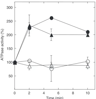
CCCP addition to a pmc1D mutant triggered a greater H1-ATPase activation, while the absence of a functional
Snf3p caused only a partial inhibition of the CCCP-induced activation of the plasma membrane ATPase (Fig. 4a).
Although the vacuole is the principal calcium internal store in yeast cells, and Pmc1p is the main pump involved in its internalization into the vacuole, we also investigated the CCCP-induced effects in a strain lacking a functional Pmr1p, a Ca21-ATPase involved in calcium pumping inside the Golgi apparatus. Surprisingly, the results shown in Fig. 4b demonstrate that while the glucose-induced activation of the enzyme in thepmr1Dmutant was only partially inhib-ited, the CCCP-induced activation of the pump was com-pletely absent in this mutant.
Considering that CCCP also acts as an uncoupler inhibit-ing the synthesis of ATP, it would not trigger the activation
ATPase activity
(%)
ATPase activity
(%)
Time (min) 0
50 100 150 200 250 (a)
(b) 300 350
0 2 4 6 8 10
0 50 100 150 200 250 300
of the plasma membrane H1-ATPase, because its presence
would affect ATP availability. Therefore, we also performed an experiment by adding 5 mM glucose 5 min before the addition of CCCP to increase the cellular ATP levels. The results shown in Fig. 4a and c demonstrate that, in spite of
its uncoupling effect, the addition of CCCP to yeast cells can still activate the H1-ATPase, suggesting the existence of a
specific effect that leads to this activation process.
Influence of external calcium in the CCCP-induced effects
Considering the above results, we investigated if external calcium would also influence the magnitude of
Time (s) 0
50 100 150 200 250 300 350 400 450 500
–30 0 30 60 90 120 150 180
0 2 4 6 8 10
100 200 300 400 500
ATPase activity
(%
)
Time (min)
ATPase activity (%
)
0 2 4 6 8 10
100 200 300 400 500
Time (min)
RLU s
(a)
(b)
(c)
Fig. 3. Relationship between CCCP-induced calcium signaling and acti-vation of the plasma membrane H1-ATPase. CCCP-induced activation of plasma membrane ATPase (a) and CCCP-induced calcium signaling (b) in the 3700 wild-type and the correspondingplc1Dstrains. CCCP-induced plasma membrane H1-ATPase activation in strains presenting a single deletion in theARG82gene or double deletions inPLC1andARG82
genes (c). Wild-type (circles);plc1D (triangles);arg82D (squares) and
plc1Darg82D(inverted triangles) strains. Closed symbols: 0.5 mM CCCP in ethanol (a and c) or in DMSO (b); open symbols: corresponding volume of ethanol or DMSO used with the CCCP solution.
ATPase activity (%
)
ATPase activity
(%
)
ATPase activity
(%
)
Time (min) 0
150 300 450 600 (a)750
–5 0 5 10
100 200 300 400 500
0 2 4 6 8 10
150 300 450 600
Time (min) (b)
(c)
the CCCP-induced phenomena. Therefore, we measured the effect of calcium deprivation, using the calcium che-lator EGTA. As shown in Fig. 5, extracellular calcium appeared to be essential for the CCCP-induced activation of the ATPase, because preincubation of wild-type cells with EGTA completely inhibited this activation process. On the other hand, when an equimolecular calcium con-centration was added together with EGTA, the CCCP-induced activation of the ATPase was nearly normal (Fig. 5).
Finally, we measured the CCCP-induced activation of the plasma membrane ATPase in a strain lacking Mid1p activity. This protein is functionally associated with Cch1p, a homologue of the mammalian high-affinity voltage-gated calcium channel component (Iidaet al., 1994; Locke
et al., 2000; Tok´es-Fuzesi et al., 2002). Surprisingly, and
as previously observed in the glucose-induced activation of the plasma membrane ATPase (Tr ´opia et al., 2006), Mid1p seems not to be essential for the CCCP-induced activation of the ATPase, because the mutant showed a nearly normal activation under these circumstances. Similar results were also observed with a strain presenting a deletion in the geneFIG1that encodes for a low affinity calcium transport system (Mulleret al., 2001) (data not shown).
Discussion
Comparison between CCCP- and glucose -induced effects
It was previously shown that the addition of protonophores, such as DNP and CCCP, triggers anin vivoactivation of the plasma membrane H1-ATPase (Becker dos Passos et al.,
1992; Branda˜oet al., 1992). Although the addition of these compounds also stimulates the cAMP-PKA pathway (Thevelein & Beullens, 1985; Thevelein, 1991) it was demon-strated that the cAMP-PKA pathway was not involved in H1-ATPase activation (Becker dos Passoset al., 1992).
Nevertheless, the mechanism by which protonophores/ depolarizing compounds would activate this enzyme has not been further investigated during the last 15 years. Curiously, the unique related effect reported for these compounds on yeast was an increase of calcium uptake triggered by CCCP (Eilamet al., 1990).
A more consistent hypothesis about the mechanism by which the plasma membrane ATPase is regulated at post-translational level has been proposed on the basis of data showing a relationship between the glucose-induced activa-tion of plasma membrane H1-ATPase and the
phosphati-dylinositol metabolism, in connection with the availability of cytosolic calcium (Tr ´opia et al., 2006). Considering all these data, it seemed logical to propose an involvement of calcium metabolism in the CCCP-induced activation of the plasma membrane ATPase in yeast cells.
Involvement of calcium in the signaling process In spite of the fact that there are clear differences between the intensities of the CCCP- and glucose-induced activation of the H1-ATPase, the results presented here indicate that
calcium metabolism is a common factor in both cases. Support for this hypothesis came from the experiments demonstrating that proteins directly involved in both the sugar-induced activation of the plasma membrane H1-ATPase and calcium signaling (phospholipase C and
Arg82p) were also involved in the CCCP-induced activation of the enzyme as well as in calcium signaling.
The Gpa2p G protein, that is partially required for the glucose-induced activation of the plasma membrane H1
-ATPase (Souzaet al., 2001) and calcium signaling (Tı¨siet al., 2002), was not involved in the CCCP-induced activation of the enzyme. This was not surprising if one considers that Gpa2p is normally activated by the glucose receptor Gpr1p and it is well known that this system normally responds directly to glucose. The nature of the internal signal gener-ated by addition of sugar (sugar phosphates?) or CCCP (drop in the internal pH) (Theveleinet al., 1987) would be different: while glucose-induced activation requires Gpa2p, the decrease in the intracellular pH triggered by CCCP
Time (min)
ATPase activity
(%)
0 2 4 6 8 10
50 100 150 200 250 300
Fig. 5.The role of external calcium on the CCCP-induced activation of plasma membrane H1-ATPase in Saccharomyces cerevisiae strains. CCCP-induced ATPase activation in glucose-grown 3700 wild-type cells in different conditions: in the absence of additional concentration of CaCl2(
); in the presence of 12 mM EGTA (n); or in the presence of12 mM CaCl2and 12 mM EGTA (m); control: corresponding volume of
would be enough to directly activate the phospholipase C generating IP3. Interestingly, it was already demonstrated
that Ras (a G protein) is specifically involved in the intracellular acidification-induced cAMP signaling in the yeastS. cerevisiae(Colomboet al., 1998). Thus, if intracel-lular calcium is the internal signal involved in the CCCP-induced effects, Ras protein could also be involved in the Plc1p activation leading to a IP3 signal and an increase of
calcium availability in the cytosol.
Another set of results supporting the involvement of calcium in the CCCP-induced effects came from the experi-ments with apmr1D mutant. Deletion of thePMR1 gene provokes an overexpression of PMC1 gene, leading to a stronger accumulation of calcium into the vacuole (Keller-mayeret al., 2003). As demonstrated here, in this mutant both the glucose- and the CCCP-induced activation of the H1-ATPase were clearly affected. However, the fact that in
thepmr1Dmutant the plasma membrane H1-ATPase was
only partially activated by glucose and nearly not activated by CCCP suggests that the protonophore-induced effects are indeed weaker or more confined regarding calcium signaling.
Finally, the requirement of extracellular calcium in order to get a CCCP-induced activation of plasma membrane ATPase is also a clear indication that external calcium is involved in this activation process equally suggesting that calcium metabolism is indeed important for the enzyme activation.
Framework of a signaling pathway for plasma membrane H1-ATPase activation
The idea emerging from our results (Souza et al., 2001; Tr ´opia et al., 2006; this paper) is that a pathway with two branches regulates activation of the H1-ATPase. In the first,
glucose (sugar) uptake, followed by phosphorylation, gen-erates a signal, probably mediated by phosphorylated sugars. Besides, this signal would stimulate the G protein Gpa2p, affecting phospholipase C activity and leading to the hydrolysis of PIP2with subsequent generation of DAG
and IP3. The increase in the intracellular calcium
concentra-tion would activate a calcium-dependent protein kinase that, in turn, would phosphorylate and activate plasma membrane ATPase.
According to our results (this paper, Tr ´opiaet al., 2006), protein kinase C would be involved in this activation process. Therefore, we searched for the existence of possible Pkc1 p phosphorylation sites on the plasma membrane ATPase using the available tools in the website NETPHOSK
(Blom et al., 2004). At least 30 putative sites with scores ranging between 0.5 and 0.92 were found. Two sites localized in the C-terminal domain were previously suggested as those involved in the enzyme activation (Eraso & Portillo, 1994).
Very recently Lecchiet al. (2007) demonstrated that tandem phosphorylations of Ser-911 and Thr-912 at the carboxy terminus of yeast plasma membrane H1-ATPase are
respon-sible for the glucose-induced activation of the enzyme. Interestingly, these two amino acids can be considered as potential targets of Pkc1 p according the website NET-PHOSK (data not shown).
In the second branch of this model, the glucose sensor Snf3p would also detect the sugar phosphates, and in some way would transduce a signal controlling the Pmc1p activity. The balance of these two branches would control the actual availability of calcium in the cytosol (Tr ´opiaet al., 2006).
The CCCP-induced H1-ATPase activation would be
weaker by the fact that it does not use the G protein Gpa2p for this activation process and it is also difficult to imagine a mechanism controlling the Snf3p activity as suggested in the glucose-induced activation process. Thus, only one branch of this mechanism would be directly affected by CCCP or DNP.
Although the results shown in this paper provide a better understanding of the mechanism by which the activity of the plasma membrane ATPase is regulated, there are many questions that deserve further investigation. First, and as already observed in the glucose-induced activation, the apparent dependence on external calcium seems to be very complex: in both cases (Tr ´opiaet al., 2006, this paper), the activation of the enzyme seems not to be affected in strains lacking proteins (Mid1p and Fig1p) involved in calcium uptake in yeast cells (Iidaet al., 1994; Lockeet al., 2000; Tok´es-Fuzesiet al., 2002). Otherwise, as we already men-tioned before (Tr ´opiaet al., 2006), it is possible that the right channel has not been identified or that a combination of different uptake systems may be involved.
Secondly, which is the mechanism responsible for the Plc1p activation by CCCP? Is this protein sensitive to membrane depolarization or to pH decrease? If so, how can one combine this mechanism with that of the glucose-induced phosphatidylinositol turnover already observed in yeast cells (Coccettiet al., 1998)? The G protein, Ras, could be involved in this specific process? These and other aspects are currently under investigation in our laboratories.
Acknowledgements
References
Becker dos Passos J, Vanhalewyn M, Branda˜o RL, Castro IM, Nicoli JR & Thevelein J (1992) Glucose-induced activation of plasma membrane H1-ATPase in mutants of the yeast Saccharomyces cerevisiaeaffected in cAMP metabolism, cAMP-dependent protein phosphorylation and the initiation of initiation of glycolisis.Biochim Biophys Acta1136: 57–67. Blom N, Sicheritz-Ponten T, Gupta R, Gammeltoft S & Brunak S
(2004) Prediction of post-translational glycosylation and phosphorylation of proteins from the amino acid sequence. Proteomics1633–1649.
Branda˜o RL, Castro IM, Becker dos Passos J, Nicoli JR & Thevelein J (1992) Glucose-induced activation of the plasma membrane ATPase inFusarium oxysporum.J Gen Microbiol
138: 1579–1586.
Branda˜o RL, de Magalha˜es-Rocha NM, Alijo R, Ramos J & Thevelein MJ (1994) Possible involvement of a phosphatidylinositol-type signaling pathway in glucose-induced activation of plasma membrane H1-ATPase and
cellular proton extrusion in the yeastSaccharomyces cerevisiae. Biochim Biophys Acta1223: 117–124.
Carmelo V, Santos H & Sa-Correia I (1997) Effect of extracellular acidification on the activity of plasma membrane ATPase and on the cytosolic and vacuolar pH ofSaccharomyces cerevisiae. Biochim Biophys Acta1325: 63–70.
Coccetti P, Tisi R, Martegani E, Teixeira LS, Branda˜o RL, Castro IM & Thevelein JM (1998) ThePLC1encoded phospholipase C in the yeastSaccharomyces cerevisiaeis essential for glucose-induced phosphatidylinositol turnover and activation of plasma membrane H1-ATPase.Biochim Biophys Acta1405:
147–154.
Colombo S, Ma P, Cauwenberg L, Winderickx J, Crauwels M, Teunissen A, Nauwelaers D, de Winde JH, Gorwa M & Thevelein JM (1998) Involvement of distinct G-proteins, Gpa2 and Ras, in glucose- and intracellular acidification-induced camp signaling in the yeastSaccharomyces cerevisiae.EMBO J
17: 3326–3341.
Eilam Y & Chernichovsky D (1987) Uptake of Ca21driven by the membrane potential in energy-depleted yeast cells.J Gen Microbiol133: 1641–1649.
Eilam Y & Othman M (1990) Activation of Ca21by metabolic
substrates inSaccharomyces cerevisiae: role of membrane potential and cellular ATP levels.J Gen Microbiol136: 861–866. Eilam Y, Othman M & Halachmi D (1990) Transient increase in
Ca21influx inSaccharomyces cerevisiaein response to glucose: effects of intracellular acidification and cAMP levels.J Gen Microbiol136: 2537–2543.
Eraso P & Gancedo C (1987) Activation of yeast plasma membrane ATPase by acid pH during growth.FEBS Lett224: 187–192.
Eraso P & Portillo F (1994) Molecular mechanism of regulation of yeast plasma membrane H1-ATPase by glucose.J Biol Chem
269: 10393–10399.
Iida H, Nakamura H, Okumura MS & Ankaru Y (1994) MID1, a novelSaccharomyces cerevisiaegene encoding a plasma
membrane protein, is required for Ca12influx and mating. Mol Cell Biol14: 8259–8271.
Ito H, Fukuda Y, Murata K & Kimura A (1983) Transformation of yeast cells treated with alkali cations.J Bacteriol153: 163–168.
Kellermayer R, Aiello DP, Miseta A & Bedwell DM (2003) Extracellular Ca21sensing contributes to excess Ca21 accumulation and vacuolar fragmentation in apmr1Dmutant ofS. cerevisiae.J Cell Sci116: 1637–1646.
Lecchi S, Nelson CJ, Allen KE, Swaney DL, Thompson KL, Coon JJ, Sussman MR & Slayman CW (2007) Tandem
phosphorylation of Ser-911 and Thr-912 at the C terminus of yeast plasma membrane H1-ATPase leads to
glucose-dependent activation.J Biol Chem282: 35471–35481. Locke EG, Bonilla M, Liang L, Takita Y & Cunnigham KW (2000)
A homolog of voltage-gated Ca12channels stimulated by depletion of secretory Ca12in yeast.Mol Cell Biol20: 6686–6694.
Lowry OH, Rosenbrough NJ, Farr AL & Randall RJ (1951) Protein measurement with the Folin phenol reagent.J Biol Chem193: 265–275.
Mazon MJ, Behrens M, Portillo F & Pinon P (1989) cAMP- and RAS-independent nutritional regulation of plasma-membrane H1-ATPase activity inSaccharomyces cerevisiae.J Gen Microbiol135: 1453–1460.
Monteiro GA & S´a-Correia I (1998)In vivoactivation of yeast plasma membrane H1-ATPase by ethanol: effect on the kinetic
parameters and involvement of the carboxyl-terminus regulatory domain.Biochim Biophys Acta1370: 310–316. Muller EM, Locke EG & Cunningham KW (2001) Differential
regulation of two Ca(21) influx systems by pheromone signaling inSaccharomyces cerevisiae.Genetics159: 1527–1538. Portillo F (2000) Regulation of plasma membrane H1-ATPase in
fungi and plants.Biochim Biophys Acta1469: 31–42.
Portillo F & Mazon MJ (1987) TheSaccharomyces cerevisiaestart mutant carrying the cdc25 mutation is defective in activation of plasma membrane ATPase by glucose.J Bacteriol168: 1254–1257.
Purwin C, Nicolay K, Scheffers WA & Holzer H (1986) Mechanism of control of adenylate cyclase activity in yeast by fermentable sugars and carbonyl cyanidem
-chlorophenylhydrazone.J Biol Chem261: 8744–8749. Sambrook J, Fritsch EF & Maniatis T (1989)Molecular Cloning: a
Laboratory Manual, 2nd edn. Cold Spring Harbor, New York. Serrano R (1983)In vivoglucose activation of the yeast plasma
membrane ATPase.FEBS Lett156: 11–14.
Sherman F, Fink GR & Hicks JB (1986)Methods in Yeast Genetics. Cold Spring Harbor, New York.
Souza MAA, Tr ´opia MJ & Branda˜o RL (2001) New aspects of glucose activation of the H1-ATPase in the yeast
Saccharomyces cerevisiae.Microbiol147: 2849–2855. Thevelein JM (1984) Regulation of trehalose mobilization in
fungi.Microbiol Rev48: 42–59.
cyclase signalling pathway in yeast: the relationship to nutrient-induced cell cycle control.Mol Microbiol5: 1301–1307.
Thevelein JM & Beullens M (1985) Cyclic AMP and the stimulation of trehalase activity in the yeastSaccharomyces cerevisiaeby carbon sources, nitrogen sources and inhibitors of protein synthesis.J Gen Microbiol131: 3199–3209.
Thevelein JM, Beullens M, Honshoven F, Hoebeeck G, Detremerie K, Den Hollander JA & Jans AWH (1987)J Gen Microbiol133: 2191–2196.
Tı¨si R, Baldassa S, Belotti F & Martegani E (2002) Phospholipase C is required for glucose-induced calcium influx in budding yeast.FEBS Lett520: 133–138.
Tı¨si R, Belotti F, Winderickx J, Thevelein JM & Martegani E (2004) Evidence for inositol triphosphate as a second messenger for glucose-induced calcium signaling in budding yeast.Curr Genet45: 83–89.
Tok´es-Fuzesi M, Bedwell DM, Repa I, Sipos K, Sumegi B, Rab A & Miseta A (2002) Hexose phosphorylation and the putative calcium channel component Mid1p are required for the hexose-induced transient elevation of cytosolic calcium response inSaccharomyces cerevisiae.Mol Microbiol44: 1299–1308.
Trevillyan JM & Pall ML (1979) Control of cyclic adenosine 30,50
-monophosphate levels by depolarizing agents in fungi.J Bacteriol138: 397–403.
Tr ´opia MJM, Cardoso AS, Tisi R, Fietto LG, Fietto JLR, Martegani E, Castro IM & Branda˜o RL (2006) Possible relationship between transient elevation of cytosolic calcium and sugar-induced activation of plasma membrane H1
-ATPase in the yeastSaccharomyces cerevisiae.Bioch Biophys Res Comm343: 1234–1243.
Ulaszewski S, Hielger F & Goffeau A (1989) Cyclic AMP controls the plasma membrane H1-ATPase activity from