Role of Yap8 and Yap1 b-ZIP transcription
factors in arsenic stress
Catarina Sá Almeida Amaral
Dissertation presented to obtain a Ph.D. degree in Biology
Instituto de Tecnologia Química e Biológica
Universidade Nova de Lisboa
Supervisor Prof. Dr. Claudina Rodrigues-Pousada
Oeiras, 22nd of January
Com o apoio financeiro da Fundação para a Ciência e Tecnologia (FCT) e do Fundo Social Europeu(FSE) no âmbito do Quadro Comunitário de Apoio, bolsa de Doutoramento
iii
Members of the JuryProfessor José Artur Martinho Simões
(President of ITQB Scientific Council, President of the Jury)
Professor Margarida Casal
(Department of Biology, CBMA, Universidade do Minho, Braga)
Professor Vitor da Costa
(Departamento: Biologia Molecular,ICBAS, Universidade do Porto)
Professor Miguel Teixeira
(Institute for Biotechnology and Bioengineering, Instituto Superior Técnico)
Professor Emmanouil Matzapetakis
(Biomolecular NMR ITQB, Universidade Nova de Lisboa,)
Dr. Regina Menezes
(Genomics and Stress, ITQB, Universidade Nova de Lisboa,)
Professor Claudina Rodrigues-Pousada
(Genomics and Stress, ITQB, Universidade Nova de Lisboa,
iv
The work reported in this thesis was performed in the
Genomics and Stress Laboratory,
Instituto de Tecnologia Química e Biológica
v
vi
AcknowledgmentsI would like to acknowledge Professor Claudina
Rodrigues-Pousada for accepting me in her laboratory, for her support in the
difficult times and accepting me with my imperfections and even so,
always allow me another opportunity. I also like to acknowledge Dr.
Regina Menezes for the knowledge and ideas she transmitted,
support and time spent teaching me. I wish also to thank Regina
and Professor for the help and support during the writing of this
thesis.
I must also add a special thanks to:
Dr. Pimentel for her constant ideas, incentive and support,
Dr.Pereira for his help in protein work and good advices in time
management, that I still have difficulty to follow. Dr. Ribeiro for
introducing me in the laboratory, Dr. Azevedo and Dr.Nevitt for the
techniques taught. Thank you to Liliana, Fabio, Ana, Joana,
Cristina and Ana Rita for their good mood and good environment in
the laboratory and Ana for her kindness. Eugénia is also greatly
acknowledged for allowing us to start our work always in the best
conditions and on time.
I am also thankful to the Laboratoire de Génétique Moléculaire,
Ecole Normale Supérieure, Paris and Professor Frédéric Devaux
for receiving me in their laboratory and for teaching me the
transcriptional arrays.
And outside the laboratory to Sofia, Ulla, Dinora, Iulia and Patricia
for the hot cup of tea and stories told.
And a special thanks to my family, for their unconditional and
constant support. Muito obrigada Mãe, Pai, Rita, Carlos e Vó, por
viii
Foreword:The work of Bobrowicz et al pointed toward a role of Acr1/Yap8 as
a regulator of the arsenic detoxification genes in yeast S.
cerevisiae. The work on Chapter 2 provides molecular evidence
that Yap8 is the transcriptional regulator responsible for the
activation of ACR2 and ACR3 in the response to arsenic stress and
elucidates the mechanisms by which Yap8 is activated under this
condition. Also the involvement of Yap1 as a regulator of YCF1 is
presented in this chapter. In Chapter 3 it is shown that arsenic
exposure generates oxidative stress in yeast cells and that Yap1
has a role in activating the expression of the genes involved in
antioxidant defences. In Chapter 4, the DNA binding properties of
Yap8 were studied as well as the importance of the Mediator
complex in Yap8 transmission of the stress signal to the basal
ix
Thesis publicationsMenezes R., Amaral, C., Batista-Nascimento, L., Santos, C., Ferreira, R.B., Devaux, F., Eleutherio, E. C.A. and
Rodrigues-Pousada, C. (2008)Contribution of Yap1 towards S. cerevisiae
adaptation to arsenic mediated oxidative stress Biochem.
J.414: 301-311
Rodrigues-Pousada, C., Nevitt, T., Menezes, R., Azevedo, D.
Pereira, J., (2004) Amaral, C.; Yeast activator proteins and
stress response: an overview . FEBS Letters 567: 80-85
Menezes, R., Amaral, C. Delaunay, A., Toledano, M., and
Rodrigues-Pousada, C.(2004) Yap8p activation in
Saccharomyces cerevisiae under arsenic conditions. FEBS
Letters 566: 141-146
Amaral, C., Ropio, J., Silva, A. Menezes, R., and
Rodrigues-Pousada, C. Yap8-DNA interaction and Mediator requirement
for Yap8 activity in preparation
Other publications not included in the thesis:
Pereira,J., Pimentel,C. Amaral, C., Menezes R.,
Rodrigues-Pousada (2009) Yap4 PKA-and Gsk3-dependent
phosphorylation affects its stability but not its nuclear
x
Abstract:
Arsenic compounds are highly toxic substances; nevertheless they
are used in the treatment of acute promyelocytic leukaemia.
Therefore it is pressing to gain knowledge on its toxicity and
detoxification mechanisms. The cellular entry pathways have been
discovered and by transcriptome analysis it is known that arsenic
activates the transcription of genes activated by, among others,
Rpn4, Met4 and Yap1.
Yap1 and Yap8 belong to the Yap family, which is composed of
eight transcription factors, each with a determined function in the
response to stress. Nevertheless Yap1 is involved in the response
to oxidative stress and multi-drug stress, including arsenic, Yap2 in
the response to cadmium, Yap4 may have a role in the response to
several stresses and Yap8 is involved in the response to arsenic,
as its major regulator. Its role is due to the fact that Yap8 is
important for the expression of the detoxification genes ACR2 and
ACR3. In this work we show that its deletion abrogates the
expression of these two genes. We also studied Yap8 activation
mechanism, proving that having conserved cysteines with Yap1
allows it to be regulated by a similar mechanism. Yap8 is modified
in these conserved cysteines, which masks the nuclear localization
signal preventing it from interacting with the nuclear exportin Crm1,
becoming nuclear. The study of Yap1 nuclear localization in an
orp1 strain, responsible for the signal transduction under oxidative
stress, indicates that arsenic behaves as a thiol-reactive chemical.
We also prove, by mutation to alanine that the cysteines are
relevant for Yap8 nuclear localization and are also relevant for
xi
In chapter3 we demonstrate that a mechanism of arsenic toxicity is
through the production of Reactive Oxygen Species (ROS) since
there is accumulation of lipidic peroxidation by-products, protein
carbonylation and intracellular ROS production after arsenic stress.
Genes related to anti-oxidative defences are up-regulated. The
glutathione levels decrease after one hour of arsenic stress which
may be a consequence of arsenic stress, because arsenic
detoxification requires the use of glutathione by Arr2 and Ycf1, this
disturbs the redox homeostasis causing oxidative stress. To
compensate it, expression analysis show an up-regulation of the
sulphur metabolism related genes. The oxidative stress responsive
genes related to oxidative stress that are dependent on Yap1, are
up-regulated, particularly in a yap8 strain. The particular
sensitivity of a yap8 strain, and higher anti-oxidative gene
response, may be explained due to a higher accumulation of
arsenic as shown by atomic absorption, which occurs because
ACR3 is not transcribed. It was shown that Yap1 and Yap8 have
distinct roles in the response to stress and Yap1 is the mediator of
the response to oxidative stress caused by arsenic.
In Chapter 4 we studied the amino acids mutations in Yap8: K21A,
R22A, A23T, Q25A, L26N R27A, S29A, N31A, K35A, K35E, R36A,
and K37A and show that K21A, R22A, A23T, R27A, K35E, R36A
and Q25A are relevant for Yap8 interaction with the DNA. The
double mutation Yap8L26N-N31R conferred a slight capacity for
Yap8 to recognize a Yap1 binding site despite the fact that the
respective single mutations are not capable of doing so.
After the binding to DNA the signal transduction from Yap8 to the
transcription machinery occurs through the Mediator complex.
xii
medium with arsenic compounds which revealed that the subunits
Med2, Med3, Med15 and Med16 and Med14 are required for the
yeast response to arsenic stress. The assays of Yap8
transactivation potential show that all the subunits are required for
its full activity. It was also studied the expression of Yap8 target
gene ACR2 and the results indicate that Med16 is the subunit more
relevant for the transcription of this target gene followed by the
xiii
Resumo:
O elemento arsénico nos seus diferentes compostos, é altamente
tóxico sendo, no entanto, usado no tratamento de leucemia
promielocítica aguda. A sua presença no ambiente como
contaminante e o seu uso como medicamento leva a necessidade
do estudo dos mecanismos relevantes para a sua destoxificação e
quais os seus mecanismos de acção. As vias de entrada nos
diferentes tipos celulares são já conhecidos e análises do
transcriptoma mostraram que genes regulados por factores de
transcrição como o Rpn4, Yap1 e Met4 estão activados.
O Yap1 e Yap8 pertencem a família Yap, família de factores de
transcrição, composta por oito membros. Cada membro regula a
resposta a diferentes tipos de stress, no entanto o Yap1 está não
só envolvido na resposta ao stress oxidativo mas também a
variadas drogas incluindo o arsénico. Yap2 está envolvido na
resposta ao cádmio, enquanto a actividade do Yap4 deverá estar
relacionada com diversos stresses. O Yap8 está envolvido na
resposta ao arsénico sendo o seu principal regulador. O seu papel
deve-se ao facto de ser o regulador da transcrição de ACR2 e
ACR3. Neste estudo é demonstrado que a deleção do YAP8
impede a transcrição destes dois genes. Foi também estudado o
mecanismo de activação desta proteína, provando-se que as
cisteinas conservadas com o Yap1 permitem um mecanismo de
regulação semelhante ao deste. O que significa que a modificação
destas cisteinas mascara o sinal de localização nuclear impedindo
a sua interacção com a exportina nuclear Crm1, tornando-o
nuclear. O estudo da localização sub-celular do Yap1 numa estirpe
orp1, proteína envolvida na transdução do sinal de stress
xiv
reage com tiols. A mutação destas cisteinas para alaninas impede
a localização nuclear do Yap8 e é também determinante para o
potencial de transactivação deste.
No capítulo 3 demonstramos que um dos mecanismos de
toxicidade do arsénico ocorre via produção de espécies reactivas
de oxigénio. O que é verificado devido a acumulação de produtos
da peroxidação lipídica, carbonilação de proteínas e produção de
espécies reactivas de oxigénio intra-celularmente. Análises do
transcriptoma indicam o aumento de expressão de genes
envolvidos na resposta ao stress oxidativo. Os níveis de glutationa
diminuem após uma hora de stress por arsénico o que pode ser
uma consequência dos processos de eliminação do arsénico uma
vez que Acr2 e Ycf1 precisam de glutationa. O gasto de glutationa
na forma reduzida causa desequilíbrio da homeostase redox
podendo causar stress oxidativo. Os genes alvo do Yap1 são
transcritos activamente, particularmente numa estirpe yap8. A
maior sensibilidade desta estirpe deve-se ao facto de acumular
mais arsenito como é mostrado por absorção atómica, esta
acumulação ocorre porque nesta estirpe o gene ACR3 não é
expresso. Foi demonstrado que Yap1 e Yap8 têm papéis distintos
na resposta ao stress e Yap1 regula a resposta ao stress oxidativo
causado por arsénico.
No capitulo 4 é mostrado que das mutações no Yap8: K21A,
R22A, A23T, Q25A, L26N, R27A, S29A, N31A, K35A, K35E, R36A
e K37A sendo as mais relevantes para a interacção com o DNA:
K21A, R22A, A23T, R27A, K35E, R36A e Q25A. No entanto as
únicas mutações com fenótipo de sensibilidade a compostos de
arsénico são R22A, R27A, R36A e Q25A. A mutação dupla
xv
ligeiramente a sequencia de DNA reconhecida pelo Yap1. Isto
apesar das mutações isoladas não terem qualquer efeito no Yap8.
Após ligação ao DNA, o Yap8 interage com a maquinaria basal de
transcrição, o que ocorre através do complexo Mediador. Estirpes
mutantes nas subunidades da cauda do Mediador foram
estudadas por crescimento em meio com compostos de arsénico
revelando que as subunidades Med2, Med3, Med15, Med16 e
Med14 são necessárias para a resposta da levedura ao stress por
arsénico. O estudo do potencial de transactivação mostra que
todas as subunidades são importantes para a total actividade de
Yap8. As subunidades mais importantes são as reveladas pelos
estudos fenotípicos com excepção da Med14, o que indica que
esta subunidade está envolvida na resposta ao arsénico de forma
independente do Yap8. Foi também estudada a expressão do
gene ACR2 e os resultados indicam que a subunidade que afecta
mais pronunciadamente a transcrição do seu gene alvo é Med16,
mas havendo também um papel relevante por parte das
xvi
Table of contents
Symbols and Abbreviations 0
Chapter 1 Introduction
1.1 Yeast as a biological model-scientific relevance 2
1.2 Transcriptional factors involved in Yeast Stress
response to arsenic stress 3
1.3 Arsenic Stress 4
1.3.1 Arsenic Stress in Yeast 4
1.3.2 Arsenic Stress in other organisms 9
1.4 Oxidative Stress defences 11
1.5 Yap family history 13
1.6 The role played by Yap family members in stress
response 15
1.7 References 19
Chapter 2 Yap8p activation in
Saccharomyces
cerevisiae
under arsenic conditions
2.1 Author’s contribution to the article 29
2.2 Abstract 31
xvii
2.4 Materials and Methods 32
2.5 Results and Discussion 36
2.6 Acknowledgments 46
2.7 References 47
Chapter 3 Contribution of Yap1 towards
S.
cerevisiae
adaptation to arsenic mediated
oxidative stress
3.1 Author’s contribution to the article 49
3.2 Abstract 51
3.3 Introduction 52
3.4 Materials and Methods 55
3.5 Results 62
3.6 Discussion 78
3.7 Acknowledgments 82
3.8 References 82
Chapter 4 Yap8-DNA interaction and Mediator
requirement for Yap8 activity
4.1 Author’s contribution to the article 89
4.2 Abstract 90
4.3 Introduction 91
4.4 Materials and Methods 94
xviii
4.6 Conclusion 121
4.7 Acknowledgments 122
4.8 References 122
Chapter 5 Discussion and Perspectives
1250
Symbols and abbreviations
AP-1 activator protein 1 ARE AP-1 recognition element As(V) arsenate
As(III) arsenite
AT 3-amino-1,2,4-triazole bp base pairs
b-ZIP basic DNA-binding domain and leucine zipper
c-CRD C-terminal cysteine rich domain
DABCO
1,4-Diazabicyclo[2.2.2]octaneAPI 4’,6’ -diamidino-2-phenylindole, dihydrochloride
DEPC di-ethyl pyrocarbonate DNA deoxyribonucleic acid EDTA ethylenediaminetetraacetic acid
11bp 11bp sequence of the ACR2/ACR3 promoter
GFP green fluorescent protein GPXs glutathione peroxidases GSH reduced glutathione HSE heat shock element HSF1 heat shock factor GSSG oxidized glutathione kb kilo base pairs kDa kiloDalton
M molar
Min minutes RNA ribonucleic acid n-CRD N-terminal cysteine rich domain
NES nuclear export signal NLS nuclear localization signal nm nanometers
O.D.600nm optical density at 600nm
ORF open reading frame ºC degree Celsius
PCR polymerase chain reaction PKA protein kinase A
RNA ribonucleic acid
ROS reactive oxygen species S. cerevisiae Saccharomyces cerevisiae
S. pombe Schizosaccharomyces pombe SC synthetic complete medium SD synthetic minimal medium STRE stress responsive element 13bp 13bp sequence of the ACR2/ACR3 promoter
Y Yap recognition element
Amino acids
A Ala alanine B Asx asparagine or
aspartic acid C Cys cysteine D Asp aspartic acid E Glu glutamic acid F Phe phenylalanine G Gly glycine H His histidine I Ile isoleucine K Lys lysine L Leu leucine
M Met methionine
N Asn asparagine P Pro proline Q Gln glutamine R Arg arginine S Ser serine T Thr threonine V Val valine W Trp tryptophan Y Tyr tyrosine
Z Glx glutamine or glutamate
X X any of the amino
acids residues listed
Nucleotide bases
A adenine C cytosine G guanine T thymine
1
Chapter I
Introduction
1.1 Yeast as a biological model - scientific relevance 2 1.2 Transcriptional factors involved in Yeast Stress
responses 3
1.3. Arsenic stress 4
1.3.1 Arsenic stress in Yeast 4
1.3.2 Arsenic stress in other organisms 9
1.4 Oxidative stress defences. 11
1.5 Yap family history 13
1.6 The role played by Yap family members in stress
Response 15
2
Introduction
1.1 Yeast as a biological model - scientific relevance
The budding yeast Saccharomyces cerevisiae is a simple eukaryote model organism that has the advantage of robust growth on minimal media and easy handling due to the fact that it does not cause pathogenesis in the health of Humans. Its genome was completely sequenced by an international consortium in which Claudina Rodrigues-Pousada’s laboratory actively participated in [2-5]. It was the first eukaryotic genome to be sequenced, showing the presence of approximately 6,000 genes.
It contains several genes with high homology to human genes, approximately 30% human disease map-based genes have yeast homologues [6]. One such example is the yeast cadmium factor, Ycf1, that possesses similarity to human Mrp1 (42.6% identity, 63.3% similarity) and the cystic fibrosis transmembrane conductance regulator (Cftr) (31% identity, 56.7% similarity), members of the ATP binding cassette (ABC) [7]. Mutations in CFTR1 that modulates chloride ion transport across epithelial
membranes lead to cystic fibrosis disease [8]. Another example of yeast genes with similarity to the ones of humans is the PAT1/PXA2 and PAT2/PXA1, each encodes two peroxisomal half
3 Yeast is in itself also a tool since it has been used to rapidly study protein-protein interaction, by the designated two hybrid technique [13]. This application has suffered several modifications that allowed the study of transactivation potential of transcription factors and their interaction with DNA, for example. Another important use of yeast as a tool is for heterologous expression of relevant human proteins, allowing again, rapid test of various candidate drugs [14].
1.2 Transcriptional factors involved in Yeast Stress
responses
All organisms must have the resources to adapt to an unbalanced environment. The cellular mechanisms are divided in general and specific stress response. The general stress response has conserved mechanisms across nature. S. cerevisiae developed also such a basic response common to all stresses. Two of the intervenient transcription factors are heat shock factor, Hsf1 and Msn2/Msn4. When cells are subjected to a mild stress they develop resistance to a subsequent lethal stress, e.g., cells become less sensitive to the imposed stronger stress due to the accumulation of compatible solutes [15]. HSF1 is an essential gene since its deletion causes lethality [16]. In yeast it is transcribed constitutively and forms homotrimers, unlike in higher eukaryotes where the trimerization occurs only after stress [17]. Each monomer binds one Heat Shock element (HSE) 5’nGAAn, and the yeast HSF1 binds constitutively to the sequence but does not activate transcription [18].
4 is nuclear localized and if hyperphosphorylated localizes in the cytoplasm [19]. They recognize the DNA sequence 5’CCCT designated Stress Responsive Element (STRE)[20].
1.3. Arsenic stress
1.3.1 Arsenic stress in Yeast
Arsenic (As) is a transition metal and a well-known carcinogen, due to its ability to induce oxidative stress, to diminished DNA repair, alter DNA methylation patterns, enhance cell proliferation and possible suppression of p53 [21]. Nevertheless, the arsenic-containing drugs are used as chemotherapeutic agents to combat infectious diseases caused by pathogenic parasites as well as cancer, including arsenic trioxide in the treatment of acute promyelocytic leukaemia [22,23]. Given the toxicity effects posed by these compounds it is pressing to elucidate the mechanisms that constitute the basis of their tolerance.
In the yeast, Saccharomyces cerevisiae, arsenate enters the cells through the phosphate transporters Pho84 and Pho87 [24] and the arsenite transport is facilitated through hexose permeases [25] although when glucose is present it is mainly transported through the aquaglyceroporin Fps1 [26]. In the absence of Fps1, the 17 hexose permeases and Acr3 (to eliminate the extrusion system) there is only residual uptake of arsenite. The transport is however resumed when the permeases Hxt1, Hxt3, Hxt4, Hxt5, Hxt7 and Hxt9 are expressed individually [25].
5 high levels of arsenic. It is composed by the transcription factor YAP8 (ARR1/ACR1) that controls the expression of the other two
genes of the cluster [27, 28]. These genes are ACR2 and ACR3 [29] which encode an arsenate reductase [30] responsible for the reduction of arsenate to arsenite, and an efflux protein that detoxifies arsenite respectively [31]. Ycf1 catalyses the ATP driven uptake of several drugs conjugated with glutathione, into the vacuole, including arsenic in the arsenite form (Fig.1.2) [32]
Schizosaccharomyces pombe resistance mechanisms to
metals such as Cd2+ and Cu2+ rely on the formation of phytochelatins, peptides synthesized from glutathione, enzymatically [33], in plant these phytochelatins also confer resistance to arsenic (see section 1.3.3). A screen of S. pombe mutants revealed two genes that confer sensitivity to arsenic and cadmium: the p38 orthologue Spc1/Sty1 and the Cnt5 a novel
Figure1.1 Arsenic entry and detoxification pathways in
6 member of the centaurin ADP ribosylation factor GTPase activating protein (Arf GAP). This protein accumulates in the membrane and septum after arsenic stress where it may regulate membrane trafficking or maintain membrane integrity [34].
The S. cerevisiae orthologue of the human p38 is Hog1, the MAPkinase (mitogen-activated protein kinase) of the HOG (high osmolarity glycerol) pathway that is activated under arsenic exposure. As(III) leads to the Hog1 activation by phosphorylation in a Ssk1 and Pbs2 dependent way. Deletion of hog1 leads to the increase uptake of arsenite which may be due to the deregulation of the As(III) transporter, Fps1. Activated Hog1 is one of the kinases responsible for Fps1 phosphorylation in the residue thr231, which reduces As(III) influx, when compared to a un-phosphorylated Fps1 [35]. Nevertheless Sotelo et al determined, by RT-PCR, that ACR3 and YCF1 are expressed at lower levels in a hog1 mutant when compared to the wild type [36]. In contradiction, results from Thorsen et al indicate that Hog1 does not interfere with As(III) efflux by Acr3 [35].
8 of iron-sulphur clusters as arsenic may remove iron from proteins [41,42]. As was observed before, there is also an up-regulation of genes involved in sulphur uptake and glutathione biosynthesis [38, 39] There is also an enrichment in genes involved in protein proteolysis, protein folding, detoxification of oxygen reactive species and telomere maintenance. The activator MAPK Hog1, together with other proteins from the HOG pathway, was also reported as required for the response to arsenic through analysis of the growth of deleted mutant strains [41] .
The Target of Rapamycin (TOR) is a central controller of the response to nutrients at the growth and metabolic level. Tor proteins are Ser/Thr kinases, conserved in eukaryotes, found in two complexes TORC1 and TORC2. TORC1 regulates several mechanisms such as protein synthesis, ribosome biogenesis, transcription and autophagy [43]. TOR kinase1, part of the TOR complex1, is inhibited under arsenic stress. TORC1 inactivation leads to down regulation of several genes, among them genes that code for ribosomal proteins. The TORC1 deactivation also prevents inhibition, of Msn2/Msn4. There is a fine regulation by PKA and TORC in order to maintain the required levels of Msn2/Msn4 under arsenite stress. PKA is active but the zinc fingers are localized in the nucleus due to down regulation of TORC1 [44].
9 reductase, involved in maintaining the deoxynucleotide triphosphate pool, therefore this enzyme is relevant to DNA repair and synthesis, this study reveals that mutation in this gene sensitizes the cells to arsenic [45].
1.3.2 Arsenic stress in other organisms
In bacteria, resistance to arsenic is mediated by the resistance operons ars. It has a conserved organization and regulation. It is composed by three genes ArsRBC. Each operon is regulated by an arsenite/antimonite responsive repressor which is encoded by the first gene of the operon, ArsR. The structure of the repressor is modified by the binding of arsenic or antimonite, therefore releasing the promoter [46]. The operon also contains: arsB and arsC. The first is highly conserved and confers resistance
to arsenite and antimonite, transporting it to the extracellular milieu [47]. The ArsC is an arsenate reductase and there are two unrelated families of this gene [48]. Operons containing 5 coding units were reported that include two additional genes arsD and arsA, as in Escherichia coli plasmid R773. ArsA is allosterically modified by arsenite or antimonite and binds to ArsB converting it in a primary ATP-driven arsenite pump [49]. The metalochaperone ArsD transports to ArsA, increasing its efficiency to extrude arsenite [50]. Entry of arsenate occurs through the phosphate transporters Pit and Pst [51] whereas arsenite is transported into the cell by the aquaglyceroporin, GIpF [47].
10 studied, AQP7 and AQP9 are the most efficient transporters [53]. A null mice in AQP9 is more sensitive to arsenic and excretes lower amounts of this compound showing that it is also involved in arsenite extrusion [54]. Arsenate transport occurs, with low efficiency, through the intestinal sodium phosphate carrier NaPi-IIb, other transporters must therefore exist [55]. Arsenite export in mammalian cells occurs through Mrp1, an ABC binding cassette that transports several drugs, probably as GSH conjugates [56]. Another protein that may be involved in the resistance to arsenic is a multi-drug resistance P-glycoprotein of the superfamily of ATP binding cassette transporters Mdr1 [57]. Mouse mutants in this protein show accumulation of arsenite (Fig.1.2) [58].
The transport of arsenite in plants is made through Nodulin 26-like Intrinsic Proteins (NIPs). The expression of some of these proteins in the yeast fps1 mutant confers sensitivity to As(III) and Sb(III) to the mutant strain indicating that Fps1 and nodulins have
11 the same role, e.g. transport of As(III) into the cells, although they also participate in extrusion [59]. To study arsenate sensitivity in Arabidopsis thaliana several mutants were tested and a gene
conferring resistance in a GSH-independent way was identified, ARS1. The mutant has higher phosphate uptake than the wild type
[60]. In plants arsenite is captured inside the cell by metallothionein protein and phytochelatins that accumulate in the vacuole. Phytochelatins are synthesized from glutathione by phytochelatin synthetase [61]. Plants also have an ABC transporter, without homology to Ycf1, that carries As(III)-GSH conjugates into the vacuole [62] and an arsenate reductase with moderate homology with the yeast Acr2 is also present [63].
Genome microarray analysis of Arabidopsis thaliana showed that As(V) stress represses the genes involved in phosphate starvation as expected because As(V) can behave as a phosphate analogue. Other genes were shown to be involved in antioxidative stress defences as for example the superoxide dismutases Cu/ZnSOD although the Fe/SOD was repressed [64].
1.4 Oxidative stress defences
Arsenic stress may cause oxidative stress as shown in several transcriptome studies that indicate that there is up-regulation of oxidative stress related genes [38, 39, 65]. This may be caused by the removal of iron from proteins that will then participate in Fenton reaction producing hydroxyl radical and anion [42]. Another cause of oxidative stress derived from arsenic is the consumption of glutathione by Acr2 and Ycf1 (fig.1).
12 defences. In each case there are the nonenzymatic mechanisms such as glutathione and enzymatic ones such as superoxide dismutases, catalases, and peroxidases.
The tripeptide glutamate-cysteine-glycine GSH is synthesized by the action of γ-glutamylcysteine synthetase (Gsh1) which catalyses the limiting step of GSH formation: the condensation of Cys onto the γ-Carbon of Glu. GSH1 expression is regulated by Yap1 and Met4 [66]. The addition of Gly is made by glutathione synthetase (Gsh2) [67]. GSH redox properties come from its Cys residue which low redox potential allows it to donate electrons to other Cys, therefore becoming oxidized enabling it to dimerize to the disulfide GSSG. This disulfide is reduced back to GSH by the action of the NADPH dependent glutathione reductase (Glr1) which expression is also regulated by Yap1 [68].
S. cerevisiae possesses two apparently redundant isoforms
from the thioredoxin system, Trx1 and Trx2 and a thioredoxin reductase Trr1 in the cytoplasm, there is also a mitochondrial system composed of Trx3 and Trr2 [69]. Thioredoxins are small proteins with two redox active Cys within the conserved active site motif, Cys-Gly-Pro-Cys, that becomes oxidized to an intramolecular disulfide, typically in a thiol-dissulfide exchange reaction with a dissulfide bonded substrate. The flavoenzyme NADPH dependent Trr reduces thioredoxin back to the active dithiol form [70]. Trx2 expression is controlled by Yap1 [71] and Skn7 [72].
13 the Orp1/Gpx3 that catalyzes the formation of the regulatory disulphide bond in Yap1 (see point 1.6 ).
Glutaredoxins are small proteins that are reduced by glutathione unlike thioredoxins that are reduced by NADPH. This protein family may have two domains, the thioredoxin-like domain and a glutaredoxin one, as is the case of Grx3 and Grx4, or a single glutaredoxin domain as is the case of Grx5 [75]. They are divided in two families according to its active site. It may have two Cys (Grx1 and Grx2) or one in the active site (Grx3, Grx4 and Grx5) [76].
Non-thiol containing antioxidative defences is for example the superoxide dismutase and catalase. Yeast, like other eukaryotes, has two superoxide dismutases, (SOD1 and SOD2) that catalyse the dismutation of superoxide to oxygen and hydrogen peroxide. Both enzymes are encoded in the nucleus being the MnSOD2 localized in the mitochondrial matrix [77] and the CuZnSOD1 localizes in the cytoplasm and in the mitochondrial inter-membrane space [78]. Mutations of SOD1, in humans are associated with a subset of familial amyotrophic lateral sclerosis (ALS) [79].
1.5. Yap family history
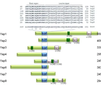
14 from Gcn4 are the amino acids that make contact with the DNA. Indeed, Ala239 is replaced by a Gln and Ser242 is replaced by Phe or Tyr. Furthermore, there are two family-specific residues, Gln234 and Ala241 (Fig.1.3). The consensus binding site of the members of the Yap family binding site was characterized as TTAC/GTAA for Yap1-Yap4 and termed the Yap site or Yap Response Element (YRE) [80]. So far the corresponding binding site for Yap5-Yap7 has not been characterized, and although Yap8 binds to the variant TGATTAATAATCA [81 and Amaral et al., unpublished results], the existence of other binding sequences cannot be excluded. Yap1 has high homology with Yap2; Yap4 with Yap6 and Yap5 with Yap7, being Yap8 the most divergent member at the level of the DNA binding domain [82].
15 Cad1 due to the acquisition of cadmium resistance in cells overexpressing this gene.
1.6. The role played by Yap family members in stress
response
Yap1, the best studied member of the family, is known to respond to oxidative stress [90]. Its expression is mainly accomplished at the level of sub-cellular localization [91]. Indeed, Yap1 shifts to the nucleus upon oxidative stress in a mechanism that is not regulated at the level of nuclear import because interaction with the importin PseI is not stress-dependent [92]. Kuge et al. demonstrated that nuclear retention of Yap1 is mediated by the cysteine-rich domain located at the C-terminus of the protein (c-CRD). Removal of this region generates a constitutively nuclear and active protein. In addition, three conserved cysteine residues (Cys598, Cys620 and Cys629) were identified as being important for this post-translational regulation [91]. Delaunay et al. showed in vivo that the Cys, Cys303 and Cys598 located in the N-terminus (named as n-CRD) and in the c-CRD respectively, are oxidized by in response to H2O2, forming an intramolecular disulfide bond that compromises the binding of the exportin Crm1 to the non-canonical NES (SDIDVDGLCSEL) leading to Yap1 nuclear retention [90].
16 Cys598 that is then resolved into the previously described Yap1 Cys303-Cys598 intramolecular bridge [93]. In contrast, Yap1 response to diamide is Orp1/Gpx3-independent and does not involve the n-CRD [94]. This is consistent with the notion that it possesses two redox centers. Indeed, the electrophile N-ethylmaleimide (NEM) and the quinone menadione, both an electrophile and a superoxide anion generator, have been shown to modify c-CRD cysteines independently of Gpx3, leading to Yap1 nuclear accumulation. Mass spectrometry analyses further revealed that NEM and possibly menadione binds directly to the Yap1 c-CRD thus occluding the NES [95].
The family member with highest homology with Yap1 is Yap2. In the laboratory, taking advantage of the homology in the CRD domain, that contains four conserved cysteines, the Yap1 c-CRD was swapped by Yap2 c-c-CRD. This constructs were fused to GFP and were used to transform yeast strains to monitor the sub-cellular localization as well as the specificity to activate Yap1 target genes. Despite the similarities of the Yap2 CRD with Yap1 c-CRD it was shown that the Yap1 with Yap2 c-c-CRD is only capable of responding to Cd but not to hydrogen peroxide. These data showed a specificity of Yap2 c-CRD towards the cadmium response. The determinants for cadmium response are in the c-CRD of Yap2 that has a role in the transcriptional activation of FRM2 the only Yap2 target that was found so far [96]. The Yap2
transactivation domain is also stimulated upon treatment with cadmium but not H2O2 [80].
YAP4 is induced under osmotic stress and this does not
17 transcription factor Msn2. YAP4 gene expression is also induced under several other stress conditions. Under oxidative stress conditions YAP4 expression is regulated by Yap1 and Msn2 [98]. It was also shown that Yap4 is phosphorylated under several stress conditions by the protein kinases PKA and Gsk3 in residues Thr192 and Ser196. Moreover Yap4 protein levels are reduced in the gsk3 mutant [99]. ChIP-chip assays showed that Yap4 binds to several targets upon osmotic stress, some of them are also recognized by Yap6 and Sko1 [100].
Yap5 regulates, the transcription of Ccc1 a vacuolar transporter responsible for accumulation of iron in the vacuole, in this way controlling the cytoplasmic iron levels [101]. It was reported that this protein is nuclear resident. However, the authors used a multicopy vector to overexpress the GFP-Yap5 fusion protein and this can lead to artefactual localizations. Indeed we found a canonic Nuclear Export Signal (NES) in this protein. Yap5 contains two cyteines rich domains in the C-terminus both with 4 cysteines. The mutation of 4 in the n-CRD and 3 residues in the C-CRD abrogates its transcriptional activity [101]. Cell cycle studies by ChIP-chip showed that Yap5 might have several other possible targets namely YCL033C a methionine-R-sulfoxide reductase that protects iron-sulphur clusters from oxidative inactivation. They also show that Yap5 is regulated by SBF (Swi4-Swi6 cell cycle box binding factor) [102].
18 Yap7 is described as nuclear resident protein since it has no NES or Cys residues [104].
19
1.8 References:
1. Rosen, B.P. and Z. Liu, Transport pathways for arsenic and selenium: a minireview. Environ Int, 2009. 35(3): p. 512-5. 2. Dujon, B., et al., Complete DNA sequence of yeast
chromosome XI. Nature, 1994. 369(6479): p. 371-8.
3. Goffeau, A., et al., Life with 6000 genes. Science, 1996.
274(5287): p. 546, 563-7.
4. Guerreiro, P., et al., Sequencing of a 9.9 kb segment on the right arm of yeast chromosome VII reveals four open reading frames, including PFK1, the gene coding for
Figure 1.3 Structural features of the Yap family members
20 succinyl-CoA synthetase (beta-chain) and two ORFs sharing homology with ORFs of the yeast chromosome VIII. Yeast, 1997. 13(3): p. 275-80.
5. Guerreiro, P. and C. Rodrigues-Pousada, Disruption and phenotypic analysis of six open reading frames from chromosome VII of Saccharomyces cerevisiae reveals one essential gene. Yeast, 2001. 18(9): p. 781-7.
6. Foury, F., Human genetic diseases: a cross-talk between man and yeast. Gene, 1997. 195(1): p. 1-10.
7. Szczypka, M.S., et al., A yeast metal resistance protein similar to human cystic fibrosis transmembrane conductance regulator (CFTR) and multidrug resistance-associated protein. J Biol Chem, 1994. 269(36): p. 22853-7. 8. Collins, F.S., Cystic fibrosis: molecular biology and therapeutic implications. Science, 1992. 256(5058): p. 774-9.
9. Shani, N. and D. Valle, A Saccharomyces cerevisiae homolog of the human adrenoleukodystrophy transporter is a heterodimer of two half ATP-binding cassette transporters. Proc Natl Acad Sci U S A, 1996. 93(21): p. 11901-6.
10. Hettema, E.H., et al., The ABC transporter proteins Pat1 and Pat2 are required for import of long-chain fatty acids into peroxisomes of Saccharomyces cerevisiae. Embo J, 1996. 15(15): p. 3813-22.
11. Bossier, P., et al., The yeast YKL741 gene situated on the left arm of chromosome XI codes for a homologue of the human ALD protein. Yeast, 1994. 10(5): p. 681-6.
12. Mosser, J., et al., Putative X-linked adrenoleukodystrophy gene shares unexpected homology with ABC transporters. Nature, 1993. 361(6414): p. 726-30.
13. Fields, S. and O. Song, A novel genetic system to detect protein-protein interactions. Nature, 1989. 340(6230): p. 245-6.
14. Outeiro, T.F. and F. Giorgini, Yeast as a drug discovery platform in Huntington's and Parkinson's diseases. Biotechnol J, 2006. 1(3): p. 258-69.
15. Lewis, J.G., R.P. Learmonth, and K. Watson, Induction of heat, freezing and salt tolerance by heat and salt shock in Saccharomyces cerevisiae. Microbiology, 1995. 141 ( Pt 3): p. 687-94.
21 Saccharomyces cerevisiae. FEBS Lett, 1996. 399(3): p. 259-63.
17. Wu, C., Clos, J., Giorgi, G., Haroun, R., Kim, S.-J., Rabidran, S., Westwood, T., Wisniewski and Yim G., Structure and regulation of heat shock transcription factor. The biology of heatshock proteins and molecular chaperones. , ed. R.I. Morimoto, Tissiere, A., and Georgopoulos ,C. and 1994, Cold Spring Harbor, N.Y.: Cold Spring Harbor Laboratory Press.
18. Jakobsen, B.K. and H.R. Pelham, Constitutive binding of yeast heat shock factor to DNA in vivo. Mol Cell Biol, 1988.
8(11): p. 5040-2.
19. Gorner, W., et al., Nuclear localization of the C2H2 zinc finger protein Msn2p is regulated by stress and protein kinase A activity. Genes Dev, 1998. 12(4): p. 586-97.
20. Martinez-Pastor, M.T., et al., The Saccharomyces cerevisiae zinc finger proteins Msn2p and Msn4p are required for transcriptional induction through the stress response element (STRE). Embo J, 1996. 15(9): p. 2227-35.
21. Schoen, A., et al., Arsenic toxicity at low doses: epidemiological and mode of action considerations. Toxicol Appl Pharmacol, 2004. 198(3): p. 253-67.
22. Borst, P. and M. Ouellette, New mechanisms of drug resistance in parasitic protozoa. Annu Rev Microbiol, 1995.
49: p. 427-60.
23. Waxman, S. and K.C. Anderson, History of the development of arsenic derivatives in cancer therapy. Oncologist, 2001. 6 Suppl 2: p. 3-10.
24. Bun-ya, M., et al., Two new genes, PHO86 and PHO87, involved in inorganic phosphate uptake in Saccharomyces cerevisiae. Curr Genet, 1996. 29(4): p. 344-51.
25. Liu, Z., E. Boles, and B.P. Rosen, Arsenic trioxide uptake by hexose permeases in Saccharomyces cerevisiae. J Biol Chem, 2004. 279(17): p. 17312-8.
26. Wysocki, R., et al., The glycerol channel Fps1p mediates the uptake of arsenite and antimonite in Saccharomyces cerevisiae. Mol Microbiol, 2001. 40(6): p. 1391-401.
27. Menezes, R.A., et al., Yap8p activation in Saccharomyces cerevisiae under arsenic conditions. FEBS Lett, 2004.
566(1-3): p. 141-6.
22 AP-1-like proteins Yap1p and Yap8p. Mol Biol Cell, 2004.
15(5): p. 2049-60.
29. Bobrowicz, P., et al., Isolation of three contiguous genes, ACR1, ACR2 and ACR3 involved in resistance to arsenic compounds in the yeast Saccharomyces cerevisiae. Yeast, 1997. 13(9): p. 819-823.
30. Mukhopadhyay, R., J. Shi, and B.P. Rosen, Purification and Characterization of Acr2p, the Saccharomyces cerevisiae Arsenate Reductase. J. Biol. Chem., 2000. 275(28): p. 21149-21157.
31. Wysocki, R., P. Bobrowicz, and S. Ulaszewski, The Saccharomyces cerevisiae ACR3 gene encodes a putative membrane protein involved in arsenite transport. J. Biol. Chem., 1997. 272(48): p. 30061-30066.
32. Ghosh, M., J. Shen, and B.P. Rosen, Pathway of AS(III) detoxification in Saccharomyces cerevisiae. Proc. Natl. Acad. Sci. USA, 1999. 96: p. 5001-5006.
33. Clemens, S., et al., Tolerance to toxic metals by a gene family of phytochelatin synthases from plants and yeast. Embo J, 1999. 18(12): p. 3325-33.
34. Vashisht, A.A., P.J. Kennedy, and P. Russell, Centaurin-like protein Cnt5 contributes to arsenic and cadmium resistance in fission yeast. FEMS Yeast Res, 2009. 9(2): p. 257-69. 35. Thorsen, M., et al., The MAPK Hog1p modulates
Fps1p-dependent arsenite uptake and tolerance in yeast. Mol Biol Cell, 2006. 17(10): p. 4400-10.
36. Sotelo J, R.-G.M., Mitogen-activated protein kinase Hog1 is essential for the response to arsenite in Saccharomyces cerevisiae. Eukaryot Cell., 2006. 10.
37. Fauchon, M., et al., Sulfur sparing in the yeast proteome in response to sulfur demand. Mol Cell, 2002. 9(4): p. 713-23. 38. Haugen, A.C., et al., Integrating phenotypic and expression
profiles to map arsenic-response networks. Genome Biol, 2004. 5(12): p. R95.
39. Thorsen, M., et al., Quantitative transcriptome, proteome, and sulfur metabolite profiling of the Saccharomyces cerevisiae response to arsenite. Physiol Genomics, 2007.
30(1): p. 35-43.
23 41. Jin, Y.H., et al., Global transcriptome and deletome profiles of yeast exposed to transition metals. PLoS Genet, 2008.
4(4): p. e1000053.
42. Ahmad, S., K.T. Kitchin, and W.R. Cullen, Arsenic species that cause release of iron from ferritin and generation of activated oxygen. Arch Biochem Biophys, 2000. 382(2): p. 195-202.
43. Martin, D.E. and M.N. Hall, The expanding TOR signaling network. Curr Opin Cell Biol, 2005. 17(2): p. 158-66.
44. Hosiner, D., et al., Arsenic toxicity to Saccharomyces cerevisiae is a consequence of inhibition of the TORC1 kinase combined with a chronic stress response. Mol Biol Cell, 2009. 20(3): p. 1048-57.
45. Vujcic, M., M. Shroff, and K.K. Singh, Genetic determinants of mitochondrial response to arsenic in yeast Saccharomyces cerevisiae. Cancer Res, 2007. 67(20): p. 9740-9.
46. Shi, W., et al., The role of arsenic-thiol interactions in metalloregulation of the ars operon. J Biol Chem, 1996.
271(16): p. 9291-7.
47. Meng, Y.L., Z. Liu, and B.P. Rosen, As(III) and Sb(III) uptake by GlpF and efflux by ArsB in Escherichia coli. J Biol Chem, 2004. 279(18): p. 18334-41.
48. Bennett, M.S., et al., Bacillus subtilis arsenate reductase is structurally and functionally similar to low molecular weight protein tyrosine phosphatases. Proc Natl Acad Sci U S A, 2001. 98(24): p. 13577-82.
49. Hsu, C.M. and B.P. Rosen, Characterization of the catalytic subunit of an anion pump. J Biol Chem, 1989. 264(29): p. 17349-54.
50. Lin, Y.F., J. Yang, and B.P. Rosen, ArsD: an As(III) metallochaperone for the ArsAB As(III)-translocating ATPase. J Bioenerg Biomembr, 2007. 39(5-6): p. 453-8. 51. Rosenberg, H., R.G. Gerdes, and K. Chegwidden, Two
systems for the uptake of phosphate in Escherichia coli. J Bacteriol, 1977. 131(2): p. 505-11.
52. Liu, Z., et al., Mammalian glucose permease GLUT1 facilitates transport of arsenic trioxide and methylarsonous acid. Biochem Biophys Res Commun, 2006. 351(2): p. 424-30.
53. Liu, Z., et al., Arsenic trioxide uptake by human and rat aquaglyceroporins. Biochem Biophys Res Commun, 2004.
24 54. Carbrey, J.M., et al., Reduced arsenic clearance and increased toxicity in aquaglyceroporin-9-null mice. Proc Natl Acad Sci U S A, 2009. 106(37): p. 15956-60.
55. Villa-Bellosta, R. and V. Sorribas, Role of rat sodium/phosphate cotransporters in the cell membrane transport of arsenate. Toxicol Appl Pharmacol, 2008.
232(1): p. 125-34.
56. Leslie, E.M., A. Haimeur, and M.P. Waalkes, Arsenic transport by the human multidrug resistance protein 1 (MRP1/ABCC1). Evidence that a tri-glutathione conjugate is required. J Biol Chem, 2004. 279(31): p. 32700-8.
57. Ambudkar, S.V., et al., Biochemical, cellular, and pharmacological aspects of the multidrug transporter. Annu Rev Pharmacol Toxicol, 1999. 39: p. 361-98.
58. Liu, J., et al., Multidrug-resistance mdr1a/1b double knockout mice are more sensitive than wild type mice to acute arsenic toxicity, with higher arsenic accumulation in tissues. Toxicology, 2002. 170(1-2): p. 55-62.
59. Bienert, G.P., et al., A subgroup of plant aquaporins facilitate the bi-directional diffusion of As(OH)3 and Sb(OH)3 across membranes. BMC Biol, 2008. 6: p. 26. 60. Lee DA, C.A., Schroeder JI., ars1, an Arabidopsis mutant
exhibiting increased tolerance to arsenate and increased phosphate uptake. Plant J. :, 2003. 35(5): p. 637-46.
61. Cobbett, C. and P. Goldsbrough, Phytochelatins and metallothioneins: roles in heavy metal detoxification and homeostasis. Annu Rev Plant Biol, 2002. 53: p. 159-82. 62. Verbruggen, N., C. Hermans, and H. Schat, Mechanisms to
cope with arsenic or cadmium excess in plants. Curr Opin Plant Biol, 2009. 12(3): p. 364-72.
63. Dhankher, O.P., et al., Hyperaccumulation of arsenic in the shoots of Arabidopsis silenced for arsenate reductase (ACR2). Proc Natl Acad Sci U S A, 2006. 103(14): p. 5413-8.
64. Abercrombie, J.M., et al., Transcriptional responses of Arabidopsis thaliana plants to As (V) stress. BMC Plant Biol, 2008. 8: p. 87.
65. Menezes, R.A., et al., Contribution of Yap1 towards Saccharomyces cerevisiae adaptation to arsenic-mediated oxidative stress. Biochem J, 2008. 414(2): p. 301-11.
25 transcription factors. J Biol Chem, 2003. 278(50): p. 49920-8.
67. Grant, C.M., F.H. MacIver, and I.W. Dawes, Glutathione synthetase is dispensable for growth under both normal and oxidative stress conditions in the yeast Saccharomyces cerevisiae due to an accumulation of the dipeptide gamma-glutamylcysteine. Mol Biol Cell, 1997. 8(9): p. 1699-707. 68. Grant, C.M., et al., Yeast glutathione reductase is required
for protection against oxidative stress and is a target gene for yAP-1 transcriptional regulation. Mol Microbiol, 1996.
21(1): p. 171-9.
69. Trotter, E.W. and C.M. Grant, Overlapping roles of the cytoplasmic and mitochondrial redox regulatory systems in the yeast Saccharomyces cerevisiae. Eukaryot Cell, 2005.
4(2): p. 392-400.
70. Arner, E.S. and A. Holmgren, Physiological functions of thioredoxin and thioredoxin reductase. Eur J Biochem, 2000. 267(20): p. 6102-9.
71. Kuge, S. and N. Jones, YAP1 dependent activation of TRX2 is essential for the response of Saccharomyces cerevisiae to oxidative stress by hydroperoxides. Embo J, 1994. 13(3): p. 655-64.
72. Morgan, B.A., et al., The Skn7 response regulator controls gene expression in the oxidative stress response of the budding yeast Saccharomyces cerevisiae. Embo J, 1997.
16(5): p. 1035-44.
73. Rhee, S.G., et al., Peroxiredoxin, a novel family of peroxidases. IUBMB Life, 2001. 52(1-2): p. 35-41.
74. Netto, L.E.S., et al., Removal of hydrogen peroxide by thiol-specific antioxidant enzyme (TSA) is involved with its antioxidant properties. TSA possesses thiol peroxidase activity. J Biol Chem, 1996. 271(26): p. 15315-21.
75. Molina, M.M., et al., Nuclear monothiol glutaredoxins of Saccharomyces cerevisiae can function as mitochondrial glutaredoxins. J Biol Chem, 2004. 279(50): p. 51923-30. 76. Carmel-Harel, O. and G. Storz, Roles of the glutathione-
and thioredoxin-dependent reduction systems in the Escherichia coli and saccharomyces cerevisiae responses to oxidative stress. Annu Rev Microbiol, 2000. 54: p. 439-61.
26 made in the cytoplasm. J Biol Chem, 1982. 257(5): p. 2713-8.
78. Sturtz, L.A., et al., A fraction of yeast Cu,Zn-superoxide dismutase and its metallochaperone, CCS, localize to the intermembrane space of mitochondria. A physiological role for SOD1 in guarding against mitochondrial oxidative damage. J Biol Chem, 2001. 276(41): p. 38084-9.
79. Shi, P., et al., Mitochondrial dysfunction in amyotrophic lateral sclerosis. Biochim Biophys Acta, 2009.
80. Fernandes, L., C. Rodrigues-Pousada, and K. Struhl, Yap, a novel family of eight bZIP proteins in Saccharomyces cerevisiae with distinct biological functions. Mol Cell Biol, 1997. 17(12): p. 6982-93.
81. Ilina, Y., et al., Characterization of the DNA-binding motif of the arsenic-responsive transcription factor Yap8p. Biochem J, 2008. 415(3): p. 467-75.
82. Tan, K., et al., A systems approach to delineate functions of paralogous transcription factors: role of the Yap family in the DNA damage response. Proc Natl Acad Sci U S A, 2008. 105(8): p. 2934-9.
83. Rodrigues-Pousada, C., T. Nevitt, and R. Menezes, The yeast stress response. Role of the Yap family of b-ZIP transcription factors. The PABMB Lecture delivered on 30 June 2004 at the 29th FEBS Congress in Warsaw. Febs J, 2005. 272(11): p. 2639-47.
84. Harshman, K.D., W.S. Moye-Rowley, and C.S. Parker, Transcriptional activation by the SV40 AP-1 recognition element in yeast is mediated by a factor similar to AP-1 that is distinct from GCN4. Cell, 1988. 53(2): p. 321-30.
85. Schnell, N. and K.D. Entian, Identification and characterization of a Saccharomyces cerevisiae gene (PAR1) conferring resistance to iron chelators. Eur J Biochem, 1991. 200(2): p. 487-93.
86. Haase, E., J. Servos, and M. Brendel, Isolation and characterization of additional genes influencing resistance to various mutagens in the yeast Saccharomyces cerevisiae. Curr Genet, 1992. 21(4-5): p. 319-24.
87. Hussain, M. and J. Lenard, Characterization of PDR4, a Saccharomyces cerevisiae gene that confers pleiotropic drug resistance in high-copy number: identity with YAP1, encoding a transcriptional activator [corrected]. Gene, 1991.
101(1): p. 149-52.
27 alleviates growth inhibition caused by 1,10-phenanthroline. J Biol Chem, 1993. 268(31): p. 23640-5.
89. Hirata, D., K. Yano, and T. Miyakawa, Stress-induced transcriptional activation mediated by YAP1 and YAP2 genes that encode the Jun family of transcriptional activators in Saccharomyces cerevisiae. Mol Gen Genet, 1994. 242(3): p. 250-6.
90. Delaunay, A., A.D. Isnard, and M.B. Toledano, H2O2 sensing through oxidation of the Yap1 transcription factor. Embo J, 2000. 19(19): p. 5157-66.
91. Kuge, S., N. Jones, and A. Nomoto, Regulation of yAP-1 nuclear localization in response to oxidative stress. Embo J, 1997. 16(7): p. 1710-20.
92. Isoyama, T., et al., Nuclear import of the yeast AP-1-like transcription factor Yap1p is mediated by transport receptor Pse1p, and this import step is not affected by oxidative stress. J Biol Chem, 2001. 276(24): p. 21863-9.
93. Delaunay, A., et al., A thiol peroxidase is an H2O2 receptor and redox-transducer in gene activation. Cell, 2002. 111(4): p. 471-81.
94. Kuge, S., et al., Regulation of the yeast Yap1p nuclear export signal is mediated by redox signal-induced reversible disulfide bond formation. Mol Cell Biol, 2001. 21(18): p. 6139-50.
95. Azevedo, D., et al., Two redox centers within Yap1 for H2O2 and thiol-reactive chemicals signaling. Free Radic Biol Med, 2003. 35(8): p. 889-900.
96. Azevedo, D., et al., The S. cerevisiae Yap1 and Yap2 transcription factors share a common cadmium-sensing domain. FEBS Lett, 2007. 581(2): p. 187-95.
97. Nevitt, T., et al., Expression of YAP4 in Saccharomyces cerevisiae under osmotic stress. Biochem J., 2004. 379: p. 367-374.
98. Nevitt, T., J. Pereira, and C. Rodrigues-Pousada, YAP4 gene expression is induced in response to several forms of stress in Saccharomyces cerevisiae. Yeast, 2004. 21(16): p. 1365-74.
99. Pereira, J., et al., Yap4 PKA- and GSK3-dependent phosphorylation affects its stability but not its nuclear localization. Yeast, 2009.
28 101. Li, L., et al., Yap5 is an iron-responsive transcriptional activator that regulates vacuolar iron storage in yeast. Mol Cell Biol, 2008. 28(4): p. 1326-37.
102. Horak, C.E., et al., Complex transcriptional circuitry at the G1/S transition in Saccharomyces cerevisiae. Genes Dev, 2002. 16(23): p. 3017-33.
103. Yang, X., et al., Functional isolation of the Candida albicans FCR3 gene encoding a bZip transcription factor homologous to Saccharomyces cerevisiae Yap3p. Yeast, 2001. 18(13): p. 1217-25.
104. Huh, W.K., et al., Global analysis of protein localization in budding yeast. Nature, 2003. 425(6959): p. 686-91.
105. Yujun Di, M.J.T., Regulation of the arsenic-responsive transcription
factor Yap8p involves the ubiquitin-proteasome
29
Chapter 2
Yap8 activation in
Saccharomyces
cerevisiae
under arsenic conditions
Regina A. Menezes, Catarina Amaral, Agnès Delaunay,
Michel Toledano, Claudina Rodrigues-Pousada
2.1 Author’s contribution to the article 29
2.2 Abstract 31
2.3 Introduction 31
2.4 Materials and Methods 32
2.5 Results and Discussion 36
2.6 Acknowledgments 46
2.7 References 47
2.1 Author’s contribution to the article
Bobrowicz et al have shown that Acr1/Yap8 is a regulator of genes
involved in arsenic detoxification in the budding yeast S. cerevisiae
[1]. However, the regulation and activity of Yap8 in cells exposed to
arsenic compounds was not investigated. In chapter 2 the
mechanisms responsible for Yap8 regulation and activity are
detailed. I performed analysis of the RNA levels of Yap8 potential
target genes ACR2 and ACR3 encoding respectively an arsenate
reductase and an arsenite efflux extrusion pump. Using wild type,
yap8, yap1 mutant strains Northern analyses were performed in
order to evaluate ACR2 and ACR3 dependence on Yap8 and
30 contributes, to a less extent, to the full activation of these genes. I
also performed Northern analysis to reveal if the expression of
YAP1 and YAP8 is altered under arsenic stress. The Heat shock
protein HSP26 was used as an indicator of general stress response
in the cell. In contrast to YAP1 mRNAs that increase under stress,
YAP8 mRNAs levels remain constant during the treatment.
Yap1 is regulated at the level of its sub-cellular localization, and
due to the presence of conserved Cys in Yap8 it was studied its
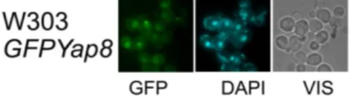
regulation using the construction GFP-Yap8. In order to do this I
performed fluorescence microscopy of cells with the GFP-Yap8
construction and of cells with GFP-Yap1. After adding arsenate or
arsenite, Yap8 accumulates in the nucleus as well as Yap1. Both
proteins are therefore regulated at the level of sub-cellular
localization. Tamás’s group showed that Yap8 is constitutively
localized in the nucleus, either in unstressed and stressed cells.
Nevertheless, in order to clarify these results I cloned the GFP
construction in a multi-copy plasmid and transformed it in a W303
strain obtaining the same result as this group. These results may
be due to the existence of an artefact because of the use of
overexpression vectors. Yap1 is regulated via its interaction of the
nuclear export signal (NES) with Crm1. When cells are subjected to
oxidative stress the C-terminal cysteines of Yap1 form a disulfide
bond masking the NES and thus the protein remains in the nucleus
upon stress. To study Yap8 localization and to evaluate if Crm1 is
also involved in Yap8 activity I have used a strain containing Crm1
under the control of the MET promoter. I show that Yap8 requires
the presence of Crm1 to be exported from the nucleus. I also
demonstrated that the cysteines conserved in Yap1 and Yap8 are
important for nuclear localization of both proteins. The sub-cellular
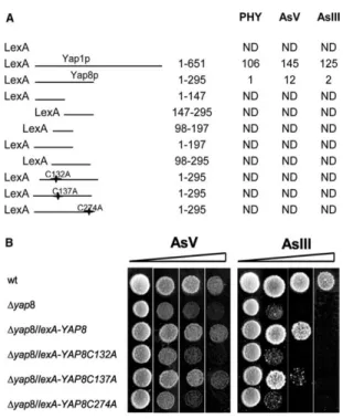
31 mutated cysteines GFP-Yap8 versions. The overall results show
that i.) like Yap1, Yap8 is only localized in the nucleus after stress
ii.) Crm1 is the exportin responsible for Yap1 and Yap8 shuttle
control between nucleus and cytoplasm and iii.) the Yap8 Cys
residues are relevant for Yap8 activity.
2.2 Abstract
Yap8, a member of the Saccharomyces cerevisiae Yap family, is
activated in response to arsenic. Both the mechanisms by which
this activation takes place and its regulation have not yet been
identified. In this report, we show that Yap8 is not activated at the
transcriptional level but, rather, its nuclear transport is actively
regulated and dependent on the exportin chromosome region
maintenance protein. In addition, it is shown that Cys132, Cys137
and Cys274 are essential for Yap8 localization and transactivation
function both of which are required for its biological activity.
2.3 Introduction
Saccharomyces cerevisiae arsenic compounds resistance (ACR)
cluster was isolated as loci that increase tolerance to arsenite [1].
The three contiguous genes identified as ACR1 (YAP8),ACR2 and
ACR3 encode a positive regulator, an arsenate-reductase and a
plasma membrane arsenite efflux protein, respectively [2-5]. ACR1,
also designated YAP8, belongs to the yeast AP-1 like factor (YAP)
family of transcription factors [6]. Members of the YAP family (Yap1
to Yap8) share a conserved b-ZIP DNA-binding domain, each
32 [7]. Yap8 could mediate arsenic stress responses by regulating the
expression of ACR2 and ACR3. Yeast cadmium factor 1 (Ycf1p) is
also involved in the arsenite detoxification and known for conferring
resistance to a variety of drugs through sequestering the
glutathione S-conjugates into the vacuole [8]. Yap1 [9], the
prototypical YAP gene family, which regulates the yeast oxidative
stress response regulon, also regulates the expression of YCF1.
Yap1 primary control lies in a redox-regulated Crm1-dependent
nuclear export [10-12]. Redox signals disrupt the Yap1–
chromosome region maintenance protein (Crm1p) interaction
through a redox dependent alteration of the Yap1 C-terminal
nuclear export signal (NES), thereby promoting Yap1 nuclear
accumulation. Data presented here shows that arsenic compounds
activate Yap8 at the level of both its nucleo-cytoplasmic shuttling
and its transactivation potential. Regulation of Yap8 nucleo
cytoplasmic shuttling involves arsenic-sensitive Crm1p-dependent
nuclear export and the Yap8 Cys residues located in positions 132,
137 and 274.
2.4 Materials and methods
2.4.1. Growth conditions
Yeast strains were grown in complete YPD (1% yeast extract, 2%
bactopeptone, 2% glucose) or selective media (SC or SD: 0.67%
ammonium sulfate-yeast nitrogen base without amino acids [Difco],
2% glucose), supplemented with the appropriate selective amino
acids. Early exponential phase cells (A600 = 0:4–0:5) were induced
by the addition of 2 mM Na2HAsO4 or NaAsO2 and samples
collected at the indicated time point. Samples for RNA and protein
![Figure 1.2 Arsenic entry and detoxification pathways in mammals. Adapted from [1]](https://thumb-eu.123doks.com/thumbv2/123dok_br/15769986.641182/29.748.243.562.473.810/figure-arsenic-entry-detoxification-pathways-mammals-adapted.webp)
![Table 3.1 Gene-ontology analyses of the arsenate transcriptional response T-profiler [38] was used to conduct gene-ontology analyses from the global set of microarray data (see the Experimental values)](https://thumb-eu.123doks.com/thumbv2/123dok_br/15769986.641182/83.748.121.638.282.849/ontology-analyses-arsenate-transcriptional-response-profiler-microarray-experimental.webp)