Research Report
AT1
receptor blockade in the lateral parabrachial nucleus
reduces the effects of muscimol on sodium intake
Camila Zambone C. Da Silva
a, José V. Menani
b, João C. Callera
a,⁎
aDepartment of Basic Science, School of Dentistry, UNESP, Univ. Estadual Paulista, Rodovia Marechal Rondom, km 527, 16018-805, Araçatuba, São Paulo, Brazil
bDepartment of Physiology and Pathology, School of Dentistry, UNESP, Araraquara, SP, Brazil
A R T I C L E I N F O A B S T R A C T
Article history: Accepted 2 June 2011 Available online 12 June 2011
The blockade of the lateral parabrachial nucleus (LPBN) with GABAA receptor agonist muscimol induces robust hypertonic NaCl and water intake by rats. In the present study we investigated the effects of previous injections of losartan (AT1 angiotensin receptor antagonist) into the LPBN on 0.3 M NaCl and water intake induced by muscimol injected bilaterally in the same area in fluid replete rats and in rats treated with the diuretic furosemide combined with a low dose of the angiotensin-converting enzyme inhibitor captopril injected subcutaneously. Male Wistar rats with stainless steel cannulas implanted bilaterally into the LPBN were used. Bilateral injections of muscimol (0.5 nmol/0.2μl, n = 8) into the LPBN in fluid replete rats induced 0.3 M NaCl intake (23.4 ± 4.1 vs. saline: 0.4 ± 0.4 ml/3 h) and water intake (9.3 ± 1.9 vs. saline: 0.7 ± 0.4 ml/3 h) and pre-treatment of the LPBN with losartan (50μg/0.2μl) reduced 0.3 M NaCl intake (3.3 ± 2.5 ml/3 h) and water intake (4.0 ± 2.9 ml/3 h) induced by muscimol. In rats treated with furosemide + captopril, pre-treatment with losartan into the LPBN attenuated the increase of 0.3 M NaCl intake produced by muscimol (12.8 ± 5.3, vs. saline + muscimol: 36.7 ± 6.7 ml/3 h) without changing water intake. Therefore, the results suggest that deactivation of LPBN inhibitory mechanisms by muscimol injections into the LPBN is facilitated by endogenous angiotensin II acting on AT1 receptors in the LPBN, which drives rats to ingest large amounts of hypertonic NaCl.
© 2011 Elsevier B.V. All rights reserved. Keywords:
GABA receptor Angiotensin II Losartan Sodium appetite Thirst
Lateral parabrachial nucleus
1.
Introduction
Important mechanisms for the control of sodium and water intake are present in the lateral parabrachial nucleus (LPBN), a pontine structure located dorsolaterally to the superior cerebellar peduncle (Andrade et al., 2006; Callera et al., 2005; De Luca et al., 2003; De Oliveira et al., 2007; Menani et al., 2002; Menani and Johnson, 1995). The LPBN is reciprocally
con-nected to forebrain areas, such as the paraventricular nucleus of the hypothalamus (PVN), the central nucleus of the amygdala (CeA) and the median preoptic nucleus (MnPO), and to medullary regions, like the area postrema (AP) and the medial portion of the nucleus of the solitary tract (mNTS) (Ciriello et al., 1984; Fulwiler and Saper, 1984; Herbert et al., 1990; Jhamandas et al., 1992, 1996; Norgren, 1981). Therefore, the LPBN may convey signals that ascend from AP/mNTS to
⁎Corresponding author at:Department of Basic Science, School of Dentistry, Universidade Estadual Paulista (UNESP), Rodovia Marechal Rondom, km 527, Building 31, CEP: 16018-805, Araçatuba, São Paulo, Brazil. Fax: +55 18 3636 3332.
E-mail address:[email protected](J.C. Callera).
0006-8993/$–see front matter © 2011 Elsevier B.V. All rights reserved. doi:10.1016/j.brainres.2011.06.004
a v a i l a b l e a t w w w . s c i e n c e d i r e c t . c o m
the forebrain areas that regulate fluid and electrolyte balance and related behaviors like water and sodium intake.
Numerous neurotransmitter systems have implicated the LPBN in the control of sodium intake. For example, bilateral LPBN injections of methysergide, a serotonergic receptor antagonist, increase hypertonic NaCl intake induced by angiotensin II (ANG II) administered intracerebroventricularly (i.c.v.) or into the subfornical organ (SFO), by 24 h of water deprivation, by 24 h of sodium depletion or by deoxycorticos-terone acetate (DOCA) (De Gobbi et al., 2001; Menani et al., 1996, 1998a, 2000; Menani and Johnson, 1995). Blockade of cholecystokinin (CCK) or serotonin receptors or activation of α2-adrenergic receptors in the LPBN enhances NaCl intake by rats injected subcutaneously with the diuretic furosemide (FURO) combined with the angiotensin converting enzyme inhibitor captopril (CAP) (Andrade et al., 2004; De Gobbi et al., 2001; Menani et al., 1996, 1998b). The blockade of LPBN neurons with bilateral injections of the GABAA agonist muscimol induces robust ingestion of hypertonic NaCl and slight ingestion of water in fluid replete rats and increases FURO + CAP- and 24 h sodium depletion-induced sodium intake, suggesting that a GABAergic mechanism present in LPBN is involved in the control of sodium intake (Callera et al., 2005; De Oliveira et al., 2007).
The cardiovascular, neuroendocrine and ingestive effects of ANG II acting centrally are mediated mainly by angiotensin type 1 (AT1) receptors located in different areas of the central nervous system, such as the LPBN, anterior hypothalamic area (AHA), amygdala, SFO, rostral and caudal ventrolateral me-dulla and NTS (Fitzsimons, 1998; Fregly and Rowland, 1991; Mckinley et al., 1996; Rowland et al., 1992; Thunhorst and Fitts, 1994). The nonpeptide antagonist losartan selectively binds on AT1receptors (Chiu et al., 1989).
Studies using whole cell voltage-clamp techniques have suggested that ANG II acting on AT1receptors may modulate GABAergic synaptic transmission and produce opposite ef-fects, depending on whether pre- or post-synaptic AT1 re-ceptors are activated (Henry et al., 2009; Li et al., 2003; Li and Pan, 2005; Xing et al., 2009). It has been suggested that ANG II acting on pre-synaptic AT1 receptors reduces GABA release and decreases the amplitude of evoked GABAergic inhibitory post-synaptic currents (IPSCs) (Li et al., 2003; Li and Pan, 2005; Xing et al., 2009). In contrast, it was shown that endogenous ANG II acting on post-synaptic AT1receptors increases IPSCs in sodium-sensitive neurons in the median preoptic nucleus (MnPO) (Henry et al., 2009). According to these studies, treatment with ANG II increased the firing of PVN neurons that project to the rostroventrolateral medulla (RVLM) and decreased the amplitude of evoked GABAergic IPSCs and the frequency of miniature IPSCs, effects blocked by the AT1 receptor antagonist losartan (Li et al., 2003; Li and Pan, 2005). Treatment with ANG II also decreased the amplitude of evoked IPSCs and the frequency of miniature IPSCs in neurons of the dorsolateral periaqueductal gray, an effect blocked by losartan, but not by the AT2antagonist PD123319 (Xing et al., 2009). Treatment with ANG II had no effect on excitatory post synaptic currents in the PVN neurons or in the dorsolateral periaqueductal gray (Li et al., 2003; Li and Pan, 2005; Xing et al., 2009). In contrast, another study showed that the amplitude of the IPSCs in the MnPO was reduced by the treatment with
losartan, suggesting a post synaptic action of endogenous ANG II that facilitated the effect of the GABAergic input to the MnPO (Henry et al., 2009).
Considering the effects of the activation of GABAA re-ceptors in the LPBN on hypertonic NaCl and water intake (Callera et al., 2005; De Oliveira et al., 2007) and the results of previous studies showing that AT1 receptor activation may modulate the action of the GABAergic mechanisms (Henry et al., 2009; Li et al., 2003; Li and Pan, 2005; Xing et al., 2009), in the present study we investigated the effects of injections of the specific AT1receptor antagonist, losartan, into the LPBN on water and hypertonic NaCl intake induced by the activation of GABAAreceptors by muscimol injections into the LPBN in fluid replete or FURO + CAP-treated rats.
2.
Results
2.1. Histological analysis
Fig. 1 is a photomicrograph of a transverse section of the brainstem of one rat, representative of the groups tested, showing the typical bilateral injection sites in the LPBN. The injections were centered in the central lateral and dorsal lateral portions of the LPBN (seeFulwiler and Saper, 1984for definitions of LPBN subnuclei). In some rats, LPBN injections reached the ventral lateral and external lateral portions, as well as the Kölliker–Fuse nucleus. The sites of injections were similar to those in previous studies that showed the effects of LPBN injections of methysergide, proglumide, moxonidine or muscimol on water and 0.3 M NaCl intake (Andrade et al., 2006; Callera et al., 2005; De Gobbi et al., 2001; De Luca et al., 2003; De Oliveira et al., 2007). In some rats, injections spread to the brachium (superior cerebellar peduncle), or slightly ventral to this structure, reaching the dorsal portions of the medial parabrachial nucleus (MPBN) uni- or bilaterally. There was no difference in the effects if the injections were restricted to the LPBN or if they spread to the ventral structures described above.
Fig. 1 – Photomicrograph of a brain slice from one rat
2.2. Effects of combined injections of losartan and muscimol into the LPBN on water and 0.3 M NaCl intake in fluid replete rats
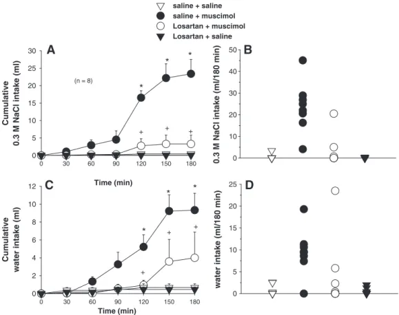
ANOVA showed significant differences between treatments for 0.3 M NaCl [F(3, 21) = 20.7;P< 0.001] and water intake [F(3, 21) = 9.05;P< 0.001] in fluid replete rats that received injections of saline or losartan combined with injections of saline or muscimol into the LPBN (Fig. 2).
In two-bottle tests, bilateral injections of muscimol (0.5 nmol/0.2μl at each site, n = 8) into the LPBN in fluid replete rats induced 0.3 M NaCl intake (23.4± 4.1 ml/3 h, vs. saline + saline: 0.4 ± 0.4 ml/3 h,Figs. 2A and B) and water intake (9.3 ± 1.9 ml/3 h, vs. saline + saline: 0.7 ± 0.4 ml/3 h,Figs. 2C and D). Previous injections of the AT1 receptor antagonist losartan (50μg/0.2μl each site) into the LPBN reduced the effects of muscimol (0.5 nmol/0.2μl) injected in the same area on 0.3 M NaCl intake (3.3 ± 2.5 ml/3 h,Figs. 2A and B) and water intake (4.0 ± 2.9 ml/3 h,Figs. 2C and D).
The ingestion of 0.3 M NaCl and water after bilateral injections of muscimol into the LPBN in replete rats was significantly different from those after saline injected into the LPBN (control) from 120 min to the end of the test (180 min) and the pre-treatment with losartan injected into the LPBN reduced the ingestion of 0.3 M NaCl and water in the same period (Figs. 2A and C).
Losartan injected alone into the LPBN did not affect water or 0.3 M NaCl intake.
2.3. Effects of combined injections of losartan and muscimol into the LPBN on FURO + CAP-induced 0.3 M NaCl and water intake
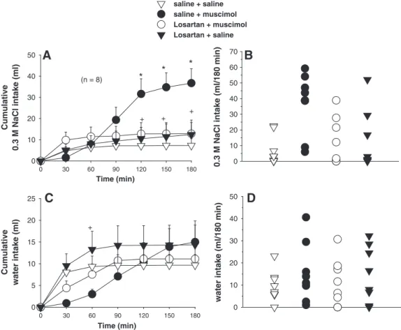
ANOVA showed significant interactions between treatments and times for 0.3 M NaCl intake [F(18, 126)= 9.5;P< 0.001] and for water intake [F(18, 126)= 4.1;P<0.001] induced by FURO+CAP in rats that received injections of saline or losartan combined with injections of saline or muscimol into the LPBN (Fig. 3).
Bilateral injections of muscimol (0.5 nmol/0.2μl at each site, n = 8) into the LPBN increased FURO + CAP-induced 0.3 M NaCl intake from 120 min to the end of the test (36.7 ± 6.7 ml/3 h, vs. saline + saline: 7.2 ± 3.3 ml/3 h) (Figs. 3A and B). Losartan (50μg/0.2μl at each site) injected into the LPBN reduced the effects of muscimol on 0.3 M NaCl intake from 120 min to the end of the test (12.8± 5.3 ml/3 h) (Figs. 3A and B). Losartan injected alone into the LPBN produced no change in FURO + CAP-induced 0.3 M NaCl intake.
A tendency toward the reduction of FURO + CAP-induced water intake at 30 and 60 min of the test occurred after injections of muscimol into the LPBN, an effect partially reversed by pre-treatment with losartan (Fig. 3C). Losartan injected alone into the LPBN produced no significant change in
Time (min)
0 30 60 90 120 150 180
Cumulative
0.3 M NaCl intake (ml)
0 5 10 15 20 25 30
(n = 8)
Time (min)
0 30 60 90 120 150 180
Cumulative
water intake (ml)
0 2 4 6 8 10 12
*
+
A
C
0.3 M NaCl intake (ml/180 min) 0
10 20 30 40 50
saline + saline saline + muscimol Losartan + muscimol Losartan + saline
B
water intake (ml/180 min)
0 5 10 15 20
25
D
*
+
+
+
*
*
*
+
*
+
Fig. 2–(A) Cumulative 0.3 M NaCl intake; (B) individual 0.3 M NaCl intakes (180 min); (C) cumulative water intake; (D) individual water intakes (180 min) by fluid replete rats treated with losartan (50μg/0.2μl) or saline combined with muscimol
(0.5 nmol/0.2μl) or saline bilaterally into the LPBN. (A and C) Values are means ± S.E.M. n = number of rats. * = different from
FURO + CAP-induced water intake compared to the treatment with saline (Figs. 3C and D). However, opposite effects on water intake after losartan and muscimol were injected alone into the LPBN resulted in a significant difference between these treatments at 60 min of the test (Fig. 3C).
2.4. Specificity of injections into the LPBN on water and 0.3 M NaCl intake
Results from rats that received injections outside the LPBN (misplaced injections) were analyzed to show that the effects Time (min)
0 30 60 90 120 150 180
Cumulative
0.3 M NaCl intake (ml)
0 10 20 30 40 50
Time (min)
0 30 60 90 120 150 180
Cumulative
water intake (ml)
0 5 10 15 20 25
*
+
A
C
0.3 M NaCl intake (ml/180 min) 0
10 20 30 40 50 60 70
saline + saline saline + muscimol Losartan + muscimol Losartan + saline
B
water intake (ml/180 min)
0 10 20 30 40
50
D
*
+
*
+ (n = 8)
+
Fig. 3–(A) Cumulative 0.3 M NaCl intake; (B) individual 0.3 M NaCl intakes (180 min); (C) cumulative water intake; (D) individual water intakes (180 min) by FURO + CAP-treated rats that received losartan (50μg/0.2μl) or saline combined with muscimol
(0.5 nmol/0.2μl) or saline bilaterally into the LPBN. (A and C) Values are means ± S.E.M. n = number of rats. * = different from
saline + saline. + = different from saline + muscimol.
Table 1–Ingestion of water and 0.3 M NaCl by fluid replete rats or FURO + CAP-treated rats that received saline or losartan combined with saline or muscimol in sites outside the LPBN (misplaced injections).
Fluid replete rats 0.3 M NaCl intake Water intake
(ml/3 h) (ml/3 h)
Saline + saline 1.7 ± 0.3 3.4 ± 0.3
Saline + muscimol 3.2 ± 0.7 2.5 ± 0.5
Losartan + muscimol 2.7 ± 0.6 2.8 ± 0.5
Losartan + saline 1.6 ± 0.2 1.5 ± 0.3
FURO + CAP-treated rats
Saline + saline 4.3 ± 1.3 7.1 ± 2.5
Saline + muscimol 7.8 ± 2.8 6.0 ± 2.3
Losartan + muscimol 9.4 ± 5.1 5.4 ± 2.5
Losartan + saline 3.6 ± 1.8 2.6 ± 1.0
on 0.3 M NaCl and water intake were due to a specific activation of GABAAreceptors in the LPBN. Bilateral injections of muscimol or losartan alone or of muscimol combined with losartan in sites outside the LPBN did not affect water and 0.3 M NaCl intake in fluid replete rats or FURO + CAP-treated rats (Table 1). ANOVA showed no significant differences between treatments for 0.3 M NaCl intake [F(3, 12) = 3.0; P> 0.05] or water intake [F(3, 12) = 4.0;P> 0.05] in fluid replete rats or between treatments for 0.3 M NaCl intake [F(3, 15) = 0.9; P> 0.05] or water intake [F(3, 15) = 3.3;P> 0.05] in FURO + CAP-treated rats that received injections in sites outside the LPBN. Misplaced injections were ventral (MPBN), dorsal or rostral to the LPBN. Some rats had unilateral injections partially into the LPBN.
3.
Discussion
Similar to previous results (Callera et al., 2005; De Oliveira et al., 2007), the present study shows that bilateral injections of muscimol (GABAA receptor agonist) into the LPBN induce hypertonic NaCl and water ingestion in fluid replete rats (untreated rats) and increase 0.3 M NaCl intake in FURO + CAP-treated rats. The involvement of GABAAreceptors of the LPBN in the control of water and NaCl intake is supported by a previous study showing that the GABAAreceptor antagonist bicuculline injected into the LPBN completely blocked water and hypertonic NaCl intake induced by muscimol, which suggests that muscimol activates LPBN GABAAreceptors to increase sodium intake (Callera et al., 2005). The present results extend the conclusions of the previous study by showing that pretreatment of the LPBN with bilateral in-jections of the nonpeptide AT1receptor antagonist losartan reduce water and 0.3 M NaCl intake caused by muscimol injected into the same site in fluid replete rats, as well as the increase in 0.3 M NaCl produced by muscimol injected bilaterally into the LPBN in FURO + CAP-treated rats. Injections of losartan alone into the LPBN did not change water or 0.3 M NaCl intake by untreated rats or FURO + CAP-treated rats. Results from rats with misplaced injections confirm that muscimol effects on water and 0.3 M NaCl intake are specific to the LPBN. The results also suggest that angiotensinergic mechanisms in the LPBN are essential for the dipsogenic and natriorexigenic responses induced by the blockade of LPBN neurons with muscimol in fluid replete rats or the increase in the natriorexigenic responses produced by muscimol injected into the LPBN in FURO + CAP-treated rats.
Pretreatment with losartan into the LPBN reduced musci-mol effects on water and/or NaCl intake by fluid replete or FURO + CAP-treated rats. Therefore, if endogenous GABA release in the LPBN was important for FURO + CAP-induced water and sodium intake, similar effects would be expected when losartan alone was injected in FURO + CAP-treated rats. However, injections of losartan alone did not modify FURO + CAP-induced water or NaCl intake, suggesting that GABA release or its interaction with activated AT1receptors in the LPBN is not essential for sodium or water intake induced by FURO + CAP. Perhaps any reduction of GABA effects by losartan was compensated for by changes in the release of other neurotransmitters in the LPBN like serotonin, CCK,
cortico-tropin-releasing hormone (CRF), glutamate, opioids or nor-adrenaline that also modulate water and sodium intake (De Castro e Silva et al., 2006; De Oliveira et al., 2008; De Gobbi et al., 2009; Gasparini et al., 2009; Menani et al., 1996; Menani and Johnson, 1998).
AT1receptors and ANG II terminals are present in the LPBN (Lenkei et al., 1997; Mckinley et al., 2002); however, we found no clear evidence in the literature of ANG II effects in the LPBN. The present results suggest that ANG II acting on AT1 receptors in LPBN is necessary for the full effects of muscimol injected into the LPBN on water and sodium intake. The treatment with FURO + CAP increases ANG II centrally ( Thun-horst and Johnson, 1994). However, it is possible that activation of AT1 receptors by baseline levels of ANG II in fluid replete rats is sufficient to facilitate the increase in water and sodium intake produced by muscimol in the LPBN. On the other hand, although there is no evidence that injections of muscimol into the LPBN increase ANG II levels, the present results do not allow us to exclude the possibility of an increase in central or peripheral levels of ANG II due to muscimol injections into the LPBN. The ingestion of sodium after muscimol injections into the LPBN takes at least 1 h to start, which is time enough for changes in the levels of ANG II within the LPBN that may intensify the effects of muscimol on LPBN neurons, a step necessary for the release of sodium intake.
The cardiovascular, neuroendocrine and ingestive effects of ANG II acting centrally are mediated mainly by AT1 receptors (Fitzsimons, 1998; Kirby et al., 1992; Mckinley et al., 1996; Rowland et al., 1992; Saavedra, 1994; Thunhorst and Fitts, 1994). For example, ingestion of water and NaCl is suggested to depend on the action of circulating ANG II on circumventricular organs like the SFO and OVLT (Krause et al., 2008; Morris et al., 2002; Thunhorst and Fitts, 1994). At the same time, AT1 receptors have a role in mediating an enhanced sodium intake produced by blockade of LPBN inhibitory mechanisms with injections of the serotonergic antagonist methysergide (Colombari et al., 1996; Menani et al., 1998b). More specifically, injections of methysergide into the LPBN combined with treatments that increase ANG II centrally or peripherally, such as FURO + CAP sc, isoproterenol or acute (1 h previous) treatment with FURO, also produce robust ingestion of 0.3 M NaCl (Menani et al., 1998a, 2000). Whereas treatment with FURO + CAP alone induces significant ingestion of NaCl, sc treatments with isoproterenol or acute furosemide do not produce significant ingestion of NaCl, despite increases in ANG II signaling, unless LPBN inhibitory mechanisms are deactivated. Therefore, sodium intake does not always increase even with increased levels of ANG II. However, if the LPBN inhibitory mechanisms are deactivated, then ANG II-induced sodium and water intake is strongly facilitated.
which might suggest that sodium intake easily arises only when facilitatory mechanisms are activated and inhibitory mechanisms are simultaneously deactivated. However, in contrast to the blockade of the other neurotransmitters orα2 adrenoceptor activation, either opioid (β endorphin) or GABAergic (muscimol) activation of the LPBN induces robust ingestion of water and 0.3 M NaCl in fluid replete rats, suggesting that the deactivation of LPBN inhibitory mecha-nisms alone is sufficient to drive rats to ingest hypertonic NaCl (Callera et al., 2005; De Oliveira et al., 2007, 2008). Substantial ingestion of sodium starts ~2–3 h after muscimol injections into the LPBN in untreated rats (Callera et al., 2005, present results). The present results also show an increased sodium intake 2–3 h after injections of muscimol into the LPBN in FURO + CAP-treated rats.
Injections of muscimol into the LPBN produces a small increase on arterial pressure and non-significant effects on renal excretion in fluid replete rats (Callera et al., 2005; De Oliveira et al., 2007), which suggests that sodium intake produced by muscimol into the LPBN is not secondary to decreases in blood pressure or an increase in urinary sodium excretion. Rather, ingestion of hypertonic NaCl solutions increases the activity of LPBN neurons, suggesting that the LPBN can be activated by taste and/or visceral stimuli (Franchini and Vivas, 1999; Yamamoto et al., 1993). Signals from volume, taste and other visceral receptors that may participate in the control of water and sodium intake reach the AP/mNTS before ascending to the LPBN which, in turn, sends projections to forebrain areas involved in the control of fluid and electrolyte balance, such as the SFO, MnPO, PVN and amygdala (Ciriello et al., 1984; Jhamandas et al., 1992; Krukoff et al., 1993; Norgren, 1981; Shapiro and Miselis, 1985). A recent study showed that bilateral lesions of the CeA abolished water and 0.3 M NaCl intake produced by the blockade of LPBN neurons with muscimol in fluid replete rats, suggesting that facilitatory mechanisms present in the CeA are essential for the dipsogenic and natriorexigenic responses induced by muscimol injected into the LPBN (Andrade-Franzé et al., 2010). Consistent with this idea, the CeA contains AT1receptors and has been proposed as a possible site of interaction between ANG II and mineralocorticoids to stimulate sodium appetite (Galaverna et al., 1992; McKinley et al., 2002).
AT1 receptors are present in different areas of the brain, including the LPBN (Fitzsimons, 1998; Mckinley et al., 1996). Modulation of GABAergic neurotransmission by ANG II de-pends upon whether the AT1receptors are located pre- or post-synaptically. Activation of pre-synaptic AT1receptors reduce the effects of GABAergic activation, whereas activation of post-synaptic AT1receptors increase the effects (Henry et al., 2009; Li et al., 2003; Li and Pan, 2005; Xing et al., 2009). The present results show that blockade of AT1receptors by the injection of losartan into the LPBN reduces hypertonic NaCl and water intake stimulated by the activation of LPBN GABAAreceptors with muscimol injected in the same area in fluid replete or in FURO+CAP-treated rats. Thus, it appears that ANG II acts on post-synaptic AT1 receptors in the LPBN to enhance the activation of GABA receptors with muscimol via a mechanism similar to that described in the MnPO (Henry et al., 2009). Taken together, these results suggest that interactions of angiotensi-nergic and GABAergic mechanisms in the LPBN are important
to stimulate sodium intake. In other words, the action of ANG II on AT1receptors in the LPBN is important for the inhibition of LPBN neurons, thereby facilitating sodium intake produced by activation of GABAergic mechanisms in the LPBN.
4.
Experimental procedures
4.1. Animals
Male Wistar rats weighing 290–310 g were used. The animals were housed in individual stainless steel cages with free access to standard sodium diet (Guabi Rat Chow, Paulinia, SP, Brazil), water and 0.3 M NaCl solution. The positions of the bottles containing water and 0.3 M NaCl were rotated daily to avoid place preference. Room temperature was maintained at 23 ± 2 °C and humidity was maintained at 55 ± 10% on a 12:12 light–dark cycle with light onset at 07:30 AM.
The procedures were approved by the Institutional Ethical Committee for Animal Care from the School of Dentistry, UNESP, Araçatuba, Brazil (Proc. CEEA no. 986/2007) and followed the recommendations from the Brazilian College of Animal Experimentation (COBEA) and the American National Institute of Health Guide for the Care and Use of Laboratory Animals (NIH publications No. 80–23, 1996, USA).
All efforts were made to minimize animal discomfort and the number of animals used.
4.2. Cerebral cannulas
Rats were anesthetized with subcutaneous (sc) ketamine (80 mg/kg of body weight, Cristália, Brazil) combined with xylazine (7 mg/kg of body weight, Agener, Brazil) and placed in a stereotaxic instrument (Kopf, USA). The skull was leveled between bregma and lambda. Stainless steel guide-cannulas (12 × 0.6 mm o.d.) were implanted bilaterally into the LPBN using the following coordinates: 9.2 mm caudal to bregma, 2.2 mm lateral to the midline, and 3.8 mm below the dura mater (Paxinos and Watson, 1997). The tips of the cannulas were positioned 2 mm above each LPBN. The cannulas were fixed to the cranium using dental acrylic resin and jeweler screws and were filled with 30-gauge metal obturators between tests. After the surgery, the rats received intramus-cular injections of the analgesic cetoprophen 1% (0.03 ml) and a prophylactic dose of the antibiotic penicillin (30,000 IU). Rats were allowed to recover for 5 days before starting ingestion tests and during this period they had free access to standard sodium diet, water and 0.3 M NaCl solution.
4.3. Injections into the LPBN
injection volume into the LPBN was 0.2μl on each site. The obturators were replaced after the injections, and the rats were placed back into their cages.
4.4. Drugs
Furosemide (FURO) (Sigma-Aldrich, Saint Louis, MO, USA) was dissolved in alkaline saline (pH adjusted to 9.0) and adminis-tered sc at the dose of 10 mg/kg of body weight (bw). Captopril (CAP) (Sigma-Aldrich, Saint Louis, MO, USA), was dissolved in 0.15 M NaCl and administered sc at the dose of 5 mg/kg of bw. Muscimol HBr and losartan potassium (Sigma-Aldrich, Saint Louis, MO, USA) were dissolved in 0.15 M NaCl. The dose of muscimol used in the present study was the same as that used in previous studies that investigated the effects of muscimol injected into the LPBN on water and 0.3 M NaCl intakes (Callera et al., 2005; De Oliveira et al., 2007). This dose of muscimol produces a long-lasting action (at least for 1 h) when injected into the LPBN (Callera et al., 2005). The dose of losartan was based on previous studies that have tested the effects of central injections of losartan on water and sodium intake and on the pressor response to ANG II (Grippo et al., 2002; Menani et al., 2004). The dose of losartan used is effective for at least 2 h (Menani et al., 2004).
4.5. Water and 0.3 M NaCl intake by fluid replete rats
The rats were tested in their home cages. Water and 0.3 M NaCl were provided from burettes with 0.1-ml divisions that were fitted with metal drinking spouts. Food was not available during the tests. Measurements were taken at 30-min in-tervals for 180 min, starting 10 min after bilateral injections of muscimol (0.5 nmol/0.2μl) or saline (0.2μl) into the LPBN.
Fluid replete rats that received no pre-treatment (n = 14), were tested for the effects of the combination of losartan and muscimol injections into the LPBN on water and 0.3 M NaCl intake. Losartan (50μg/0.2μl) was injected into the LPBN 10 min before muscimol (0.5 nmol/0.2μl). These rats were submitted to four tests and received the following combina-tions of treatments into the LPBN: saline + saline, saline + muscimol, losartan + muscimol and losartan+ saline. In each test, the group of rats was divided in two and half of the group received one of the combination of treatments listed above, while the remaining animals received another combination of treatments into the LPBN. The sequence of the treatments was randomized for each rat so that, at the end of testing, rats had received all four treatments. A recovery period of at least 2 days was allowed between tests.
4.6. Water and 0.3 M NaCl intake by FURO + CAP-treated rats
Another group of rats (n = 14) was used to test water and 0.3 M NaCl intake induced by treatment with FURO + CAP sc. On the day of the experiment, food, water and 0.3 M NaCl were removed and the cages were rinsed with water. Rats received sc injections of the diuretic FURO (10 mg/kg bw) plus CAP (5 mg/kg bw) as described previously (Callera et al., 2005; De Gobbi et al., 2001; Menani et al., 1996; Thunhorst and Johnson, 1994). One hour after FURO + CAP treatment, burettes with water and 0.3 M NaCl solution were returned and
measure-ments were taken at 30-min intervals for 180 min (sodium appetite test). Ten minutes before access to water and 0.3 M NaCl, rats received bilateral injections of muscimol (0.5 nmol/0.2μl) or saline into the LPBN. Bilateral injections of losartan (50μg/0.2μl) or saline into the LPBN were performed 10 min before the injections of muscimol or saline into the LPBN. In each experimental session, the group of rats was divided in two and each half of the group received one of the four treatments in the LPBN: saline + saline, saline + muscimol, losartan + muscimol and losartan + saline. The sequence of the treatments was in a randomized order so that at the end of testing, rats had received all four treatments. A recovery period of at least 3 days was allowed between experimental sessions. The order of treatments was random-ized because repeated FURO + CAP injections enhances stim-ulated and spontaneous NaCl intake (Pereira et al., 2010).
4.7. Histology
At the end of the experiments, the animals received bilateral injections of 2% Evans blue dye solution (0.2μl/injection site) into the LPBN. They were then deeply anesthetized with sodium thiopental (CRISTALIA, Itapira, SP, Brazil, 80 mg/kg of body weight) and perfused transcardially with saline followed by 10% formalin. The brains were removed, fixed in 10% formalin, frozen, cut in 60μm sections, stained with Giemsa, and analyzed by light microscopy to confirm the injection sites in the LPBN.
4.8. Data analysis
The results are reported as means± S.E.M. Water and 0.3 M NaCl intake was analyzed by two-way analysis of variance (ANOVA) with repeated measures for both factors (treatments and times), followed by Newman–Keuls post hoc test. Differences were considered significant at P< 0.05. The soft-ware used for the analysis was SigmaStat for Windows, version 2.03 from SPSS Inc.
Acknowledgments
The authors thank Arnaldo Cesar dos Santos for animal care. This research was supported by Brazilian public funding from Sao Paulo State Research Foundation (FAPESP) and Fundação para o Desenvolvimento da UNESP (FUNDUNESP). Camila Zambone C. Da Silva was a recipient of graduate fellowships from FAPESP (grant 07/56280-0).
R E F E R E N C E S
Andrade, C.A., Barbosa, S.P., De Luca Jr., L.A., Menani, J.V., 2004. Activation of alpha2-adrenergic receptors into the lateral parabrachial nucleus enhances NaCl intake in rats Neuroscience 129, 5–34.
Andrade-Franzé, G.M.F., Andrade, C.A., De Luca Jr., L.A., De Paula, P.M., Colombari, D.S.A., Menani, J.V., 2010. Lesions in the central amygdala impair sodium intake induced by the blockade of the lateral parabrachial nucleus. Brain Res. 21, 57–64.
Callera, J.C., De Oliveira, L.B., Barbosa, S.P., Colombari, D.S.A., De Luca Jr., L.A., Menani, J.V., 2005. GABAAreceptor activation in the lateral parabrachial nucleus induces water and hypertonic NaCl intake. Neuroscience 134 (3), 725–735.
Chiu, A.T., Herblin, W.F., Mccall, D.E., Ardecky, R.J., Carini, D.J., Duncia, J.V., Pease, L.J., Wong, P.C., Wexler, R.R., Johnson, A.L., Timmermans, P.B.M.W.M., 1989. Identification of angiotensin II receptor subtypes. Biochem. Biophys. Res. Commun. 165, 196–203.
Ciriello, J., Lawrence, D., Pittman, Q.J., 1984. Electrophysiological identification of neurons in the parabrachial nucleus projecting directly to the hypothalamus in the rat. Brain Res. 322, 388–392.
Colombari, D.S.A., Menani, J.V., Johnson, A.K., 1996. Forebrain angiotensin type 1 receptors and parabrachial serotonin in the control of NaCl and water intake. Am. J. Physiol. Regul. Integr. Comp. Physiol. 271, R1470–R1476.
De Castro e Silva, E., Fregoneze, J.B., Johnson, A.K., 2006. Corticotropin-releasing hormone in the lateral parabrachial nucleus inhibits sodium appetite in rats. Am. J. Physiol. Regul. Integr. Comp. Physiol. 290, R1136–R1141.
De Gobbi, J.I.F., De Luca Jr., L.A., Johnson, A.K., Menani, J.V., 2001. Interaction of serotonin and cholecystokinin in the lateral parabrachial nucleus to control sodium intake. Am. J. Physiol. Regul. Integr. Comp. Physiol. 280, R1301–R1307.
De Gobbi, J.I.F., Beltz, T.G., Johnson, R.F., Menani, J.V., Thunhorst, R.L., Johnson, A.K., 2009. Non-NMDA receptors in the lateral parabrachial nucleus modulate sodium appetite. Brain Res. 1301, 44–51.
De Luca Jr., L.A., Barbosa, S.P., Menani, J.V., 2003. Brain serotonin blockade and paradoxical salt intake in rats. Neuroscience 121, 1055–1061.
De Oliveira, L.B., Callera, J.C., De Luca Jr., L.A., Colombari, D.S.A., Menani, J.V., 2007. GABAergic mechanisms of the lateral parabrachial nucleus on sodium appetite. Brain Res. Bull. 73, 238–247.
De Oliveira, L.B., De Luca Jr, L.A., Menani, J.V., 2008. Opioid activation in the lateral parabrachial nucleus induces hypertonic sodium intake. Neuroscience 155, 350–358.
Fitzsimons, J.T., 1998. Angiotensin, thirst, and sodium appetite. Physiol. Rev. 78, 583–686.
Franchini, L.F., Vivas, L., 1999. Distribution of Fos immunoreactivity in rat brain after sodium consumption induced by peritoneal dialysis. Am. J. Physiol. Regul. Integr. Comp. Physiol. 276, R1180–R1187.
Fregly, M.J., Rowland, N.E., 1991. Effect of a nonpeptide angiotensin II receptor, DuP 753, on angiotensin-related water intake in rats. Brain Res. Bull. 27, 97–100.
Fulwiler, C.E., Saper, C.B., 1984. Subnuclear organization of the efferent connections of the parabrachial nucleus in the rat. Brain Res. Rev. 7, 229–259.
Galaverna, O.G., De Luca Jr., L.A., Schulkin, J., Yao, S.Z., Epstein, A.N., 1992. Deficits in NaCl ingestion after damage to the central nucleus of the amygdala in the rat. Brain Res. Bull. 28, 89–98.
Gasparini, S., De Luca Jr., L.A., Colombari, D.S.A., De Paula, P.M., Barbosa, S.P., Johnson, A.K., 2009. Adrenergic mechanisms of the Kölliker–Fuse/A7 area on the control of water and sodium intake. Neuroscience 164, 370–379.
Grippo, A.J., Kirby, R.F., Beltz, T.G., Johnson, A.K., 2002. Angiotensin II-induced drinking and pressor responses to central or systemic irbesartan and losartan. Pharmacol. Biochem. Behav. 71, 139–146.
Henry, M., Grob, M., Mouginot, D., 2009. ANG II facilates GABA(A) receptor-mediated responses via the activation of postsynaptic AT1 receptors. Am. J. Physiol. Regul. Integr. Comp. Physiol. 297, R783–R792.
Herbert, H., Moga, M.M., Saper, C.B., 1990. Connections of the parabrachial nucleus to the solitary tract and the medullary reticular formation in the rat. J. Comp. Neurol. 293, 540–580.
Jhamandas, J.H., Harris, K.H., Petrov, T., Krukoff, T.L., 1992. Characterization of the parabrachial nucleus input to the hypothalamic paraventricular nucleus in the rat. J. Endocrinol. 4, 461–471.
Jhamandas, J.H., Petrov, T., Harris, K.H., Vu, T., Krukoff, T.L., 1996. Parabrachial nucleus projection to the amygdala in the rat. Electrophysiological and anatomical observations. Brain Res. Bull. 39, 115–126.
Kirby, R.F., Thunhorst, R.L., Johnson, A.K., 1992. Effects of a non-peptide angiotensin receptor antagonist on drinking and blood pressure responses to centrally administered angiotensins in the rat. Brain Res. 576, 348–350.
Krause, E.G., Melhorn, S.J., Davis, J.F., Scott, K.A., Ma, L.Y., De Kloet, A.D., Benoit, S.C., Woods, S.C., Sakai, R.R., 2008. Angiotensin type 1 receptors in the subfornical organ mediate the drinking and hypothalamic–pituitary–adrenal response to systemic isoproterenol. Endocrinology 149, 6416–6424.
Krukoff, T.L., Harris, K.H., Jhamandas, J.H., 1993. Efferent projections from the parabrachial nucleus demonstrated with the anterograde tracerPhaseolus vulgarisleucoaglutinin. Brain Res. Bull. 30, 163–172.
Lenkei, Z., Palkovits, M., Corvol, P., Llorens-Cortès, C., 1997. Expression of angiotensin type-1 (AT1) and type-2 (AT2) receptor mRNAs in the adult rat brain: a functional
neuroanatomical review. Front. Neuroendocrinol. 18, 383–439.
Li, D.P., Pan, H.L., 2005. Angiotensin II attenuates synaptic GABA release and excites paraventricular–rostral ventrolateral medulla output neurons. J. Pharmacol. Exp. Ther. 313, 1035–1045.
Li, D.P., Chen, S.R., Pan, H.L., 2003. Angiotensin II stimulates spinally projecting paraventricular neurons through presynaptic disinhibition. J. Neurosci. 23, 5041–5049.
Mckinley, M.J., McAllen, R.M., Pennington, G.L., Smardencas, A., Weisinger, R.S., Oldfield, B.J., 1996. Physiological actions of angiotensin II mediated by AT1and AT2receptors in the brain. Clin. Exp. Pharmacol. Physiol. Suppl. 3, 99–104.
McKinley, M.J., Albiston, A.L., Allen, A.M., Mathai, M.L., May, C.N., McAllen, R.M., Oldfield, B.J., Mendelsohn, F.A.O., Chai, S.Y., 2002. The brain renin–angiotensin system: location and physiological roles. Int. J. Biochem. Cell Biol. 35, 901–918.
Menani, J.V., Johnson, A.K., 1995. Lateral parabrachial serotonergic mechanisms: angiotensin-induced pressor and drinking responses. Am. J. Physiol. Regul. Integr. Comp. Physiol. 269, R1044–R1049.
Menani, J.V., Johnson, A.K., 1998. Cholecystokinin actions in the parabrachial nucleus: effects on thirst and salt appetite. Am. J. Physiol. Regul. Integr. Comp. Physiol. 275, R1431–R1437.
Menani, J.V., Thunhorst, R.L., Johnson, A.K., 1996. Lateral parabrachial nucleus and serotonergic mechanisms in the control of salt appetite in rats. Am. J. Physiol. Regul. Integr. Comp. Physiol. 270, R162–R168.
Menani, J.V., De Luca Jr, L.A., Johnson, A.K., 1998a. Lateral parabrachial nucleus serotonergic mechanisms and salt appetite induced by sodium depletion. Am. J. Physiol. Regul. Integr. Comp. Physiol. 274, R555–R560.
Menani, J.V., Colombari, D.S.A., Beltz, T.G., Thunhorst, R.L., Johnson, A.K., 1998b. Salt appetite: interaction of forebrain angiotensinergic and hindbrain serotonergic mechanisms. Brain Res. 801, 29–35.
Menani, J.V., De Luca Jr, L.A., Thunhorst, R.L., Johnson, A.K., 2000. Hindbrain serotonin and the rapid induction of sodium appetite. Am. J. Physiol. Regul. Integr. Comp. Physiol. 279, R126–R131.
Menani, J.V., Barbosa, S.P., McKinley, M.J., Wade, J.D., De Luca Jr, L.A., 2004. Serotonergic mechanism of the lateral parabrachial nucleus and relaxin-induced sodium intake. Brain Res. 1030, 74–80.
Morris, M.J., Wilson, W.L., Starbuck, E.M., Fitts, D.A., 2002. Forebrain circumventricular organs mediate salt appetite induced by intravenous angiotensin II in rats. Brain Res. 949, 42–50.
Norgren, R., 1981. The central organization of the gustatory and visceral systems in the nucleus of the solitary tract. In: Katsuki, Y., Norgren, R., Sato, M. (Eds.), Brain Mechanisms of Sensation, pp. 143–160. Wiley, New York.
Paxinos, G., Watson, C., 1997. The Rat Brain in Stereotaxic Coordinates, 4 ed. Academic Press, San Diego.
Pereira, D.T.B., Menani, J.V., De Luca Jr, L.A., 2010. FURO/CAP: a protocol for sodium intake sensitization. Physiol. Behav. 99, 472–481.
Rowland, N.E., Rozzelle, A.K., Riley, P.J., Fregly, M.J., 1992. Effect of nonpeptide angiotensin receptor antagonists on water intake and salt appetite in rats. Brain Res. Bull. 29, 389–393.
Saavedra, J.M., 1994. Brain angiotensin II receptor subtypes. In: Saavedra, J.M., Timmermans, P.B.M.W.M. (Eds.), Angiotensin Receptors. Plenun Press, New York, pp. 151–175.
Shapiro, R.E., Miselis, R.R., 1985. The central neural connections of the area postrema of the rat. J. Comp. Neurol. 234, 344–364.
Thunhorst, R.L., Fitts, D.A., 1994. Peripheral angiotensin causes salt appetite in rats. Am. J. Physiol. Regul. Integr. Comp. Physiol. 36, R171–R177.
Thunhorst, R.L., Johnson, A.K., 1994. Renin–angiotensin, arterial blood pressure, and salt appetite in rats. Am. J. Physiol. Regul. Integr. Comp. Physiol. 266, R458–R465.
Xing, J., Lu, J., Li, J., 2009. Angiotensin II inhibits GABAergic synaptic transmission in dorsolateral periaqueductal gray neurons. Neurosci. Lett. 445, 8–13.