(Annals of the Brazilian Academy of Sciences)
Printed version ISSN 0001-3765 / Online version ISSN 1678-2690 www.scielo.br/aabc
Characterization of the cGMP-dependent protein kinase SmcGK1
of
Schistosoma mansoni
SILKE LEUTNER, SVENJA BECKMANN and CHRISTOPH G. GREVELDING
Institute for Parasitology, Justus-Liebig-University Giessen, Rudolf-Buchheim-Strasse 2, 35392 Giessen, Germany
Manuscript received on December 13, 2010; accepted for publication on March 14, 2011
ABSTRACT
Schistosomes are trematode parasites and of worldwide medical importance for humans and animals. Growth and development of these parasites require a specific host environment, but also permanent communication processes between the two genders. Accumulating molecular evidence indicates that the responsible interactions are mediated by signal transduction processes. Conserved signaling molecules were identified, and first approaches made for their characterization. However, no representative of the conserved family of cGMP-dependent protein kinases (cGKs) has been described in this parasite yet. Within theSchistosoma mansonigenome data-set we identified cGK homologs,
of which one was investigated in more detail in this study. We present the cloning of SmcGK1, whose sequence shows homology to cGKs of higher eukaryotes. SmcGK1 was found to be gender-independently transcribed in adult schistosomes. The occurrence of SmcGK1 sense and antisense transcripts suggests that the expression of this gene is controlled at the post-transcriptional level. In situhybridization experiments demonstrated a gonad-preferential
expression profile in both genders indicating a role of SmcGK1, at least during sexual development of schistosomes. Using a cGK-specific inhibitor to treat adult schistosomesin vitrofinally resulted in a multifaceted phenotype
includ-ing slow motion, oocyte congestion, and reduced egg production.
Key words: Schistosoma mansoni, signal transduction, cGMP-dependent protein kinases (cGKs), serine/threonine
(S/T) kinases, gonad development.
INTRODUCTION
Cyclic guanosine-3′, 5′-monophosphate
(cGMP)-de-pendent protein kinases (cGKs) belong to the group of serine/threonine (S/T) kinases. There are two genes known, coding for cGKs I and II in higher eukaryotes. The cGKI isoforms cGKIalpha and cGKIbeta are ex-pressed in smooth muscle cells, platelets, and in ronal tissue such as Purkinje cells, hippocampal neu-rons, and the lateral amygdale. cGKII is active in the secretory epithelium of the small intestine, the juxta-glomerular cells, the adrenal cortex, the chondrocytes, and in the nucleus suprachiasmaticus (Pfeifer 1999 et
Correspondence to: Christoph G. Grevelding E-mail: Christoph.Grevelding@vetmed.uni-giessen.de
a role for cGKs in regulating locomotion processes emerges as a common functional theme between pro-tozoa and higher eukaryotes (Baker and Deng 2005).
Schistosomiasis is a disease of worldwide signi-ficance for humans and animals and is caused by para-sitic helminths of the genusSchistosoma(Savioli et al.
2009, Quack et al. 2006). Ambitious efforts have been made in the last decade to develop an effective vaccine against schistosomiasis, but they are ongoing and no candidate is in sight, ready for application (McManus and Loukas 2008). In addition, there is a pressing need to develop new anthelmintics due to the emerging fear of resistance against the commonly used drug prazi-quantel (Doenhoff et al. 2008, Melman et al. 2009). Towards this end, research concentrates on the under-standing of essential physiological or developmental processes of schistosome biology, because attempts to intervene in these processes will open new ways to control the disease. Schistosome parasites reveal un-usual biological features. They are the only members of the trematodes that have evolved separate sexes. Furthermore, a continuous pairing contact is essential for the development of the reproductive organs, ovary and vitellarium, of the female (Kunz 2001, LoVerde et al. 2004, Grevelding 2004), which is a prerequisite for egg production. Approximately 30-50% of eggs reach the environment of a definitive host to complete the life cycle (Moore and Sandground 1956). The remaining eggs are deposited in the host tissue, such as spleen or liver, causing severe pathogenesis, while the worm burden itself has less impact (Ross et al. 2002). As typ-ical for trematodes schistosomes produce composite eggs consisting each of one oocyte, produced in the ovary, and 30-40 vitelline cells with nurse cell character, delivered by the vitellarium. During recent years data have been obtained indicating that developmental pro-cesses leading to female reproductive activity are con-trolled by signal transduction processes. Besides recep-tor tyrosine kinases (RTKs), a variety of cellular pro-tein tyrosine kinases (CTKs) have been identified and shown to play important roles during gonad differentia-tion in both genders (Ahier et al. 2008, LoVerde et al. 2009, Beckmann et al. 2010a).
In this study we made use of the great amount of transcriptome and genome data obtained from
differ-ent initiatives during the last decade to decipher the genetic equipment of schistosomes (Verjovski-Almeida et al. 2004, Haas et al. 2007, Oliveira et al. 2008, Ber-riman et al. 2009). By homology screening we identi-fied members of the class of cGKs in S. mansoni, of which one was characterized in this study. We provide first evidence that SmcGK1 is expressed in the adults, being controlled by antisense regulation. Localization studies pointed to a role of SmcGK1 in the gonads of both genders. Furthermore, inhibitor studies in adult schistosomesin vitro indicated further roles of cGKs,
probably associated with muscle activity.
MATERIALS AND METHODS
PARASITESTOCK
The parasite life cycle was maintained in the labor-atory using a Liberian isolate ofSchistosoma mansoni
(Grevelding 1995), Biomphalaria glabrataas
interme-diate snail host, and Syrian hamsters (Mesocricetus au-ratus) as final host. Hamsters were perfused 42-49 days after infection to obtain adult worms. To produce single sex male or female worms, snails were infected with one miracidium only to generate unisexual cercariae, which were used for final host infection.
All experiments with hamsters have been done in accordance with the European Convention for the Pro-tection of Vertebrate Animals used for Experimental and other Scientific Purposes (ETS No 123; revised Ap-pendix A) and have been approved by the Regional Council (Regierungspraesidium) Giessen (V54-19 c 20/ 15 c GI 18/10).
ISOLATION OFNUCLEICACIDS
CLONING ANDSEQUENCING
Following total RNA extraction, cDNA synthesis was performed with the Quantitect Reverse Transcription Kit (Qiagen), following the manufacturer’s protocol. Due to the length of the transcript three different primer combinations were used for PCR to amplify different parts of the transcript (1: fwd 5′
-GGTTACATTAAA-TTATGCGAT-3′ + rev 5′
-GAGAGAAAGAGGGGG-AAATGG-3′; 2: fwd 5′
-ACCTCAACAGGCCATAG-ACG-3′ + rev 5′
-TTTTGACCAATTCC-AATGTATT-TAGC-3′; 3: fwd 5′
-TTACTGAAGACGATAGCGTT-TGG-3′ + rev 5′
-TTTTAT-ATTGGTTGCATTCTTG-GT-3′). Primer sequences were based on the sequence
information available at SchistoDB for the gene predic-tion Smp_123290 (http://www.genedb.org/Homepage/ Smansoni; Berriman et al. 2009, Zerlotini et al. 2009). For amplification a standard Taq Polymerase (NEB) was used. All PCRs were performed in a final volume of 25µl using primer end concentrations of 800 nM each.
PCR products were separated on 1.0% agarose gels stained with ethidium bromide. The amplicons were cut out from the gel and the DNA extracted using the PeqGold Gel Extraction Kit (Peqlab), following the manufacturer’s protocol. Extracted fragments were cloned into pDrive (Qiagen). Sequencing was done commercially (LGC Genomics, Berlin).
RT-PCRSANALYSINGPAIRING-DEPENDENT
TRANSCRIPTION ANDANTISENSEDETECTION
Male and female worms from mixed as well as single-sex infections were used for RNA isolation. To analyze pairing-dependent transcription the following primer combinations were used in a PCR, following a strand-specific RT reaction: the latter was performed using (PKG-RNAr) 5′-CAATGGTCCATTCAATTTAACT-3′,
which was then combined with (PKG-RNAf) 5′
-GTT-ACATTAAATTATGCGATTT-3′(product size: 452 bp)
for PCR. RT was done using 200 ng total RNA and Thermoscript Reverse Transcriptase (Invitrogen) ac-cording to the manufacturer’s protocol, 1µl of cDNA
was entered into PCR reaction.
For detection of anti-sense transcripts the primer (in situ III-5′) 5′-TCACAATCAATTAACAGTAG-3′
was used for RT. For subsequent PCRin situIII-5′was
combined with (in situ III-3′) 5′
-CTGAACTGCATC-TAAATTCTTGATTAAATC-3′ (product size: 609 bp)
using 5µl of the synthesized cDNA. Further steps were
done as above.
In situ HYBRIDIZATION
In situ hybridization was done following the instruc-tions of an established protocol (Quack et al. 2009) with the following modifications: hybridization temper-ature was 42◦C, and slides were washed up to 1×SSC.
Three different probes were designed and used in sense and antisense for detection. Probe one is located at posi-tion 2572-3087 of the nucleotide sequence (FR749994), probe two at position 1851-2435 and probe three at position 656-1254. Antisense and sense probes for the eggshell protein precursor p14 (Kunz et al. 1987, Kös-ter et al. 1988) were used as positive and negative con-trols, respectively. The probes fit to the published se-quence from nucleotide position 776-1073 (accession number X05841).
INHIBITORSTUDIES ANDMORPHOLOGICALANALYSES
For first experiments to interfere with cGK activity in schistosomes, the cGMP analog (PR)-8-pCPT-cGM-PS (Rp-8-(4-chlorophenylthio)-guanosine-39,59-cyclic monophosphorothioate) (Biolog, Germany) was used for in vitro studies with adult schistosomes following
a previously described procedure (Beckmann et al. 2010b). According to studies on purified cGKs, indicat-ing a Ki of 0,5µM or IC50 of 18,3µM for cGKI and
IC50 of 0,16µM for cGKII (Butt et al. 1994, Gamm
et al. 1995) the inhibitor was used in a 100fold higher concentration of 1 mM. Before treatment, couples were left in culture for two days to allow recovery from per-fusion stress. For inhibitor treatment experiments we started with 12 couples ofS. mansoni, with 4 couples taken out for fixation and carmine-red staining every second day. Couples were maintained in a volume of 200µl medium/per couple (M199 [Gibco] including
glucose, sodium bicarbonate, 4 (2 hydroxyethyl)-1-pipe-razineethane sulfonic acid including an antibiotic/anti-mycotic mixture [1.25%, Sigma] and FCS [10%, Gibco] at 37◦C and 5% CO2), supplemented with 1 mM
−→
Fig. 1 –A, alignment section of the complete coding sequence of SmcGK1 and the partial genomic sequence of Smp_123290 showing its 3’ part
of the intron 8 and its complete exon 9 (boxed). The additional nucleotide sequence found in SmcGK1 (1896-2060) is marked in light and dark grey. The part of Smp_123290, which had been predicted as intronic, but which was found in our study to be transcribed is marked in dark grey. Nucleotides (82) found to be new in SmcGK1, but which did not occur in Smp_123290, are shown in light grey. B, part of the alignment of the
amino acid (aa) sequence of SmcGKI and Smp_123290. The additional 55 aa detected in SmcGKI are marked light grey. C, phylogenetic analysis
of SmcGK1 and cGK homologs of human cGKI-beta (CAA68810.1), human cGKI-alpha (BAA08297.1), mouse cGKI-alpha (NP_001013855), mouse cGKI-beta (NP_035290),DrosophilacGKII (NP_477487.1),Caenorhabditis elegans(NP_500141.1), human cGKII (EAX05867), mouse cGKII (NP_032952),DrosophilacGKI (AAB03405.1),Plasmodium falciparumcGK (AAN36959.1). D, part of the multialignment of the aa sequence used in C. Boxed area represents the highly conserved catalytic tyrosine kinase domain.
Morphological analyses were done according to a confocal laser scanning microscopical (CLSM) method described in earlier studies (Machado-Silva et al. 1998, Neves et al. 2005). In short, adult worms were fixed for at least 24 hours in AFA (alcohol 95%, formalin 3%, and glacial acetic acid 2%), stained for 30 minutes with 2.5% hydrochloric carmine (Certistainr, Merck),
and destained in acidic 70% ethanol. After dehydra-tion for 5 minutes in 70%, 90%, and 100% ethanol, re-spectively, worms were preserved as whole-mounts in Canada balsam (Merck) on glass slides. CLSM images were made on a Leica TSC SP2 microscope using a 488 nm He/Ne laser and a 460 nm long-pass filter us-ing reflection mode.
In silico ANALYSES
The following public domain tools were used: Schis-toDB (http://www.schistodb.net/schistodb20/; Zerlotini et al. 2009), BLAST (http://www.ncbi.nlm.nih.gov/ BLAST), ClustalW (http://www.ebi.ac.uk/Tools/msa/ clustalw2/), Clustal X, Treeview (Page 1996), and Image J (http://rsb.info.nih.gov/ij/index.html; Rasband, W.S., ImageJ, U.S. National Institutes of Health, Be-thesda, Maryland, USA, http://rsb.info.nih.gov/ij/) for densitometric analyses.
RESULTS
CLONING ANDCHARACTERIZATION OF THE CDNA SEQUENCE OFSMCGK1
In silico search in the Schistosoma genome detected
six predictions for cGKs in the Schistosoma genome:
Smp_123290, Smp_080860, Smp_151100, Smp_168-670, Smp_174820, Smp_078230. Of these we could amplify by RT-PCR Smp_151100, as well as
Smp_080-860 and Smp_078230 which could represent kinase and cGMP binding sites of the same gene. In this study we focused on the characterization of Smp_123290, whose sequence was used as template for primer design and sequence verification resulting in the cloning and characterization of SmcGK1.
After cloning and sequencing of three SmcGK1 subfragments, a full-length coding sequence (CDS) of 3105 nucleotides was reconstructed (accession number FR749994). This sequence was compared to Smp_123-290. Thein silicoprediction of the gene comprised 16
exons and 15 introns with a total of 96.390 bp. The cloned SmcGK1 sequence matched to a great extent to Smp_123290, but revealed additional 165 nucleotides at position 1896-2060, of which the last 83 align to the 3′ area of the predicted eighths intron of Smp_123290
(Fig. 1A). The remaining 82 nucleotides did not show similarity to any known sequence according to blast ana-lysis. Figure 1B shows the amino acid (aa) sequence alignment of SmcGK1 and Smp_123290 in the area of the resulting 55 additional aa (position 632-686), which are part of the cGMP binding domain of SmcGK1.
Blast analyses with the complete aa sequence of SmcGK1 indicated a greater similarity to type II cGKs of other organisms (Fig. 1C/D). However, the aa se-quence of SmcGK1 lacks a myristoylation motive typ-ically found in mammalian membrane-bound cGKII. Phylogenetic analyses indicated that the homology to mammalian cGKII is mainly due to a high similarity of the kinase domains (Fig. 1C). However, a separate ana-lysis of cGMP-binding domains only placed SmcGK1 closer toDrosophilacGKI andC. eleganscGK (result
SMCGK1 TRANSCRIPTIONALANALYSES INADULTS ANDIDENTIFICATION OFNATURALSMCGK1 ANTISENSETRANSCRIPTS INADULTSCHISTOSOMES
To confirm the presence of SmcGK1 in all adult life stages ofSchistosoma mansoniRT-PCR reactions were performed with RNAs from pairing-experienced females (ef), pairing-unexperienced females (uf) obtained from hamster infections with unisexual cercariae, pairing-ex-perienced (em) and pairing-unexpairing-ex-perienced males (um) obtained in the same way. SmcGK1-specific products were confirmed in all adult stages (Fig. 2A). Control re-actions for DNA contamination lacking the enzyme dur-ing reverse transcription reactions (RT−) did not detect
DNA in ef, em or um, but in uf, however, the resulting PCR products were unspecific.
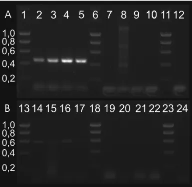
Fig. 2 –A, RT-PCR analyses detecting SmcGK1 transcripts of the
expected size (452 bp) in pairing-experienced females (ef; lane 2); pairing-unexperienced females (uf; 3); pairing-experienced males (em; 4); pairing-unexperienced males (um; 5). Controls: ef RT-negative (RT−; 7); uf RT−(8); RT without template (9); em RT−(10); um
RT−(12). The unspecific products in lane 8 originated from residual
amounts of DNA. Marker: Hyperladder I (Bioline, Germany; sizes given in kb), lanes 1, 6, 11, 13, 18, 23. B, strand-specific RT-PCR analyses confirming the existence of antisense products (609 bp) in ef (14), uf (15), em (16), and um (17). Controls (lanes 19-22, 24) as in A.
Localization studies by in situhybridization with
paired male and female worms detected SmcGK1 tran-scripts in the gonads of male and female worms as well
as in the vitellarium of the female (Fig. 3). This tran-scriptional pattern was detected with three different SmcGK1-specific probes in three independent ap-proaches. However, during these experiments we did not only detect sense RNAs, but also received signals for antisense RNAs in the same locations, when Sm-cGK1 sense probes were used for control purposes. This indicated a putative antisense regulation of this gene, a phenomenon well known for eukaryotes in-cluding schistosomes (Verjovski-Almeida et al. 2007). Therefore, as positive and negative controls we in-cludedin situ hybridizations with antisense and sense
probes of p14 (Kunz et al. 1987), which represents a high abundantly transcribed gene coding for an egg-shell precursor protein shown before to be expressed in the vitellarium (Köster et al. 1988).
Fig. 3 –In situhybridization with PKG-specific antisense probes. Pictures are representative for the three different probes as described in materials and methods as well as for the detection of sense transcripts. Detection of SmcGK1 transcript inA, ovary (o),B, testes (t),C, weakly
within the vitellarium (v);D, p14 positive control,E, p14 negative
To confirm the specificity of the SmcGK1 anti-sense signals, strand-specific RT-PCRs were performed using RNAs of ef, uf, em and um as template (Fig. 2B). A specific product could be detected in all tested stages, and no DNA contamination was found in RT−controls.
Cloning and sequencing of this PCR product finally confirmed its SmcGK1 origin. The occurrence of Sm-cGK1 sense and antisense transcripts was also con-firmed by transcriptome analyses using microarrays and SuperSAGE (Leutner et al., in preparation).
INHIBITORSTUDIES TOINFLUENCESMCGK1 ACTIVITY
To functionally test SmcGK activity and its putative role for schistosome physiology, worm couples were treated with the inhibitory cGMP analog (PR)-8-pCPT-cGMPS. Previous studies on the activity of purified human platelet or mouse intestine cGKI as well as re-combinantly expressed and purified cGKII determined a higher specificity of the inhibitor for cGKII than cGKI (Butt et al. 1994, Gamm et al. 1995). As the in-hibitory effect in these studies was reported to occur in
µmolar doses of the inhibitor, we performed an initial
experiment to determine the inhibitory dose for schisto-somes (data not shown). As the concentration of 1 mM proved to induce a behavioral phenotype in comparison to control this concentration was used for subsequent experiments. Over a treatment period of six days we observed that more couples separated following inhibitor treatment compared to an untreated control group. When couples separated in the control popula-tion, they repaired after a short time, whereas treated worms never repaired after separation. In addition, clear differences were seen in the movement pattern between treated and control group. During the first two days the movement of control worms was smooth and swift, whereas the movement of treated worms was choppy. Separation of couples occurred, and separated males started to curl in a screw-like fashion. From 3-6 days on, more couples were separated and movement slowed down progressively, which resulted in a slow motion (slomo) phenotype (supplementary data videos A, B;
http://www.unigiessen.de/cms/fbz/fb10/institute_klinikum/ institute/parasitologie/forschung/schisto/publikationen/mehr).
To investigate possible changes at the
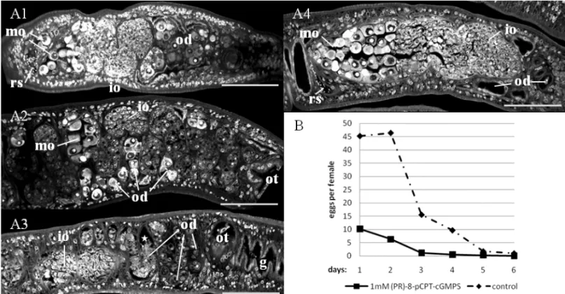
morpho-logical level, worms were stained with carmine red af-ter 2, 4 and 6 days and studied by CLSM. While we were not able to detect a morphological difference be-tween treated and control males by CLSM, we found a kind of oocyte congestion within the oviduct of the females (Fig. 4A 1-3). In untreated control worms (Fig. 4A 4), migrating oocytes are rarely detected with-in the oviduct on their way towards the ootype (Beck-mann et al. 2010a, b; this study). In inhibitor-treated females a high number of eggs were detected at the same time within the oviduct. Furthermore, an apopto-sis-like degradation of oocytes occurred. Depending on the worm batch used, this effect was already seen at day 2 following treatment or later. This effect also in-creased with the time of treatment. Correspondingly to this phenotype, egg production was affected. Already at day 1 following treatment the number of eggs of treated couples was lower compared to that of untreated control couples (Fig. 4B). Egg production of adults is generally decreasing underin vitro culture conditions, which leads to a leveling of egg numbers in treated and untreated worms after about one week (2 days ahead of treatment, 5 days treatment period).
DISCUSSION
Searching for cGK homologs in the schistosome ge-nome data set led to the identification of six predic-tions for schistosome cGMP-dependent protein kinases. Cloning and sequencing of cDNA parts of Smp_123-290 from our Liberian strain of S. mansoni
Localiza-Fig. 4 – Effects of inhibitor treatment on female morphology (analyzed by confocal laser scanning microscopy) and egg production (analyzedin
vitro).A, influence of inhibitor treatment on the ovary and oviduct of a female worm treated with inhibitor for A1: two days, A2: four days, A3:
six days; A4: untreated control after two days, which is representative for days 4-6 (scale bar: 75µm; g: gut; io: immature oocytes; mo: mature
oocytes; od: oviduct; ot: ootype; rs: receptaculum seminis).B, comparison of egg production of inhibitor-treated (1 mM) versus control females,
showing eggs per day and female. Supplementary data: videos showing treated (A) and control (B) wormsin vitroat day 4 of the experiment. http://www.unigiessen.de/cms/fbz/fb10/institute_klinikum/institute/parasitologie/forschung/schisto/publikationen/mehr
tion studies exhibited transcription in the reproductive organs of both genders. Unexpectedly, SmcGK1 tran-scripts were repeatedly detected in control reactions dur-ingin situ hybridization, for which labeled sense tran-scripts were used. This pointed to the presence of Sm-cGK1 antisense transcripts naturally occurring in these tissues, a hypothesis which was confirmed by RT-PCR detection and sequencing of SmcGK1 antisense tran-scripts from adult worms. Research of the last couple of years exhibited that post-transcriptional gene regula-tion by antisense RNA is a widespread phenomenon in eukaryotes (Ryusuke and Slack 2007, Siomi and Siomi 2010). This includes schistosomes, since in a former microarray analysis first genome-wide evidence was obtained for the antisense regulation of about 7% of schistosome genes (Verjovski-Almeida et al. 2007).
To obtain first functional evidence for the impor-tance of SmcGK1 for schistosome biology we per-formed an inhibitor approach with adult schistosomes
in vitrousing a cGMP analog, which was proven before
iso-forms of SmcGKs, which may have been affected by the inhibitor as well. Due to lower degrees of homology we were not able to classify the other SmGKs unequiv-ocally. In addition to the slomo phenotype, we observed peculiar changes at the morphological level. By CLSM major changes in ovary, ootype, or testes morphology were not detected, although the structure of the ovary seemed to vary to some extent with increasing inhibitor concentrations. Within the oviduct, however, which con-nects the ovary with the ootype allowing oocytes to be fertilized and transported, an atypical oocyte congestion was observed in treated females. This finding indicates that the transport of mature oocytes is hampered. It is tempting to speculate that this observation may coincide with the putatively decreasing motor activity of slomo males. Assumed that the constant pairing combined with the regular peristaltic, wavelike movements of the muscular male body could have a massaging effect on the female and thus contribute to the transport of oocytes and eggs, both oocyte congestion and reduction of egg production could be explained. The latter is prob-ably also influenced by apoptotic processes in oocytes starting early after treatment.
Due to their involvement in similar physiological processes cGKs have also come into focus of pro-tozoan research and are considered as drug targets (Taylor et al. 2010, Diaz et al 2005, Donald et al. 2002). In Eimeria growth was negatively affected by
trisub-stituted pyrrole, a potent cGK inhibitor (Gurnett et al. 2002). cGKs participate in gliding motility of Plasmo-dium (Moon et al. 2009), Eimeria and Toxoplasma
and is involved in the host invasion of the latter two parasites (Wiersma et al. 2004). Also, inPlasmodium
they influence blood-stage schizogony (Taylor et al. 2010), merosome egress from liver cells (Falae et al. 2010), and finally influence gametogenesis (McRobert et al. 2008). In eukaryotes cGKs play diverse physio-logical functions (Lohmann and Walter 2005), among which a cGMP/PKG pathway was hypothesized to be involved in oocyte maturation (Petr et al. 2006, Zhang et al. 2005).
To elucidate the biological roles of SmcGKs in more detail, further studies have to be performed in-cluding experiments to confirm the remaining gene predictions, the use of a variety of different inhibitors,
and – in the light of antisense regulation of this gene – a filed strategy for knock-down approaches. The find-ings of our study indicate that a cGK inhibitor exists being able to block SmcGK activity in such a way that important physiological processes in schistosomes are negatively affected: motor activity in males and egg production in females. This provides a novel perspec-tive towards the identification of target structures for the design of alternative ways to fight schistosomes, which is urgently needed in the light of the fact that praziquantel is the only widely used drug to treat schisto-somiasis evoking the fear of emerging resistance (Doenhoff et al. 2008, Stothard et al. 2009).
ACKNOWLEDGMENTS
The authors thank Christine Henrich and Gabriele Lang for their excellent technical assistance, Thomas Quack for general support as well as Anne Holz and Adrian Dorresteijn for help with confocal microscopy. This work was supported by the Studienstiftung des Deutschen Volkes (SL) and a grant of the Deutsche Forschungsgemeinschaft (GR-1549/7-1).
RESUMO
Esquistossomos são parasitas trematodos de importância mé-dica em todo o mundo para o homem e os animais. O cresci-mento e o desenvolvicresci-mento destes parasitas requerem um am-biente específico do hospedeiro, mas também um processo de comunicação permanente entre parasitas dos dois sexos. Evidência molecular tem se acumulado e indica que as in-terações são mediadas por processos de transdução de sinal. Moléculas sinalizadoras conservadas foram identificadas, e as primeiras abordagens têm sido feitas para sua caracterização. Contudo, não foi ainda descrito nenhum representante da fa-mília conservada das proteína-quinases dependentes de cGMP (cGKs) neste parasita. Analisando o genoma doSchistosoma mansoninós identificamos homólogos de cGK, dos quais um
Experimentos de hibridizaçãoin situdemonstraram uma ex-pressão preferencial nas gônadas em ambos os gêneros, in-dicando um papel para SmcGK1, pelo menos durante o de-senvolvimento de esquistossomos. Usando um inibidor es-pecífico de cGK para tratamento de esquistossomos adultos
in vitrofinalmente resultou em um fenótipo multifacetado, in-cluindo movimentos lentos, congestão dos oócitos, e redução da produção de ovos.
Palavras-chave: Schistosoma mansoni, transdução de sinal, proteína-quinases dependentes de cGMP (cGKs), serina/treo-nina (S/T) quinases, desenvolvimento gonadal.
REFERENCES
AHIERA, KHAYATHN, VICOGNEJANDDISSOUSC. 2008. Insulin receptors and glucose uptake in the human para-siteSchistosoma mansoni. Parasite 15: 573–579. BAKER DA ANDDENG W. 2005. Cyclic GMP-dependent
protein kinases in protozoa. Front Biosci 10: 1229–1238. BECKMANNS, QUACKT, BURMEISTERC, BUROC, LONG
T, DISSOUSCANDGREVELDINGCG. 2010a. Schisto-soma mansoni: signal transduction processes during the
development of the reproductive organs. Parasitology 137: 497–452.
BECKMANN S, BUROC, DISSOUS C, HIRZMANNJ AND
GREVELDING CG. 2010b. The Syk kinase SmTK4 of Schistosoma mansoni is involved in the regulation of spermatogenesis and oogenesis. PLoS Pathog 6(2): e1000769.
BERRIMAN M ET AL. 2009. The genome of the blood flukeSchistosoma mansoni. Nature 460: 352–358. BUTT E, EIGENTHALER M AND GENIESER HG. 1994.
(Rp)-8-pCPT-cGMPS, a novel cGMP-dependent protein kinase inhibitor. Eur J Pharmacol 269: 265–268. DIAZ CA, ALLOCCO J, POWLES MA, YEUNG L,
DONALD RGK, ANDERSON JW AND LIBERATOR
PA. 2005. Characterization of Plasmodium falciparum
cGMP-dependent protein kinase (PfPKG): Antiparasitic activity of a PKG inhibitor. Mol Biochem Parasitol 146: 78–88.
DOENHOFFMJ, CIOLI DANDUTZINGERJ. 2008. Prazi-quantel: mechanisms of action, resistance and new de-rivatives for schistosomiasis. Curr Opin Infect Dis 21: 659–667.
DONALDRGK, ALLOCCOJ, SINGHSB, NAREB, SALOWE
S, WILTSIE J ANDLIBERATOR P. 2002. Toxoplasma gondiiCyclic GMP-Dependent Kinase:
Chemotherapeu-tic Targeting of an Essential Parasite Protein Kinase. Eu-karyot Cell 1: 317–328.
FALAE A, COMBE A, AMALADOSS A, CARVALHO T, MENARD R AND BHANOT P. 2010. Role of Plas-modium bergheicGMP-dependent protein kinase in late liver stage development. J Biol Chem 285: 3282–3288. FUJIWARAM, SENGUPTAPANDMCINTIRESL. 2002.
Re-gulation of body size and behavioral state ofC. elegans
by sensory perception and the EGL-4 cGMP-dependent protein kinase. Neuron 36: 991–993.
GAMM DM, FRANCIS SH, ANGELOTTI TP, CORBIN JD
ANDUHLERMD. 1995. The Type II Isoform of cGMP-dependent Protein Kinase Is Dimeric and Possesses Re-gulatory and Catalytic Properties Distinct from the Type I Isoforms. J Biol Chem 270: 27380–27388.
GREVELDINGCG. 1995. The female-specific W1 sequence of the Puerto Rican strain ofSchistosoma mansonioccurs in both genders of a Liberian strain. Mol Biochem Para-sitol 71: 269–272.
GREVELDINGCG. 2004. Schistosoma. Curr Biol 14: R545. GURNETT AM ET AL. 2002. Purification and molecular
characterization of cGMP-dependent protein kinase from Apicomplexan parasites. A novel chemotherapeutic tar-get. J Biol Chem 277: 15913–15922.
HAAS BJ, BERRIMAN M, HIRAI H, CERQUEIRA GG, LOVERDEPTANDEL-SAYEDNM. 2007.Schistosoma mansoni genome: closing in on a final gene set. Exp Parasitol 117: 225–228.
HOFMANN F, BERNHARD D, LUKOWSKI R ANDWEIN -MEISTER P. 2009. cGMP regulated protein kinases
(cGK). Handb Exp Pharmacol 191: 137–162.
KÖSTER B, DARGATZ H, SCHRÖDER J, HIRZMANN J, HAARMANN C, SYMMONS P AND KUNZ W. 1988. Identification and localisation of the products of a puta-tive eggshell precursor gene in the vitellarium of Schisto-soma mansoni. Mol Biochem Parasitol 31: 183–198. KUNZ W. 2001. Schistosome male-female interaction:
in-duction of germ-cell differentiation. Trends Parasitol 17: 227–231.
KUNZ W, OPATZK, FINKEN MANDSYMMONSP. 1987.
Sequences of two genomic fragments containing an iden-tical coding region for a putative egg-shell precursor pro-tein ofSchistosoma mansoni. Nucl Acids Res 15: 5894.
LEE JI, O’HALLORAN DM, EASTHAM-ANDERSON J, JUANG BT, KAYE JA, SCOTT HAMILTON O, LESCH
LOHMANNSMANDWALTERU. 2005. Tracking functions of cGMP-dependent protein kinases (cGK). Front Biosci 10: 1313–1328.
LOVERDE PT, ANDRADE LF AND OLIVEIRA G. 2009.
Signal transduction regulates schistosome reproductive biology. Curr Opin Microbiol 12: 422–428.
LOVERDEPT, NILESEG, OSMANAANDWUWJ. 2004.
Schistosoma mansoni male-female interactions. Can J
Zool 82: 357–374.
MACHADO-SILVA JR, PELAJO-MACHADOM, LENZI HL
AND GOMESDC. 1998. Morphological study of adult male worms ofSchistosoma mansoni Sambon, 1907 by confocal laser scanning microscopy. Mem Inst Oswaldo Cruz 93(Suppl 1): 303–307.
MCMANUSDPANDLOUKASA. 2008. Current Status of Vaccines for Schistosomiasis. Clin Microbiol Rev 21: 225–242.
MCROBERT L, TAYLOR CJ, DENG W, FIVELMAN QL, CUMMINGS RM, POLLEYSD, BILLKEROAND BA
-KER DA. 2008. Gametogenesis in malaria parasites is mediated by the cGMP-dependent protein kinase. PLoS Biol 6(6): e139.
MELMANSDET AL. 2009. Reduced susceptibility to pra-ziquantel among naturally occurring Kenyan isolates of
Schistosoma mansoni. PLoS Negl Trop Dis 3(8): e504. MOONRW, TAYLORCJ, BEXC, SCHEPERSR, GOULDING
D, JANSECJ, WATERSAP, BAKERDAANDBILLKER
O. 2009. A cyclic GMP signalling module that regulates gliding motility in a malaria parasite. PLoS Pathog 5(9): e1000599.
MOOREDVANDSANDGROUNDJH. 1956. The relative egg production capacity ofSchistosoma mansoniand Schis-tosoma japonicum. Am J Trop Med Hyg 5: 831–1382. NEVESRH,DELAMAREBC, MACHADO-SILVAJR, CAR
-VALHO JJ, BRANQUINHO TB, LENZI HL, HULSTIJN
MANDGOMESDC. 2005. A new description of the re-productive system ofSchistosoma mansoni(Trematoda:
Schistosomatidae) analyzed by confocal laser scanning microscopy. Parasitol Res 95: 43–49.
OLIVEIRA G, FRANCO G ANDVERJOVSKI-ALMEIDA S. 2008. The Brazilian contribution to the study of the
Schistosoma mansoni transcriptome. Acta Trop 108: 179–182.
PAGER DM. 1996. TREEVIEW: An application to display phylogenetic trees on personal computers. Comp Appl Biosci 12: 357–358.
PETR J, RAJMON R, CHMELÍKOVÁ E, TOMÁNEK M, LÁNSKÁV, PRIBÁNOVÁMANDJÍLEKF. 2006.
Nitric-oxide-dependent activation of pig oocytes: the role of the cGMP-signalling pathway. Zygote 14: 9–16.
PFEIFER A, RUTH P, DOSTMANN W, SAUSBIER M,
KLATTPANDHOFMANNF. 1999. Structure and
func-tion of cGMP-dependent protein kinases. Rev Physiol Biochem Pharmacol 135: 105–149.
QUACK T, BECKMANN S ANDGREVELDING CG. 2006. Schistosomiasis and the molecular biology of the male-female interaction ofS. mansoni. Berl Munch Tierarztl Wochenschr 119: 365–372.
QUACK T, KNOBLOCH J, BECKMANN S, VICOGNE J, DISSOUSCANDGREVELDINGCG. 2009. The formin-homology protein SmDia interacts with the Src kinase SmTK and the GTPase SmRho1 in the gonads of Schisto-soma mansoni. PloS One 4: e6998.
RAIZENDM, CULLISONKM, PACKAIANDSUNDARAM
MV. 2006. A novel gain-of-function mutant of the cyc-lic GMP-dependent protein kinase egl-4 affects multiple physiological processes inCaenorhabditis elegans. Gen-etics 173: 177–187.
RAIZENDM, ZIMMERMANJE, MAYCOCKMH, TAUD,
YOUYJ, SUNDARAMMVANDPACKAI. 2008.
Lethar-gus is aCaenorhabditis eleganssleep-like state. Nature
451: 569–572.
ROSS AG, BARTLEY PB, SLEIGH AC, OLDS GR, LIY, WILLIAMSGMANDMCMANUSDP. 2002. Schistoso-miasis. N Engl J Med 346: 1212–1220.
RYUSUKENIWA RANDSLACK FJ. 2007. The evolution of animal microRNA function. Curr Opin Genet Dev 17: 1–6.
SAVIOLI L, GABRIELLI AF, MONTRESOR A, CHITSULO
L AND ENGELS D. 2009. Schistosomiasis control in Africa: 8 years after World Health Assembly Resolution 54.19. Parasitology 136: 1677–1681.
SCHLOSSMANN J, FEIL R AND HOFMANN F. 2003.
Sig-naling through NO and cGMP-dependent protein kinases. Ann Med 35: 21–27.
SIOMI HANDSIOMI MC. 2010. Posttranscriptional regu-lation of microRNA biogenesis in animals. Mol Cell 38: 323–332.
STOTHARD JR, CHITSULO L, KRISTENSEN TK AND
UTZINGERJ. 2009. Control of schistosomiasis in sub-Saharan Africa: progress made, new opportunities and remaining challenges. Parasitology 136: 1665–1675. TAYLOR HM, MCROBERT L, GRAINGER M, SICARD
A, DLUZEWSKI AR, HOPP CS, HOLDER AA AND
GMP-de-pendent protein kinase plays a central role in blood-stage schizogony. Eukaryot Cell 9: 37–45.
VERJOVSKI-ALMEIDA S, LEITE LC, DIAS-NETO E,
MENCK CF AND WILSON RA. 2004. Schistosome
transcriptome: insights and perspectives for functional genomics. Trends Parasitol 20: 304–308.
VERJOVSKI-ALMEIDAS, VENANCIOTM, OLIVEIRAKC, ALMEIDAGTANDDEMARCOR. 2007. Use of a 44k oligoarray to explore the transcriptome of Schistosoma mansoniadult worms. Exp Parasitol 117: 236–245. WIERSMAHI, GALUSKASE, TOMLEYFM, SIBLEYLD,
LIBERATOR PSANDDONALDRGK. 2004. A role for coccidian cGMP-dependent protein kinase in motility and invasion. Int J Parasitol 34: 369–380.
ZERLOTINI A, HEIGES M, WANG H, MORAES RLV, DOMINITINI AJ, RUIZ JC, KISSINGER JC AND OLI
-VEIRA G. 2009. SchistoDB: a Schistosoma mansoni
genome resource. Nucl Acids Res 37: D579–582. ZHANG M, TAO Y, XIA G, XIE H, HONG H, WANG F
AND LEI L. 2005. Atrial natriuretic peptide negatively