RADIOLOGY PAGE
435
Unilateral renal cystic disease in the right kidney
Eun Hui Bae, Young-Hwan Hwang, Soo Wan Kim
Departments of Internal Medicine, Chonnam National University Medical School, Gwangju, and Department of Internal Medicine, Eulji General Hospital, Seoul, Korea
_______________________________________________________________________________
A 51-year-old man was referred from on-cology department with incidental detection of unilateral multiple renal cysts on computed to-mography (CT) during evaluation of colon can-cer (Figure-1). Abdominal CT revealed enlarged right kidney with variable-sized round, well--marginated multiple cysts without capsule for-mation or solid content, while the left kidney was normal. Family history was negative for kidney disease. Ultrasound imaging of the parents, five adult siblings, one son and two daughters sho-wed normal kidneys. He received chemotherapy with 5-flurouracil plus leucovorin after laparos-copic right hemicolectomy for colon cancer. The
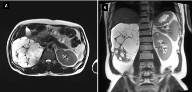
relevant laboratory data were as follows: white blood cell count, 7000/mm3; hemoglobin level, 15.9 g/dL; platelet count, 174,000/mm3; blood urea nitrogen level, 15.5 mg/dL; creatinine level, 0.8 mg/dL and no proteinuria or hematuria. We did not introduce specific therapy for the unila-teral cyst and follow-up was made with magne-tic resonance image (MRI) six month after hemi-colectomy. The exam showed numerous cysts in the right kidney with no changes according to previous CT (Figure-2).
This patient has unilateral renal cystic disease (URCD), a rare entity characterized by multiple cysts in one kidney or a portion of one
Figure 1 - Preoperative axial (A) and coronal scan (B, C) of abdominal CT showing multiple cysts in the right kidney.
A
B C B
Vol. 39 (3): 435-437, May - June, 2013
436
IBJU |RADIOLOGY PAGEkidney. The clinical importance of URCD is to make a differential diagnosis of such abnorma-lities including multilocular cystic nephroma, cystic partially differentiated Wilms’ tumor, segmental cystic dysplasia, and atypical pre-sentation of polycystic kidney disease such as asymmetric evolution and mosaicism (1,2). The pathogenesis of this cystic renal disease is unk-nown. Since there is a morphological similari-ty of this cystic change to autosomal dominant polycystic kidney disease (ADPKD), it is specu-lated that pathogenesis is similar (3). The gross and microscopic features are indistinguishable from ADPKD, and patients may present with he-maturia, pain, or a flank mass (4). However, it can be differentiated from ADPKD by its uni-lateral localization, negative family history, no progression to chronic renal insufficiency, and no extrarenal manifestation. Cystic adenosarco-ma of the kidney can present in a very simi-lar way and it can be differentiated by positive reaction to epithelial membrane antigen (EMA), vimentin and transducin-like enhancer protein 1 (TLE1), and CD99 (5). Most importantly, it must be differentiated against segmental cystic dys-plastic disease. In case of URCD, collecting
sys-Figure 2 - Postoperative axial (A) and coronal scan (B) of the kidney MRI (T2 weighted image) showing multiple cysts in the right kidney.
tem should be shown continuously though dis-torted collecting system. Otherwise it would lean to segmental cystic dysplastic disease (6,7). The collecting system of this patient was continuous (Figure-1C).
URCD is a stable disease and patients can be followed up by imaging techniques (8). In conclusion, unilateral cystic disease of kidney is a rare cystic disease of the kidney diagnosed by imaging techniques and requires nephrectomy only when suspicion of malignancy is strong.
REFERENCES
1. Reed B, Nobakht E, Dadgar S, Bekheirnia MR, Masoumi A, Belibi F, et al.: Renal ultrasonographic evaluation in children at risk of autosomal dominant polycystic kidney disease. Am J Kidney Dis. 2010; 56: 50-6.
2. Connor A, Lunt PW, Dolling C, Patel Y, Meredith AL, Gardner A, et al.: Mosaicism in autosomal dominant polycystic kidney disease revealed by genetic testing to enable living related renal transplantation. Am J Transplant. 2008; 8: 232-7.
3. Gouldesbrough DR, Fleming S: Unilateral and segmental local-ised polycystic kidney disease. J Clin Pathol. 1998; 51: 703-5.
437
IBJU |RADIOLOGY PAGE4. Hwang DY, Ahn C, Lee JG, Kim SH, Oh HY, Kim YY, et al.: Uni-lateral renal cystic disease in adults. Nephrol Dial Transplant. 1999; 14: 1999-2003.
5. Sameshima N, Marutsuka K, Tsukino H, Kamoto T, Kono S, Asada Y: So-called ‘adenosarcoma’ of the kidney a novel adult renal tumor with a cystic appearance. Pathol Int. 2011; 61: 313-8.
ARTICLE INFO
Int Braz J Urol. 2013; 39: 435-7
__________________
Submitted for publication: February 18, 2013
__________________
Accepted after revision: March 12, 2013
_____________________
Correspondence address:
Dr. Soo Wan Kim Department of Internal Medicine, Chonnam National University Medical School Hakdong 8, Dongku, Gwangju 501-757, Korea Fax : + 82 62 225-8578 E-mail: [email protected] 6. Clevert DA, Horng A, Staehler M, Haseke N, Stief C, Reiser M: Diagnostic algorithm in cystic renal masses. Urologe A. 2010; 49: 421-32.
7. Song C, Min GE, Song K, Kim JK, Hong B, Kim CS, et al.: Differ-ential diagnosis of complex cystic renal mass using multiphase computerized tomography. J Urol. 2009; 181: 2446-50. 8. Slywotzky CM, Bosniak MA: Localized cystic disease of the