Key words: Acromegaly; BPH; Prostate-specific antigen; Prostate
Int Braz J Urol. 2013; 39: 393-401
__________________ Submitted for publication: February 28, 2012
__________________ Accepted after revision: December 14, 2012 Introduction: Published data suggest that patients with acromegaly have an increased
prevalence of prostate disorders.
Objective: To evaluate prostatic disorders in acromegalic patients comparing these results after one year of treatment of acromegaly and with a group of healthy men.
Materials and Methods: This study was composed of two parts: sectional study com-paring patients with healthy controls (baseline) and prospective, longitudinal study (at baseline and after one year of treatment). Forty acromegalic patients were enrolled and evaluated at baseline and after one year with the application of international prostatic symptoms score (IPSS), digital rectal examination, measurements of growth hormone (GH), insulin-like growth factor-I (IGF-I), insulin-like growth factor-binding protein-3 (IGFBP-3), sex hormone-binding globulin (SHBG), prolactin, luteinizing hormone (LH), follicle-stimulating hormone (FSH), total testosterone, total and free prostate-specific antigen (PSA) levels and prostate ultrasonography (US). Thirty healthy men were selected as control group.
Results: We stratified patients and controls according to age, considering 40 years-old as cut off. Healthy controls under 40 had IPSS values lower than acromegalic patients. When considering only older patients and controls prostate hyperplasia and structural abnormalities were more frequent in acromegalics. After one year of treatment there was significant decrease in GH, IGF-I and prostate volume in acromegalics over 40 years-old.
Conclusions: Acromegalics under 40 have more urinary symptoms according to IPSS and above 40 years-old higher frequency of structural changes and increased prostate volume than healthy men. Significant reduction of GH and IGF-I levels during treatment of acromegaly leads to decrease in the prostate volume.
INTRODUCTION
Acromegaly is a rare disease caused by GH hyper secretion (1,2). It is well known that GH promotes a stimulatory effect on IGF-I and IGFBP-3. IGF-I stimulates cell proliferation (3),
however, IGFBP-3 stimulates apoptosis (4). There-fore, it is not well established whether acromegaly is associated or not with increased relative risk for cancer development.
Several epidemiologic studies have sug-gested that high-normal serum IGF-I levels may
Prostatic disorders in acromegalic patients experience
of a Brazilian center
_______________________________________________
Lívia L. Corrêa, Giovanna A. Balarini Lima, Suzana A. Cavallieri, Luiz Carlos D. de Miranda, Mônica
R. Gadelha
Service of Endocrinology, University Hospital Clementino Fraga Filho (HUCFF), Federal University of Rio de Janeiro (UFRJ) (LLC, GABL, MRG); Department of Urology, University Hospital Clementino Fraga Filho (HUCFF), Federal University of Rio de Janeiro (UFRJ) (LCDM) and Labs D’Or Laboratory and Imaging (SAC), Rio de Janeiro, RJ, Brazil
ABSTRACT
ARTICLE
INFO
be concordant with a higher risk of prostate can-cer in the general population, and that high-nor-mal serum IGFBP-3 levels are concordant with a lower risk (5-8). Published data suggest that the relationship between prostatic carcinoma and acromegaly is infrequent (9).
Patients with acromegaly have an incre-ased prevalence of prostatic disorders compared to age-matched healthy subjects. Increased size of the whole prostate, together with an elevated in-cidence of other structural changes, such as nodu-les, cysts, and calcifications were shown in a large proportion of patients (10,11). The presence of an enlarged prostate in acromegalic patients under 40 years suggests a possible role of GH-IGF-I axis on this gland growth (10,11). Only two studies evaluated the effects of acromegaly treatment on prostate volume (PV), and demonstrated that well controlled patients presented significant reduction in PV after treatment of acromegaly (10,12).
The IPSS is a symptom index for benign prostatic hyperplasia (BPH), developed and vali-dated by a multidisciplinary measurement com-mittee of the American Urological Association (AUA) (13). The IPSS is widely used to evaluate the severity of urinary symptoms (14,15). Until today, there is no report of the use of IPSS in the evalu-ation of acromegalic patients.
In a recent guideline for acromegaly ma-nagement (16) there was no information about the BPH evaluation. The aim of this study was to evaluate the prostate of acromegalic patients through digital rectal examination, transrectal US and IPSS, comparing these results with a group of healthy men, above and below 40 years, and after one year of acromegaly treatment.
MATERIALS AND METHODS
Patients
Forty acromegalic patients, aged 45.2 + 11.3 years (24-69 years), were recruited from the outpa-tient endocrinology clinic of the University Hospital Clementino Fraga Filho - HUCFF, of the Federal Uni-versity of Rio de Janeiro - UFRJ, over a 24-month period. The diagnosis of acromegaly was based on the following criteria: 1) a lack of suppression of GH to below 1 ng/mL after oral administration of 75g
glucose or 2) high serum IGF-I levels. Reasons for ineligibility included patients with previous treat-ment of prostate cancer or BPH. All subjects entered the study after obtaining written informed consent according to a protocol approved by the Ethics Com-mittee of HUCFF.
At baseline, 36 patients had active disease (10 were de novo patients) and four had acrome-galy cure or control for less than one year before the inclusion in this study. Twenty-six patients were previously submitted to surgery, nine to radiothe-rapy and 25 to medical treatment with octreotide LAR and/or cabergoline. Hypogonadism, based on low testosterone levels, was present in 27 (67.5%) patients. Seventeen of these patients were not recei-ving testosterone as replacement therapy because of severe sleep apnea.
As control group, thirty healthy and educa-tion-matched men were included. Fourteen of them were less than 40 years-old and were paired to the 14 acromegalic patients younger than 40 years. The other 16 were proportionally age-matched with the 26 acromegalics older than 40 years-old.
Study design
This study was composed of two parts: a) sectional study comparing patients (at baseline) with healthy controls; b) prospective, longitudinal study (at baseline and after one year of treatment). The study protocol included application of IPSS, digi-tal recdigi-tal examination, measurements of GH, IGF-I, IGFBP-3, SHBG, prolactin, LH, FSH, total testoste-rone, total and free PSA levels and transrectal US, both at baseline and after one year of treatment for patients and on baseline for controls. The free tes-tosterone and the bioavailable testes-tosterone were cal-culated according to the Vermeulen’s formula (17).
Urologic evaluation
The digital rectal examination was perfor-med by the same urologist (LCDM).
Hormone assays
Serum GH, IGF-I, IGFBP-3, SHBG, prolactin, LH, FSH and total testosterone levels were determi-ned by chemiluminescense immunometric assays. Total and free PSA serum levels were measured by electrochemiluminescense assay.
The low detection limit of GH by the Immu-lite 2000 kit (DPC- Diagnostic Products Corporation, Los Angeles, CA) is 0.01 ng/mL and its linear working range is 0.01-40 ng/mL. Standards are calibrated against the International Standard WHO 98/574. The intra-assay CVs at the respective concentrations of 1.7, 7.8 and 31.0 ng/mL are 5.3%, 6.0% and 6.5%, while the inter-assay CVs at 3.0, 9.3 and 18.0 ng/ mL are 5.7%, 6.2% and 6.1%, respectively. The low detection limit of IGF-I measured by Immulite 2000 kit DPC is 20 ng/mL and the intra and inter-assay CVs are 3.6 and 6.6%, respectively. The standards are calibrated against the first International Referen-ce Reagent WHO 87/518 and IGF-I was expressed in mass units and age-related standard deviation scores (SD-scores).
All serum samples were collected in the early morning after an eight-hour fasting period.
Transrectal US
Transrectal US was performed with a HDI 11XE, Phillips, 2008, using a 5.0-7.5 MHz transducer by the same radiologist (SAC). The prostate examina-tion included the anterior-posterior (AP), transversal (T) and longitudinal (L) diameters, the morphology of boundaries, texture, the occurrence of calcifica-tions and/or nodules, and the evaluation of seminal vesicles and bladder. Prostate volume was calculated by the elliptical shape volume formula (π/6 x APD x TD x LD). Prostate hyperplasia was defined as a PV exceeding 30 mL (19-21).
Statistical analysis
Analyses were performed by SAS System (version 6.11; SAS Institute North Caroline). The results were expressed as median (min-max). Com-parisons between categorical variables were done by χ22 test and between numerical variables were
carried out using the Mann Whitney test. Compari-sons between related samples were done by Wilco-xon test. McNemar’s test was used to compare paired proportions. Correlations were sought by calculating the Spearman’s rank correlation coefficient. P values < 0.05 were considered statistically significant.
RESULTS
Comparison between acromegalic patients and control group under 40 years at baseline
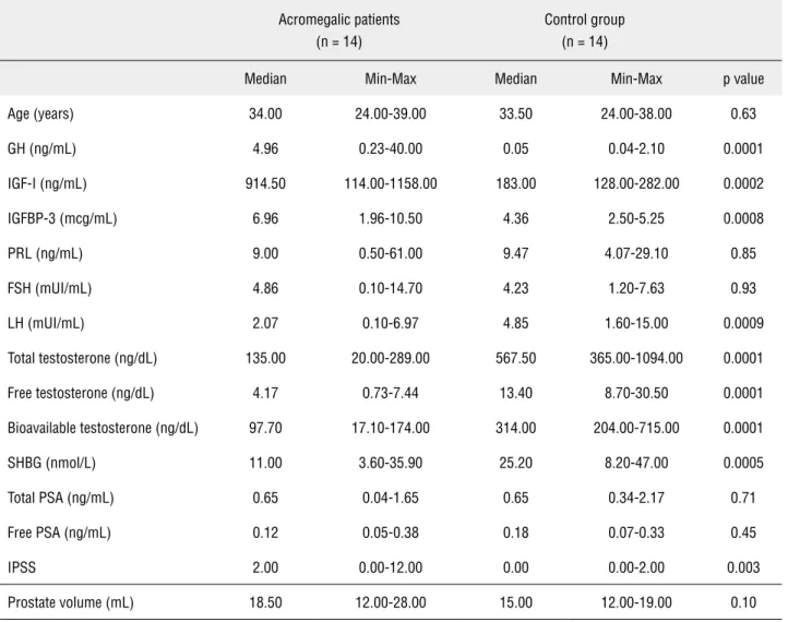
Because it is well established that prostate enlargement starts approximately at the age of 40 (22,23), we stratified patients and controls according to age, considering 40 years-old as cut off. When considering only younger patients and controls (Ta-ble-1), the acromegalics presented high levels of GH (4.96 vs 0.05, p = 0.0001), IGF-I (914.50 vs 183.00, p = 0.0002), IGFBP-3 (6.96 vs 4.36, p = 0.0008) and IPSS (2.00 vs 0.00, p = 0.003) and presented low levels of LH (2.07 vs 4.85, p = 0.0009), total tosterone (135.00 vs 567.50, p = 0.0001), free tes-tosterone (4.17 vs 13.40, p = 0.0001), bioavailable testosterone (97.70 vs 314.00, p = 0.0001) and SHBG (11.00 vs 25.20, p = 0.0005). The mean PV was not different between these two groups (18.50 vs 15.00, p = 0.10).
Prostate hyperplasia was not found in pa-tients or controls under 40. Structural abnormalities found at US were: calcifications in one patient and one control (7% vs 7%, p = 1.0) and hyperechogenic foci suggesting corpora amylacea in 4 patients and 3 controls (28.6% vs 21.4%, p = 1.0).
Comparison between acromegalic patients and control group above 40 years at baseline
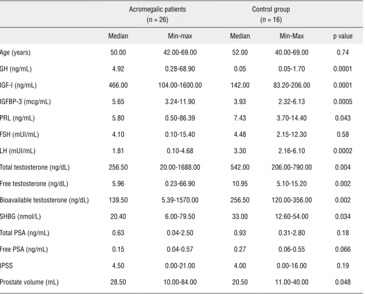
When considering only patients and controls
≥ 40 years-old (Table-2), the acromegalics presented higher levels of GH (4.92 vs 0.05, p = 0.0001), IGF--I (466.00 vs 142.00, p = 0.0001), IGFBP-3 (5.65 vs 3.93, p = 0.0005) and PV (28.50 vs 20.50, p = 0.048) and presented lower levels of LH (1.81 vs 3.30, p = 0.0002), total testosterone (256.50 vs 542.00, p = 0.004), free testosterone (5.96 vs 10.95, p = 0.002), bioavailable testosterone (139.50 vs 256.50, p = 0.002) and SHBG (20.40 vs 33.00, p = 0.034).
Table 1 - Characteristics of acromegalic patients at baseline and control group (< 40 years-old).
Acromegalic patients (n = 14)
Control group (n = 14)
Median Min-Max Median Min-Max p value
Age (years) 34.00 24.00-39.00 33.50 24.00-38.00 0.63
GH (ng/mL) 4.96 0.23-40.00 0.05 0.04-2.10 0.0001
IGF-I (ng/mL) 914.50 114.00-1158.00 183.00 128.00-282.00 0.0002
IGFBP-3 (mcg/mL) 6.96 1.96-10.50 4.36 2.50-5.25 0.0008
PRL (ng/mL) 9.00 0.50-61.00 9.47 4.07-29.10 0.85
FSH (mUI/mL) 4.86 0.10-14.70 4.23 1.20-7.63 0.93
LH (mUI/mL) 2.07 0.10-6.97 4.85 1.60-15.00 0.0009
Total testosterone (ng/dL) 135.00 20.00-289.00 567.50 365.00-1094.00 0.0001
Free testosterone (ng/dL) 4.17 0.73-7.44 13.40 8.70-30.50 0.0001
Bioavailable testosterone (ng/dL) 97.70 17.10-174.00 314.00 204.00-715.00 0.0001
SHBG (nmol/L) 11.00 3.60-35.90 25.20 8.20-47.00 0.0005
Total PSA (ng/mL) 0.65 0.04-1.65 0.65 0.34-2.17 0.71
Free PSA (ng/mL) 0.12 0.05-0.38 0.18 0.07-0.33 0.45
IPSS 2.00 0.00-12.00 0.00 0.00-2.00 0.003
Prostate volume (mL) 18.50 12.00-28.00 15.00 12.00-19.00 0.10
Normal Values: GH (0.06-5 ng/mL), IGF-I (116-358, 117-329, 115-307, 109-284, 101-267, 94-252, 87-238, 81-225, 75-212, 69-200 ng/mL for patients aged 21-25, 26-30, 31-35, 36-40, 41-45, 46-50, 51-55, 56-60, 61-65 and 66-70, respectively), IGFBP-3 (3.4-7.8, 3.5-7.6, 3.5-7.0, 3.4-6.7, 3.4-6.6, 3.3-6.7, 3.4-6.8, 3.4-6.9, 3.2-6.6, 3.0-6.2 mcg/mL for patients aged 21-25, 26-30, 31-35, 36-40, 41-45, 46-50, 51-55, 56-60, 61-65 and 66-70, respectively), PRL (2.1-17.7 ng/mL), FSH (1.4-18.1 mUI/mL), LH (1.5-9.3 mUI/mL), total testosterone (241-827 ng/dL), free testosterone (4.7-23 ng/dL), bioavailable testosterone (140-400 ng/dL), SHBG (13-71 nmol/L), total PSA (less than 4 ng/mL), free PSA (less than 0.92 ng/mL), IPSS (0-35), prostate volume (less than 30 mL).
significantly higher in the acromegalic population when compared with the control group (46.15% vs 12.50%, p = 0.015). Structural abnormalities were also more frequent in acromegalics: calcifications in 11 patients and one control (42.31% vs 6.25%, p = 0.018) and hyperechogenic foci suggesting corpora amylacea in 18 patients and 5 controls (69.23% vs 31.25%, p = 0.036). A uthricular cyst of 0.4 cm was found in one patient.
Comparison of the acromegalic patients under 40 years at baseline and after one year of treatment
The main characteristics of the acromegalic population under 40 years before and after treat-ment are presented in Table-3.
Table 2 - Characteristics of acromegalic patients at baseline and control group (≥ 40 years-old).
Acromegalic patients (n = 26)
Control group (n = 16)
Median Min-max Median Min-Max p value
Age (years) 50.00 42.00-69.00 52.00 40.00-69.00 0.74
GH (ng/mL) 4.92 0.28-68.90 0.05 0.05-1.70 0.0001
IGF-I (ng/mL) 466.00 104.00-1600.00 142.00 83.20-206.00 0.0001
IGFBP-3 (mcg/mL) 5.65 3.24-11.90 3.93 2.32-6.13 0.0005
PRL (ng/mL) 5.80 0.50-86.39 7.43 3.70-14.40 0.043
FSH (mUI/mL) 4.10 0.10-15.40 4.48 2.15-12.30 0.58
LH (mUI/mL) 1.81 0.10-4.68 3.30 2.16-6.10 0.0002
Total testosterone (ng/dL) 256.50 20.00-1688.00 542.00 206.00-790.00 0.004
Free testosterone (ng/dL) 5.96 0.23-66.90 10.95 5.10-15.20 0.002
Bioavailable testosterone (ng/dL) 139.50 5.39-1570.00 256.50 120.00-356.00 0.002
SHBG (nmol/L) 20.40 6.00-79.50 33.00 12.60-54.00 0.034
Total PSA (ng/mL) 0.63 0.04-2.50 0.93 0.31-2.80 0.18
Free PSA (ng/mL) 0.15 0.04-0.57 0.27 0.06-0.55 0.066
IPSS 4.50 0.00-21.00 4.00 0.00-16.00 0.19
Prostate volume (mL) 28.50 10.00-84.00 20.50 11.00-40.00 0.048
Normal Values: GH (0.06-5 ng/mL), IGF-I (116-358, 117-329, 115-307, 109-284, 101-267, 94-252, 87-238, 81-225, 75-212, 69-200 ng/mL for patients aged 21-25, 26-30, 31-35, 36-40, 41-45, 46-50, 51-55, 56-60, 61-65 and 66-70, respectively), IGFBP-3 (3.4-7.8, 3.5-7.6, 3.5-7.0, 3.4-6.7, 3.4-6.6, 3.3-6.7, 3.4-6.8, 3.4-6.9, 3.2-6.6, 3.0-6.2 mcg/mL for patients aged 21-25, 26-30, 31-35, 36-40, 41-45, 46-50, 51-55, 56-60, 61-65 and 66-70, respectively), PRL (2.1-17.7 ng/mL), FSH (1.4-18.1 mUI/mL), LH (1.5-9.3 mUI/mL), total testosterone (241-827 ng/dL), free testosterone (4.7-23 ng/dL), bioavailable testosterone (140-400 ng/dL), SHBG (13-71 nmol/L), total PSA (less than 4 ng/mL), free PSA (less than 0.92 ng/mL), IPSS (0-35), prostate volume (less than 30 mL).
in five patients, two of them under 40 years, and the four patients initially considered cured/controlled at baseline, persisted with cure/control criteria. The in-clusion of these four patients did not influence the outcomes of the research. No significant reduction in PV was observed after treatment (18.50 vs 18.50, p = 0.75) and there was no significant difference in the frequency of prostate hyperplasia (0% vs 7.14%, p = 0.50) neither in the frequency of structural
ab-normalities found at transrectal US. The reduction in IPSS achieved borderline statistical significance (2.00 vs 2.00, p = 0.056).
Comparison of the acromegalic patients above 40 years at baseline and after one year of treatment
After one year of treatment, there was a sig-nificant reduction in GH (4.92 vs 1.85, p = 0.015) and IGF-I (466.00 vs 362.50, p = 0.020) levels. Sig-nificant reduction in PV was observed after treat-ment (28.50 vs 25.50, p = 0.001), however there was no significant difference in the frequency of prosta-te hyperplasia (46.15% vs 30.76%, p = 0.43) neither in the frequency of structural abnormalities found at transrectal US. There was no reduction in IPSS (4.50 vs 4.50, p = 0.65).
The findings of digital rectal examination, both in acromegalics under and above 40 years, were in accordance with transrectal US.
DISCUSSION
There are three important studies in the literature, all of them European, reporting the prevalence of benign prostate hyperplasia and structural abnormalities in acromegalic patients Table 3 - Characteristics of acromegalic patients at baseline and after one year of treatment (< 40 years-old).
Baseline (n = 14)
1 year (n = 14)
p value
Median Min-Max Median Min-Max
GH (ng/mL) 4.96 0.23-40.00 3.28 0.21-20.30 0.011
IGF-I (ng/mL) 914.50 114.00-1158.00 522.50 131.00-1345.00 0.133
IGFBP-3 (mcg/mL) 6.96 1.96-10.50 6.72 3.33-9.26 0.638
PRL (ng/mL) 9.00 0.50-61.00 9.70 0.30-85.59 0.363
FSH (mUI/mL) 4.86 0.10-14.70 2.95 0.30-10.50 0.013
LH (mUI/mL) 2.07 0.10-6.97 1.23 0.07-6.95 0.03
Total testosterone (ng/dL) 135.00 20.00-289.00 170.00 31.00-424.00 0.079
Free testosterone (ng/dL) 4.16 0.73-7.44 5.14 1.10-12.80 0.035
Bioavailable testosterone (ng/dL) 97.00 17.10-174.00 120.50 25.70-301.00 0.035
SHBG (nmol/L) 11.00 3.60-35.90 12.00 5.00-28.30 0.47
Total PSA (ng/mL) 0.65 0.04-1.65 0.48 0.01-2.27 0.28
Free PSA (ng/mL) 0.12 0.05-0.38 0.16 0.01-0.41 0.78
IPSS 2.00 0.00-12.00 2.00 0.00-6.00 0.056
Prostate volume (mL) 18.50 12.00-28.00 18.50 11.00-32.00 0.753
(10-12). Two of these studies compared acromega-lics to age-matched healthy subjects (10,11). Until today, there is no report of the use of IPSS in the evaluation of acromegalic patients.
Although physiological development and growth of the prostate depend on testosterone and dihydrotestosterone (DHT), androgens action alone seems to be insufficient to explain prostatic disea-ses (22,25). The existence of important cross-talk
among androgens, growth factors and IGF binding proteins at prostatic level has been suggested (26). Patients with acromegaly are an interesting popu-lation model to study the possible involvement of IGF-I in the development of prostatic diseases. In our study, the group of acromegalic patients abo-ve 40 years presented a higher proportion of BPH (46.15 vs 12.50%) and structural prostatic changes, such as calcifications and corpora amylacea, when Table 4 - Characteristics of acromegalic patients at baseline and after one year of treatment (≥ 40 years-old).
Baseline (n=26)
1 year (n=26)
p value
Median Min-Max Median Min-Max
GH (ng/mL) 4.92 0.28-68.90 1.85 0.16-68.40 0.015
IGF-I (ng/mL) 466.00 104.00-1600.00 362.50 129.00-959.00 0.020
IGFBP-3 (mcg/mL) 5.65 3.24-11.90 5.36 2.33-9.09 0.131
PRL (ng/mL) 5.82 0.50-86.39 5.16 0.20-43.87 0.135
FSH (mUI/mL) 4.06 0.10-15.40 3.65 0.10-17.40 0.367
LH (mUI/mL) 1.81 0.10-4.68 2.53 0.07-6.30 0.191
Total testosterone (ng/dL) 256.50 20.00-1688.00 252.00 14.00-661.00 0.751
Free testosterone (ng/dL) 5.95 0.23-66.90 6.05 0.18-12.00 0.568
Bioavailable testosterone (ng/dL) 139.50 5.39-1570.00 142.00 4.15-281.00 0.568
SHBG (nmol/L) 20.40 6.00-79.50 20.00 9.00-63.20 0.696
Total PSA (ng/mL) 0.63 0.04-2.50 0.93 0.03-3.91 0.049
Free PSA (ng/mL) 0.15 0.04-0.57 0.18 0.01-0.54 0.501
IPSS 4.50 0.00-21.00 4.50 0.00-25.00 0.649
Prostate volume (mL) 28.50 10.00-84.00 25.50 12.00-69.00 0.001
compared to age-matched healthy men. Colao et al., in a study that included 30 acromegalics with active disease and a control group, demonstrated that the prevalence of BPH was significantly higher in the group of acromegalics (58% vs 26.6%) and structu-ral abnormalities were shown in a large proportion of these patients (11). Probably, we found less pros-tate hyperplasia than Colao’s group because our pa-tients were younger. Nonetheless, these data suggest a possible role of GH-IGF-I axis on prostate growth.
Only one study evaluated the effects of acromegaly treatment on prostatic disorders (12). This study included 23 acromegalics and evalua-ted PV and structural prostatic abnormalities before and after two years of acromegaly treatment with surgery and/or lanreotide. Considering the whole group of patients, there was no significant change in PV after two years of treatment (34.6 vs 32.5 mL, p = 0.3). However, when analyzing only well controlled patients (n = 16), there was a significant reduction in PV (29.3 vs 25.4 mL, p = 0.03). Regar-ding the eleven patients that presented BPH at ba-seline, four had PV < 30 mL at the end of the 2-year follow-up, all of them with controlled acromegaly. In our study, when we analyzed the acromegalics above 40 years, there was a significant reduction in the PV after one year of treatment (28.50 vs 25.50 mL, p = 0.001), independently of disease control. Of the twelve patients that had BPH at baseline, five presented PV < 30 mL at the end of the study and only one of them had the disease controlled. This finding suggests that a significant reduction in GH and IGF-I levels is sufficient to promote reversion of BPH, even though biochemical criteria of acro-megaly control was not achieved.
It is well established that prostate enlarge-ment starts approximately at the age of 40 (22). In a community-based group of 502 men aged 55 to 74 years without prostate cancer, the prevalence of BPH was 19% using the criteria of a PV above 30 mL together with a high IPSS score (15). Berry et al. (19) described that the PV in men aged 21 to 30 years is approximately 20 ± 6 mL. Analyzing our patients according to age, 14 (35%) were youn-ger than 40 years-old and prostate hyperplasia was not found at baseline. Five out of 12 patients with prostate hyperplasia (41.6%) were aged from 40 to 50 years-old. Besides, our data shows that PV in
acromegalics and the control group younger than 40 years-old is not different, however, IPSS score in the acromegalics is higher than in the control group. In contrast, probably there was no differen-ce in IPSS score in acromegalics and the control group older than 40 years-old because at this age both groups are already exposed to the onset of urinary symptoms. This is the first report of the use of IPSS in the evaluation of acromegalic patients.
CONCLUSIONS
This was the first Brazilian study that asses-sed prostate disorders in patients with acromegaly. We demonstrated that acromegalic patients above 40 years-old have a higher frequency of structural changes and increased prostate volume in compa-rison to age-matched healthy men. In this group, significant reduction of GH and IGF-I levels during treatment of acromegaly leads to decrease in the prostate volume.
ABBREVIATIONS
AUA = American Urological Association BPH = Benign Prostate Hyperplasia DHT= Dihydrotestosterone
DPC = Diagnostic Products Corporation FSH = Follicle-Stimulating Hormone GH = Growth Hormone
HUCFF = Hospital Universitário Clementino Fraga Filho
IGF-I = Insulin-Like Growth Factor-I
IGFBP-3 = Insulin-Like Growth Factor-Binding Protein-3
IPSS = International Prostatic Symptoms Score LH = Luteinizing Hormone
PSA = Prostate-Specific Antigen PV = Prostate Volume
SHBG = Sex Hormone-Binding Globulin UFRJ = Universidade Federal do Rio de Janeiro US = Ultrasonography
ACKNOWLEDGMENTS
CONFLICT OF INTEREST
None declared.
REFERENCES
1. Melmed S: Acromegaly. N Engl J Med. 1990; 322: 966-77. 2. Taboada GF, van Haute FR, Corrêa LL, Casini AF, Gadelha MR:
Etiologic aspects and management of acromegaly. Arq Bras Endocrinol Metabol. 2005; 49: 626-40.
3. Qu BH, Karas M, Koval A, LeRoith D: Insulin receptor sub-strate-4 enhances insulin-like growth factor-I-induced cell proliferation. J Biol Chem. 1999; 274: 31179-84.
4. Butt AJ, Firth SM, Baxter RC: The IGF axis and programmed cell death. Immunol Cell Biol. 1999; 77: 256-62.
5. Chan JM, Stampfer MJ, Giovannucci E, Gann PH, Ma J, Wilkin-son P, et al.: Plasma insulin-like growth factor-I and prostate cancer risk: a prospective study. Science. 1998; 279: 563-6. 6. Wolk A, Mantzoros CS, Andersson SO, Bergström R,
Signo-rello LB, Lagiou P, et al.: Insulin-like growth factor 1 and pros-tate cancer risk: a population-based, case-control study. J Natl Cancer Inst. 1998; 90: 911-5.
7. Mantzoros CS, Tzonou A, Signorello LB, Stampfer M, Tricho-poulos D, Adami HO: Insulin-like growth factor 1 in relation to prostate cancer and benign prostatic hyperplasia. Br J Cancer. 1997; 76: 1115-8.
8. Renehan AG, Zwahlen M, Minder C, O’Dwyer ST, Shalet SM, Egger M: Insulin-like growth factor (IGF)-I, IGF binding pro-tein-3, and cancer risk: systematic review and meta-regres-sion analysis. Lancet. 2004; 363: 1346-53.
9. Jenkins PJ, Besser M: Clinical perspective: acromegaly and can-cer: a problem. J Clin Endocrinol Metab. 2001; 86: 2935-41. 10. Colao A, Marzullo P, Ferone D, Spiezia S, Cerbone G, Marinò
V, et al.: Prostatic hyperplasia: an unknown feature of acro-megaly. J Clin Endocrinol Metab. 1998; 83: 775-9.
11. Colao A, Marzullo P, Spiezia S, Ferone D, Giaccio A, Cerbone G, et al.: Effect of growth hormone (GH) and insulin-like growth factor I on prostate diseases: an ultrasonographic and endocrine study in acromegaly, GH deficiency, and healthy subjects. J Clin Endocrinol Metab. 1999; 84: 1986-91. 12. Colao A, Marzullo P, Spiezia S, Giaccio A, Ferone D, Cerbone
G, et al.: Effect of two years of growth hormone and insulin-like growth factor-I suppression on prostate diseases in acro-megalic patients. J Clin Endocrinol Metab. 2000; 85: 3754-61. 13. Barry MJ, Fowler FJ Jr, O’Leary MP, Bruskewitz RC, Holtgrewe
HL, Mebust WK, et al.: The American Urological Association symptom index for benign prostatic hyperplasia. The Mea-surement Committee of the American Urological Association. J Urol. 1992; 148: 1549-57; discussion 1564.
14. Suaid HJ, Gonçalves MA, Rodrigues AA Jr, Cunha JP, Colo-gna AJ, Martins AC: Estimated costs of treatment of benign prostate hyperplasia in Brazil. Int Braz J Urol. 2003; 29: 234-7. 15. Bosch JL, Hop WC, Kirkels WJ, Schröder FH: The Internation-al Prostate Symptom Score in a community-based sample of men between 55 and 74 years of age: prevalence and correla-tion of symptoms with age, prostate volume, flow rate and residual urine volume. Br J Urol. 1995; 75: 622-30.
16. Melmed S, Colao A, Barkan A, Molitch M, Grossman AB, Kleinberg D, et al.: Guidelines for acromegaly management: an update. J Clin Endocrinol Metab. 2009; 94: 1509-17. 17. Vermeulen A, Verdonck L, Kaufman JM: A critical evaluation
of simple methods for the estimation of free testosterone in serum. J Clin Endocrinol Metab. 1999; 84: 3666-72.
18. Berger M, Luz Junior PN, Silva Neto B, Koff WJ: Statistical val-idation of the international prostatic symptom score (I-PSS) in portuguese. J Bras Urol. 1999; 25:225-34.
19. Berry SJ, Coffey DS, Walsh PC, Ewing LL: The development of human benign prostatic hyperplasia with age. J Urol. 1984; 132: 474-9.
20. Collins GN, Raab GM, Hehir M, King B, Garraway WM: Re-producibility and observer variability of transrectal ultrasound measurements of prostatic volume. Ultrasound Med Biol. 1995; 21: 1101-5.
21. McNeal JE: Normal histology of the prostate. Am J Surg Pathol. 1988; 12: 619-33.
22. Wilson JD: The pathogenesis of benign prostatic hyperplasia. Am J Med. 1980; 68: 745-56.
23. Lepor H: Pathophysiology, epidemiology, and natural history of benign prostatic hyperplasia. Rev Urol. 2004; 6(Suppl 9): S3-S10.
24. Giustina A, Chanson P, Bronstein MD, Klibanski A, Lamberts S, Casanueva FF, et al.: A consensus on criteria for cure of acromegaly. J Clin Endocrinol Metab. 2010; 95: 3141-8. 25. Cunha GR, Donjacour AA, Cooke PS, Mee S, Bigsby RM,
Hig-gins SJ, et al.: The endocrinology and developmental biology of the prostate. Endocr Rev. 1987; 8: 338-62.
26. Motta M, Dondi D, Moretti RM, Montagnani Marelli M, Pimp-inelli F, Maggi R, et al.: Role of growth factors, steroid and peptide hormones in the regulation of human prostatic tumor growth. J Steroid Biochem Mol Biol. 1996; 56: 107-11.