What is the Incidence of Kidney Stones after Chemotherapy
in Patients with Lymphoproliferative or Myeloproliferative
Disorders?
_______________________________________________
Hossein S. Mirheydar
1, Pooya Banapour
2, Rustin Massoudi
2, Kerrin L. Palazzi
1, Ramzi Jabaji
2, Erin G.
Reid
3, Frederick E. Millard
3, Christopher J. Kane
1,2,4, Roger L. Sur
1,2,4,51Department of Urology, UC San Diego Health System, San Diego CA, USA; 2UC San Diego School
of Medicine, La Jolla, CA, USA; 3Department of Medicine, Division of Hematology/Oncology, UC San Diego Health System, San Diego, CA, USA; 4VA San Diego Healthcare System, San Diego, CA, USA, 5Uniformed Services University of the Health Sciences, Bethesda, MD, USA
ABSTRACT
ARTICLE
INFO
______________________________________________________________ ______________________
Introduction: This study describes the incidence and risk factors of de novo nephrolithiasis among patients with lymphoproliferative or myeloproliferative diseases who have under-gone chemotherapy.
Materials and Methods: From 2001 to 2011, patients with lymphoproliferative or mye-loproliferative disorders treated with chemotherapy were retrospectively identified. The incidence of image proven nephrolithiasis after chemotherapy was determined. Demogra-phic and clinical variables were recorded. Patients with a history of nephrolithiasis prior to chemotherapy were excluded. The primary outcome was incidence of nephrolithiasis, and secondary outcomes were risk factors predictive of de novo stone. Comparative statistics were used to compare demographic and disease specific variables for patients who develo-ped de novo stones versus those who did not.
Results: A total of 1,316 patients were identified and the incidence of de novo nephrolithia-sis was 5.5% (72/1316; symptomatic stones 1.8% 24/1316). Among patients with nephro-lithiasis, 72.2% had lymphoproliferative disorders, 27.8% had myeloproliferative disorders, and 25% utilized allopurinol. The median urinary pH was 5.5, and the mean serum uric acid, calcium, potassium and phosphorus levels were 7.5, 9.6, 4.3, and 3.8 mg/dL, respec-tively. In univariate analysis, mean uric acid (p=0.013), calcium (p<0.001)), and potassium (p=0.039) levels were higher in stone formers. Diabetes mellitus (p<0.001), hypertension (p=0.003), and hyperlipidemia (p<0.001) were more common in stone formers. In multiva-riate analysis, diabetes mellitus, hyperuricemia, and hypercalcemia predicted stone. Conclusions: We report the incidence of de novo nephrolithiasis in patients who have un-dergone chemotherapy. Diabetes mellitus, hyperuricemia, and hypercalcemia are patient--specific risk factors that increase the odds of developing an upper tract stone following chemotherapy.
Key words:
Urolithiasis; Chemotherapy, Adju-vant; Calculi; Kidney Calculi; Ure-teral Calculi
Int Braz J Urol. 2014; 40: 772-80
_____________________
Submitted for publication: March 05, 2014
_____________________
Accepted after revision: June 23, 2014
INTRODUCTION
Tumor lysis syndrome (TLS) is an on-cologic emergency observed among patients
spontaneou-sly or after chemotherapy, leading to hyperu-ricemia, hyperkalemia, hyperphosphatemia, and hypocalcemia (1). Such metabolic sequelae are known to cause renal insufficiency, cardiac ar-rhythmias, seizures, and death.
The increased rate of cell turnover as-sociated with proliferative disorders and che-motherapy induced cell turnover may result in an increased rate of urate nephropathy and nephrolithiasis (2). This results from the rapid release of intracellular macromolecules that are metabolized to phosphorous and uric acid at a pace that may exceed the patient’s clearan-ce capacity. The hyperphosphatemia may result in precipitation of calcium phosphate crystals and lead to increased nephrolithiasis (3). Lastly, hyperuricemia may lead to increased nephroli-thiasis via intrarenal precipitation (4).
Lymphoproliferative disorders are a set of diseases characterized by the abnormal prolife-ration of lymphocytes into a monoclonal popu-lation, and include a wide spectrum of diagnos-tic entities (e.g., follicular lymphoma, chronic lymphocytic leukemia (CLL), acute lymphoblas-tic leukemia (ALL), hairy cell leukemia, lympho-mas, multiple myeloma, and Waldenstrom’s ma-croglobulinemia). Myeloproliferative disorders are bone marrow stem cell disorders and include chronic myeloid leukemia, polycythemia vera, essential thrombocythemia, and idiopathic mye-lofibrosis, which all have potential to transform into acute leukemias.
While it is well established that lympho-proliferative/myeloproliferative disorders and tu-mor lysis can result in hyperuricosuria and hype-ruricemia, there is limited literature describing the incidence of nephrolithiasis in this select popula-tion. There are case reports documenting nephro-lithiasis among patients receiving chemotherapy for these disorders (5-7), and a single study exami-ned a larger cohort (5). Moreover, risk factors for nephrolithiasis in patients undergoing chemothe-rapy have been identified in the pediatric litera-ture but not in an adult chemotherapy population (8). We therefore sought to identify the incidence and risk factors of de novo nephrolithiasis after chemotherapy in adult patients with lymphoproli-ferative or myeloprolilymphoproli-ferative disorders.
MATERIALS AND METHODS
With Institutional Review Board approval, patients with lymphoproliferative or myelopro-liferative disorders who received chemotherapy were identified through a retrospective view of the institution’s electronic pharmacy and medical re-cords (June 2001 through November 2011) at the University of California San Diego (UCSD) Health System. Searching for specific chemotherapy re-gimens in the UCSD Siemens and PCSi electro-nic pharmacy records, we identified patients with hematologic malignancies. The Siemens and PCSi retail pharmacy systems recorded each patient’s name, medical record number, start and stop date of chemotherapy, chemotherapy regimen, and location of therapy (outpatient infusion center versus inpatient chemotherapy ward). The UCSD Siemens and PCSi electronic pharmacy databa-se was databa-searched for all chemotherapy regimens for all lymphoproliferative and myeloproliferati-ve disorders treated during this time period (See Appendix 1 for a summary of these chemothera-py regimens).
After including all possible chemotherapy regimens in our query, 2,540 patients were iden-tified. Each medical record was independently reviewed using the Epic medical record system (Verona, Wisconsin) to confirm that each of these patients indeed had a lymphoproliferative or mye-loproliferative disorder. After reviewing each me-dical record and excluding patients who either did not have a hematologic malignancy or who alre-ady had a diagnosis of nephrolithiasis, we deter-mined the population at risk to be 1,316 patients.
One hundred percent of patients included underwent CT abdomen/pelvis with IV contrast studies before and after chemotherapy. Patients were followed for up to 10 years with CT abdo-men/pelvis with IV contrast studies after their last chemotherapy treatment to calculate incidence. We did not rely on patient’s history, but reviewed each CT scan independently and confirmed our review with a radiologist’s dictation. Each CT ab-domen/pelvis was independently reviewed by the same physician.
patient’s past medical as well as past surgical his-tory. Each note dictated by the patient’s oncologist and by their primary care physician was carefully reviewed to assess for prior history of nephrolithia-sis. Furthermore, each pretreatment CT study was reviewed to search for history of nephrolithiasis.
Exhaustive manual chart reviews for diag-nosis and surgery of nephrolithiasis was performed using Epic to further identify patients with sympto-matic stones. All results were confirmed by identi-fying the stone manually on imaging (abdominal/ pelvic computerized tomography (CT). We excluded any subject with a history of nephrolithiasis prior to chemotherapy initiation to determine the primary outcome, cumulative incidence of de novokidney stone formation. This was not reported as person--time incidence rate because this measure assumes that the incidence rate is constant over different pe-riods of time.
The secondary outcomes were risk factors for stone formation (expressed as odds ratio with 95% confidence intervals) derived from demogra-phic and clinical variables. Clinical variables exami-ned included age, race, gender, primary malignancy, diabetes mellitus, hypertension, hyperlipidemia, obesity, allopurinol use, other stone preventing dru-gs (potassium citrate, thiazide diuretics), peak serum values (uric acid, calcium, potassium, and phos-phorous) and trough urinary pH during chemothe-rapy. Chemotherapy regimen was not included as a clinical predictor variable because many patients were treated with multiple chemotherapy regimens as a result of relapse, making comparisons difficult. Results of 24-hour urine collections, time to stone formation, size, location, Hounsfield Units (HU) of stone, symptoms of stone presentation, and mana-gement of symptomatic stones were also recorded. Because allopurinol use was more com-monly associated with stone formers in our initial analysis, we performed a sub-analysis to deter-mine differences in allopurinol users versus non--allopurinol users.
Statistical analysis
Comparative statistics were used to compa-re demographic and disease specific variables for patients who developed de novo stones versus
tho-se who did not, and to compare patients who took allopurinol versus those who did not. Independent t-test and Mann-Whitney U test were used for continuous variables depending on distribution, and Chi-square and Fishers exact test were used for categorical variables. Proportion of all predic-tor variables forming stones was determined with Chi-square testing for significance. Multivariate analysis using binary logistic regression (with ba-ckwards log-rank model building) was performed on variables found to be statistically significant on univariate analysis, or of clinical interest to identify predictors of de novo stone formation; only the variables that remained significant on multivariate analysis were included in the final model. All reported p-values were 2-sided, with p<0.05 considered statistically significant. Statis-tical analyses were performed using SPSS softwa-re (version 18.0, SPSS Inc., Chicago IL).
RESULTS
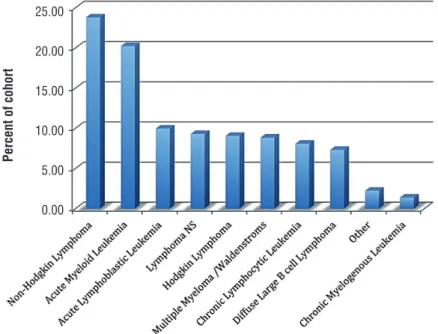
Figure 1 - Proportion of hematologic malignancies observed in cohort, n=1316.
the proportion of primary hematologic malignan-cies observed in stone formers vs. non-stone for-mers (p=0.601) (Table-2). Specifically, there were no significant differences in rates of any lymphopro-liferative disorder (diffuse large B cell lymphoma, Non-Hodgkins lymphoma, Hodgkins lymphoma, chronic lymphocytic leukemia, acute lymphoblas-tic leukemia, or multiple myeloma/Waldenstrom’s macroglobulinemia) among patients with or wi-thout stones, nor were there any significant diffe-rences observed in rates of any myeloproliferative disorder (AML or chronic myelogenous leukemia) among patients with or without stones.
Among stone formers, the median urinary pH was 5.5, the mean serum uric acid was 7.5, calcium was 9.6, potassium was 4.3, and phos-phorus level was 3.8 mg/dL. Median stone size was 3 mm, median HU was 341, median time from initial chemotherapy to incident stone for-mation was 3.9 months (1.3-10.7), and 34.8% of stones were symptomatic.
Only 30 of the 72 stone formers (43.5%) underwent very elementary 24-hour urine col-lections, and median 24-hour urinary volume was 1,873 mL. Only 1 of 30 patients undergoing 24-hour urine collection had 24-24-hour urinary uric acid
and calcium levels analyzed, and this patient had both hyperuricosuria (urinary uric acid 825 mg) and hypercalciuria (urinary calcium 334 mg). No stones were analyzed, however, spot urinalyses performed during chemotherapy demonstrated crystalluria in 11 patients (15.2%): calcium oxalate crystals obser-ved in 10 (14.3%) and uric acid crystals obserobser-ved in 1(1.4%).
Allopurinol users had different metabolic parameters compared with non-allopurinol users (mean uric acid 7.4 vs. 5.9 mg/dL, p<0.001, mean potassium 4.3 vs. 4.1 mg/dL, p=0.002, and mean phosphorus 4.0 vs. 3.7 mg/dL, p=0.018 respecti-vely). The proportions of diffuse large B cell lym-phoma (DLBCL) (11%vs. 6.6%, p=0.020) and CLL (19.2% vs. 5.8%, p<0.001) were also significantly higher in allopurinol users, while proportions of Hodgkin lymphoma (HL) (5.0% vs. 9.9%, p=0.020) and acute myeloid leukemia (AML) (14.2% vs. 21.4%, p=0.016) were significantly lower. Hyperu-ricemia (35% versus 16.1%, p<0.001, respective-ly), and hypercalcemia (17.6% vs. 12.8%, p=0.065) were observed more frequently in allopurinol users than non-allopurinol users.
In multivariate analysis, diabetes melli-tus (OR=6.38, p<0.001), hyperuricemia (OR=2.31,
Percent of cohort
25.00
20.00
15.00
10.00
5.00
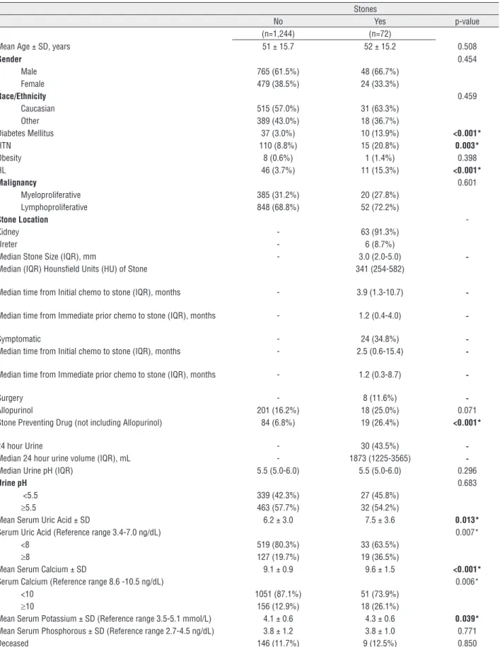
Table 1 - Demographics and clinical variables of stone formers and non-stone formers.
Stones
No Yes p-value
(n=1,244) (n=72)
Mean Age ± SD, years 51 ± 15.7 52 ± 15.2 0.508
Gender 0.454
Male 765 (61.5%) 48 (66.7%)
Female 479 (38.5%) 24 (33.3%)
Race/Ethnicity 0.459
Caucasian 515 (57.0%) 31 (63.3%)
Other 389 (43.0%) 18 (36.7%)
Diabetes Mellitus 37 (3.0%) 10 (13.9%) <0.001*
HTN 110 (8.8%) 15 (20.8%) 0.003*
Obesity 8 (0.6%) 1 (1.4%) 0.398
HL 46 (3.7%) 11 (15.3%) <0.001*
Malignancy 0.601
Myeloproliferative 385 (31.2%) 20 (27.8%)
Lymphoproliferative 848 (68.8%) 52 (72.2%)
Stone Location
-Kidney - 63 (91.3%)
Ureter - 6 (8.7%)
Median Stone Size (IQR), mm - 3.0 (2.0-5.0)
-Median (IQR) Hounsfield Units (HU) of Stone 341 (254-582)
Median time from Initial chemo to stone (IQR), months - 3.9 (1.3-10.7)
-Median time from Immediate prior chemo to stone (IQR), months - 1.2 (0.4-4.0)
-Symptomatic - 24 (34.8%)
-Median time from Initial chemo to stone (IQR), months - 2.5 (0.6-15.4)
-Median time from Immediate prior chemo to stone (IQR), months - 1.2 (0.3-8.7)
-Surgery - 8 (11.6%)
-Allopurinol 201 (16.2%) 18 (25.0%) 0.071
Stone Preventing Drug (not including Allopurinol) 84 (6.8%) 19 (26.4%) <0.001*
24 hour Urine - 30 (43.5%)
-Median 24 hour urine volume (IQR), mL - 1873 (1225-3565)
-Median Urine pH (IQR) 5.5 (5.0-6.0) 5.5 (5.0-6.0) 0.296
Urine pH 0.683
<5.5 339 (42.3%) 27 (45.8%)
≥5.5 463 (57.7%) 32 (54.2%)
Mean Serum Uric Acid ± SD 6.2 ± 3.0 7.5 ± 3.6 0.013*
Serum Uric Acid (Reference range 3.4-7.0 ng/dL) 0.007*
<8 519 (80.3%) 33 (63.5%)
≥8 127 (19.7%) 19 (36.5%)
Mean Serum Calcium ± SD 9.1 ± 0.9 9.6 ± 1.5 <0.001*
Serum Calcium (Reference range 8.6 -10.5 ng/dL) 0.006*
<10 1051 (87.1%) 51 (73.9%)
≥10 156 (12.9%) 18 (26.1%)
Mean Serum Potassium ± SD (Reference range 3.5-5.1 mmol/L) 4.1 ± 0.6 4.3 ± 0.6 0.039*
Mean Serum Phosphorous ± SD (Reference range 2.7-4.5 ng/dL) 3.8 ± 1.2 3.8 ± 1.0 0.771
p=0.007), and hypercalcemia (OR=2.14, p=0.022) at time of chemotherapy predicted de novostone.
DISCUSSION
The findings from this study spanning 10 years support the hypothesis that metabolic de-rangements during chemotherapy are associated with an increased risk of nephrolithiasis. Previous extensive reports demonstrating this include a pe-diatric study of over 2,000 children treated for ALL and a Korean study of over 900 adults who were treated for both lymphoproliferative and myelo-proliferative disorders (5,8). Both investigations demonstrated that the incidence of nephrolithiasis in these populations was significantly higher than in the general population. Until these reports, the claim of increased nephrolithiasis risk with he-matologic malignancy had been substantiated by
only case reports and the plausible pathophysiolo-gic theory of endogenous nucleotide catabolism.
The investigators of the pediatric stone stu-dy postulated that stone formation was associated with chemotherapy, but perhaps more importantly was due to glucocorticoid therapy. Steroids are used in multiple contexts for lymphoproliferative disea-ses and can increase the risk of nephrolithiasis by decreasing renal calcium absorption. The authors cited the predominance of calcium-based stones as opposed to uric acid stones as evidence supporting the steroid-nephrolithiasis hypothesis (8). However, almost half of stone analyses in the Korean adult study showed uric acid stones despite the common use of glucocorticoid therapy (5).
Our study included a predominance of cal-cium oxalate over uric acid crystals, although no stones were formally analyzed, and the median HU of de novostones was 341, suggesting a mixture of
Table 2 - Rates of primary malignancy in stone formers vs. non-stone formers.
Stones
No Yes p-value (n=1,244) (n=72)
Lymphoproliferative Disorders 947 (76.1%) 56 (77.8%) 0.601 Diffuse Large B cell Lymphoma (DLBL) 87 (7%) 9 (12.5%) 0.098 Non-Hodgkin Lymphoma (NHL) 293 (23.6%) 20 (27.8%) 0.396 Hodgkin Lymphoma (HL) 108 (8.7%) 11 (15.3%) 0.086 Lymphoma NS 120 (9.6%) 2 (2.8%) 0.057 Chronic Lymphocytic Leukemia (CLL) 100 (8%) 6 (8.3%) 0.826 Acute Lymphoblastic Leukemia (ALL) 127 (10.2%) 4 (5.6%) 0.308 Multiple Myeloma (MM)/Waldenstroms 112 (9%) 4 (5.6%) 0.397 Myeloproliferative Disorders 274 (22%) 10 (13.9%) 0.601 Acute Myeloid Leukemia (AML) 257 (20.7%) 9 (12.5%) 0.099 Chronic Myelogenous Leukemia (CML) 17 (1.4%) 1 (1.4%) 1.000 Other 23 (1.8%) 6 (8.3%) 0.003*
Other: T-cell prolymphocytic leukemia, Aplastic anemia, chronic congenital neutropenia, CVID
both calcium and uric acid stone compositions (9). These findings are consistent with our analysis of nephrolithiasis risk factors, which demonstrated in multivariate analysis that diabetes mellitus, hype-ruricemia, or hypercalcemia significantly increased risk of nephrolithiasis. The presence of hypercalce-mia and hyperuricehypercalce-mia permits heterogeneous nu-cleation and subsequent calcium stone formation (10). Diabetes is thought to be a risk factor for uric acid calculi due to insulin resistance leading to low urinary pH associated with defective ammonia syn-thesis occurring in the proximal tubule cell as well as ammonium transport into the renal tubular lu-men (11). The role of diabetes mellitus in our stu-dy is significant, as it was associated with an over 6-fold increased risk of nephrolithiasis. Allopurinol is a xanthine oxidase inhibitor frequently used pro-phylactically to decrease the uric acid production prior to instituting chemotherapy for patients at risk for TLS (12). In a double blinded randomized pros-pective trial, allopurinol utilization decreased the number of stone events while increasing the time to recurrence of stone event among calcium oxalate stone formers with hyperuricosuria on 24-hour uri-ne collections, substantiating its preventive role in patients at risk for calcium oxalate nephrolithiasis (13). Published guidelines regarding the prevention and management of TLS include specific recom-mendations for the appropriate utilization of allo-purinol (14). Risk factors for TLS include tumor type (Burkitt’s lymphoma, lymphoblastic lymphoma, diffuse large cell lymphoma, ALL), tumor burden/ extent of disease defined by elevated WBC>25,000 and/or bulky nodal disease >10 cm, preexisting re-nal failure, and baseline uric acid≥7.5 mg/dL (14).
These high-risk patients are recommended to undergo aggressive hydration, urinary alkaliniza-tion, diuresis, and allopurinol or recombinant ura-te oxidase (rasburicase) prophylaxis. Our data de-monstrated patients at higher risk for TLS received allopurinol more frequently than those at lower risk, as one would expect, but in multivariate analysis use of allopurinol did not decrease incident stone risk, presumably due to selection bias. Given this and our finding that hyperuricemia is a risk factor for stone formation in this population, recombi-nant urate oxidase (rasburicase) may offer an ad-vantage, as it catalyzes the conversion of uric acid
to allantoin, which is 5-10 times more soluble in urine (14). Rasburicase has been well studied in both pediatric and adult patient populations at risk for TLS, demonstrating a significantly more rapid lowering of serum uric acid levels compared to allopurinol (15-17). Current guidelines for ma-nagement of TLS recommend rasburicase in pe-diatric patients at high risk for TLS and in adults with hyperuricemia diagnosed with TLS or refrac-tory to allopurinol (14). Our findings suggest that prompt and effective treatment of hyperuricemia may prevent upper tract stone formation and re-duce the incidence of nephrolithiasis.
Unfortunately, our study highlights both the low number of 24-hour urine collections (43.5%) as well as the lack of necessary detail in these 24-hour urine collections (3.33%) performed in high-risk stone formers. Urological consulta-tions were infrequently obtained in this patient population, and as such, metabolic stone evalu-ations were rarely performed in these patients. While it would have been incredibly useful to have a 24-hour urine collection in every single stone former, the absence of this data provides an area of opportunity for urologists to improve the care of these patients. Urologists should be con-sulted in the care of such patients to appropriately work up the etiology of stone disease, and thereby prevent future stones from occurring.
Inherent limitations of this retrospective study are acknowledged, specifically, selection bias was observed when comparing patients re-ceiving allopurinol prophylaxis. Patients in our study attended regular office visits and underwent frequent cross-sectional imaging for oncologic management and surveillance, resulting in more opportunities to diagnose asymptomatic nephro-lithiasis. Moreover, patients may have been more likely to report symptoms of abdominal or flank pain at follow-up visits, increasing the likelihood of diagnosis of nephrolithiasis.
CONCLUSIONS
mellitus, as well as hyperuricemia and hypercal-cemia at time of chemotherapy as risk factors for nephrolithiasis that should assist oncologists in appropriately selecting patients for prophylaxis, while also providing urologists an earlier oppor-tunity to collaborate and assist in treatment and evaluation of nephrolithiasis.
ABBREVIATIONS
ALL = Acute Lymphoblastic Leukemia
AML = Acute Myeloid Leukemia
CLL = Chronic Lymphocytic Leukemia
DLBL = Diffuse Large B cell Lymphoma
HL = Hodgkins lymphoma
HU = Hounsfield Unit
MM = Multiple Myeloma
NHL = Non Hodgkins Lymphoma
TLS =
Tumor lysis syndrome
CONFLICT OF INTEREST
None declared.
REFERENCES
1. Cairo MS, Coiffier B, Reiter A, Younes A; TLS Expert Panel: Recommendations for the evaluation of risk and prophylaxis of tumour lysis syndrome (TLS) in adults and children with malignant diseases: an expert TLS panel consensus. Br J Haematol. 2010;149:578-86.
2. Abu-Alfa AK, Younes A: Tumor lysis syndrome and acute kidney injury: evaluation, prevention, and management. Am J Kidney Dis. 2010;55(5 Suppl3):S1-13.
3. Howard SC, Ribiero RC, Pui C-H. Acute complications in childhood leukemias. cambridge, UK: Cambridge University Press, 2006.
4. Shimada M, Johnson RJ, May WS Jr, Lingegowda V, Sood P, Nakagawa T, et al.: A novel role for uric acid in acute kidney injury associated with tumour lysis syndrome. Nephrol Dial Transplant. 2009;24:2960-4.
5. Kim SW, Kim SD, Yoo JM, Cho YH, Sohn DW: Urolithiasis in patients suffering from malignant hematologic diseases. Yonsei Med J. 2010;51:244-7.
6. Shiozawa Y, Sakaguchi S, Sakakibara O, Yagishita K, Saito M, Yamashiro Y: Urolithiasis in an acute lymphoblastic leukemia child during induction chemotherapy. Pediatr Hematol Oncol. 2008;25:359-63.
7. Jaing TH, Hung IJ, Lin CJ, Chiu CH, Luo CC, Wang CJ: Acute myeloid leukemia complicated with staghorn calculus. Jpn J Clin Oncol. 2002;32:365-7.
8. Howard SC, Kaplan SD, Razzouk BI, Rivera GK, Sandlund JT, Ribeiro RC: Urolithiasis in pediatric patients with acute lymphoblastic leukemia. Leukemia. 2003;17:541-6.
9. Nakada SY, Hoff DG, Attai S, Heisey D, Blankenbaker D, Pozniak M: Determination of stone composition by noncontrast spiral computed tomography in the clinical setting. Urology. 2000;55:816-9.
10. Zerwekh JE, Hwang TI, Poindexter J, Hill K, Wendell G, Pak CY: Modulation by calcium of the inhibitor activity of naturally occurring urinary inhibitors. Kidney Int. 1988;33:1005-8. 11. Maalouf NM, Sakhaee K, Parks JH, Coe FL, Adams-Huet
B, Pak CY: Association of urinary pH with body weight in nephrolithiasis. Kidney Int. 2004; 65: 1422-5.
12. Hochberg J, Cairon MS: Tumor lysis syndrome: current perspective. Haematologica. 2008; 93: 9-13.
13. Ettinger B, Tang A, Citron JT, Livermore B, Williams T: Randomized trial of allopurinol in the prevention of calcium oxalate calculi. N Engl J Med. 1986;315:1386-9.
14. Coiffier B, Altman A, Pui CH, Younes A, Cairo MS: Guidelines for the management of pediatric and adult tumor lysis syndrome: an evidence-based review. J Clin Oncol. 2008 Jun 1;26(16):2767-78. Erratum in: J Clin Oncol. 2010;28:708. 15. Pui CH, Mahmoud HH, Wiley JM, Woods GM, Leverger
G, Camitta B, et al.: Recombinant urate oxidase for the prophylaxis or treatment of hyperuricemia in patients With leukemia or lymphoma. J Clin Oncol. 2001;19:697-704. 16. Goldman SC, Holcenberg JS, Finklestein JZ, Hutchinson R,
Kreissman S, Johnson FL, et al.: A randomized comparison between rasburicase and allopurinol in children with lymphoma or leukemia at high risk for tumor lysis. Blood. 2001;97:2998-3003.
17. Coiffier B, Mounier N, Bologna S, Fermé C, Tilly H, Sonet A, et al.: Efficacy and safety of rasburicase (recombinant urate oxidase) for the prevention and treatment of hyperuricemia during induction chemotherapy of aggressive non-Hodgkin’s lymphoma: results of the GRAAL1 (Groupe d’Etude des Lymphomes de l’Adulte Trial on Rasburicase Activity in Adult Lymphoma) study. J Clin Oncol. 2003;21:4402-6.
_______________________ Correspondence address:
APPENDIX 1: LIST OF CHEMOTHERAPY REGIMENS
Lymphoma
1. Cyclophosphamide, doxorubicin, vincristine and prednisone (CHOP) with/without rituximab 2. Etoposide, prednisone, vincristine, doxorubicin, and cyclophosphamide (EPOCH) with/without
rituximab
3. Ifosfamide, carboplatin, and etoposide (ICE) with or without rituximab 4. Doxorubicin, bleomycin, vinblastine, and dacarbazine (ABVD)
5. Nitrogen mustard, doxorubicin, vinblastine, vincristine, bleomycin, etoposide, and prednisone (Stanford V)
Acute myelogenous leukemia
1. Cytarabine and idarubicin or daunorubicin (7+3)
2. Fludarabine, cytarabine, idarubicin, and filgrastim (FLAG) 3. All trans-retinoic acid (ATRA)