ORIGIN
AL RESEAR
CH
Mailing address: Lívia Pimenta Renó Gasparotto – Rua Governador Jorge Lacerda, 244 – Curitiba (PR), Brasil – CEP 81510040 – E-mail: [email protected] – Financing source: Nothing to declare – Conlict of interest: Nothing to declare – Presentation: Jan. 2016. Accepted for publication: Sept. 2016. Approved by the Ethics Committee of Unicamp under No. 387,026.
1Graduate Program in Gerontology, Universidade Estadual de Campinas (Unicamp) – Campinas (SP), Brazil.
2Professor of the Family Health Department and of the Graduate Program in Gerontology, Universidade Estadual de Campinas (Unicamp) – Campinas (SP), Brazil.
ABSTRACT | The literature about posture in older adults includes studies that measure spine angles through several strategies. This may interfere with postural diagnosis and impact on the resolution of comorbidities afecting the spine in older people. The objective of this study was to compare two lumbar spine measurement methods commonly used in research. The association between these measures and the angle of kyphosis was used as the basis for comparison. One hundred-sixty older adults were submitted to anterior-posterior lumbar spine x-ray. Two Cobb measures were used for each image (L1-L5 and L1-S1) and the conventional measure for hyperkyphosis. Only the measure based on L1-L5 was associated with the kyphosis angle. The study suggests that lumbar spine x-rays should be analyzed through the L1-L5 Cobb strategy in older people.
Keywords | Aging; Lordosis/Radiography; Posture.
RESUMO | A literatura sobre postura em idosos tem apresentado artigos que medem os ângulos da coluna por diferentes estratégias. Isso pode interferir no diagnóstico postural e impactar na resolução das comorbidades que afetam a coluna desses indivíduos. O objetivo deste estudo foi comparar dois métodos de medida da coluna lombar comumente utilizados em pesquisa. A associação dessas medidas com o ângulo da cifose foi utilizada como base para comparação. Participaram da pesquisa 160 idosos, submetidos a raio-X anteroposterior da coluna lombar. Foram realizadas duas medidas de Cobb para cada radiograia: uma com base em T12-S1 e outra em
388
L1-L5. Somente a medida com base em L1-L5 apresentou associação signiicativa com o ângulo da cifose. Não houve associação da medida T12-S1 com o ângulo da curva torácica. Nossos achados indicam que o método de Cobb mais adequado para analisar radiograias de coluna lombar em idosos é o do traçado de linhas entre L1-L5.
Descritores | Envelhecimento; Lordose/Radiograia, Postura.
RESUMEN | La literatura sobre la postura de adultos mayores está compuesta de textos que estudian la medida angular de la columna a través de distintas estrategias. Esto puede producir interferencias en el diagnóstico postural e impactar en comorbidades que les afectan la columna de los adultos mayores. El propósito de este estudio es comparar dos métodos de medición de la columna lumbar empleados frecuentemente en los estudios. Se empleó como base de comparación la asociación de las medidas con el ángulo de cifosis. A los 160 adultos mayores participantes del estudio se les sometieron al rayo X anteroposterior de la columna lumbar. Se llevó a cabo dos medidas Cobb para cada radiografía: una con base en T12-S1 y la otra en L5. La medida con base en L1-L5 fue la única que presentó la asociación signiicativa con el ángulo de cifosis. No presentó asociación la medida T12-S1 con el ángulo de la curvatura torácica. Los resultados encontrados en este estudio muestran que del método Cobb para analizar radiografías de la columna lumbar de adultos mayores el más adecuado es el de trazado de líneas entre L1-L5.
Palabras clave | Envejecimiento; Lordosis/Radiografía; Postura.
Lumbar spine angular measures in older people:
comparison between two radiographic analysis
strategies
Medidas angulares da coluna lombar em idosos: comparação entre duas estratégias de análise
radiográica
Medidas angulares de la columna lumbar en adultos mayores: comparación entre dos
estrategias de análisis radiográico
INTRODUCTION
Studies on human aging are deined under biological, behavioral and social aspects. One of the lines of investigation has been changes in body structure, such as spinal posture and its modiications during the aging
process1-5. he study of posture involves measuring the
angles of the spine. However, the diversity of strategies for measuring causes divergences of results and hinders
standardization6-8.
In the sagittal plane the lordotic and kyphotic curves of the spine are evaluated, in the anterior-posterior direction. he spine (thoracic region) is susceptible to deformities as structural changes which are characteristic of human aging occur, what causes changes in the
lumbar angle9-12. Due to this, researches on the inluence
of diferent postures on the social and physical condition of the elderly stand out. However, these analyses have diferences regarding their diferent measuring strategies,
among them of the lumbar spine7-9.
he study by Bruno et al.10 points out that when the
elderly show increase in the kyphotic angle, one of the ways to keep the body balance is the posterior rotation of the iliac crest in the pelvis. Other adjustments such as hip extension, knee lexion and ankle dorsilexion make up the structural adjustments for keeping stable the center of mass.
he radiographic evaluation, considered the gold standard for angular measurements of the spine, can be analyzed through diferent strategies. he Cobb method advocates the drawing of parallel lines over the vertebral surfaces of each point that deines the curves and, through them, perpendicular lines are crossed with each other for the identiication of the angle. However, measurements of the lumbar spine can be made from
diferent points of these lines9,11-15.
One of the methods for the measuring of the lumbar spine uses as its starting point the lines parallel to the superior surface of the vertebral body of L1 and to the inferior surface of L5. Another method analyzes the lines starting from the inferior surface of T12 and the superior surface of S1. It is unclear whether the use of either corresponds to the same clinical outcome. he places indicated as starting points of the lines (such as the superior surface of the sacrum, for example), have biomechanical peculiarities that change the direction of
the lines11,13,15-17. he use of the line starting from T12
and S1 considers the position of the sacrum as decisive in the lordosis curve. It is known that the sacrum has
varying positions from one individual to another, and can be in either horizontal or vertical inclination. herefore, it is possible that the use of the method which adopts the position of S1 does not accurately
determine the lumbar curvature in elderly people13,16,18.
In addition, there are studies that show that there are diferences between men and women regarding the curvature of the lumbar and thoracic spine.
herefore, this work aims to identify the lumbar spine angle, determined through two measures (L1-L5 and T12-S1) and evaluate which one is best associated with the angulation of the thoracic region in the elderly. he primary objective is to investigate possible divergences between measures, caused by the diferent markings and that can inluence the postural diagnosis. he secondary objective is to compare the curvatures of elderly men and women.
METHODOLOGY
he research has a descriptive and transversal character and used a probabilistic sample of a population of elders at a primary health care unit in the city of Amparo-SP.
Of the 820 active elders, 420 were randomly chosen and invited to participate in a follow-up survey with the conducting of the radiographic examination. Of these, 160 attended the examination and were positioned for the recording of the sagittal plane, in its anterior-posterior view. he inclusion criterion was to be more than 65 years old, not having gone through a spine surgery nor having a restrictive disease that would hinder walking or maintaining a standing up position.
he posture was analyzed through the Cobb method, using the two aforementioned lines strategies for the lumbar spine (T12-S1 and L1-L5, Fig. 1). he thoracic spine was also evaluated through the Cobb method based on the line on the superior surface of the vertebral body which had a more evident superior curve and another line on the inferior surface of the vertebral body of T12 (Fig. 2).
he study obtained approval of the Research Ethics Committee under number 387,026, with the signing of the Informed Consent Form by all participants.
with hyperkyphosis and hyperlordosis was described by frequency distribution, with a 95% conidence interval. he Pearson correlation coeicient was used to verify the relationship between the kyphosis angle and the two diferent measures of the lordosis angle. he association of the proportion of individuals with hyperkyphosis and hyperlordosis evaluated through two measuring strategies was tested through the Chi-square test. he odds ratio of individuals with hyperlordosis having hyperkyphosis was veriied through binary logistic regression adjusted according to gender. All analyses were performed using the SPSS 18.0 software and the established signiicance level was p<0.05.
Figure 1 – Measurements of lumbar lordosis (T12/S1 and L1/L5)
Figure 2 – Measurement of thoracic kyphosis
RESULTS
Correlation was observed between the kyphosis angle and both measures of the lordosis angles (L1-L5 and T12-S1). However, the measuring of lordosis performed through L1-L5 had a more expressive correlation. he correlation between the measures of the kyphosis and lordosis angles is described in Table 1.
Table 1. Correlation between the kyphosis angle and the lordosis angle with the two measuring strategies
Lordosis L1-L5 Lordosis T12-S1 P
Kyphosis 0.475 0.382 <0.001*
*p<0.05
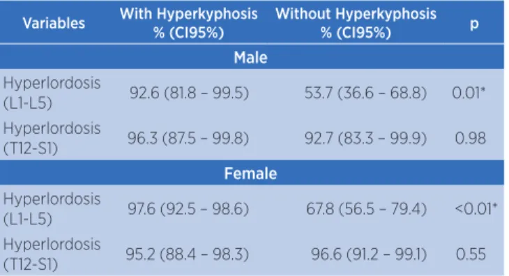
he association between the proportion of individuals with hyperkyphosis and hyperlordosis was identiied only through the measurement performed with L1-L5. he analysis of the association between the proportion of hyperkyphosis with hyperlordosis measured through the two strategies is described in Table 2.
Table 2. Association between hyperkyphosis and hyperlordosis (evaluated through two measuring strategies)
Variables With Hyperkyphosis
% (CI95%)
Without Hyperkyphosis
% (CI95%) p
Male
Hyperlordosis
(L1-L5) 92.6 (81.8 – 99.5) 53.7 (36.6 – 68.8) 0.01* Hyperlordosis
(T12-S1) 96.3 (87.5 – 99.8) 92.7 (83.3 – 99.9) 0.98
Female
Hyperlordosis
(L1-L5) 97.6 (92.5 – 98.6) 67.8 (56.5 – 79.4) <0.01* Hyperlordosis
(T12-S1) 95.2 (88.4 – 98.3) 96.6 (91.2 – 99.1) 0.55 CI: Conidence interval; p*<0.01
he adjusted logistic regression showed that hyperkyphosis was associated with hyperlordosis only when measured through L1-L5, regardless of gender. he odds ratio of individuals with hyperlordosis having hyperkyphosis is presented in Table 3 and shows that, through the L1-L5 measure for lordosis, there is an 81% chance of that same individual having hyperkyphosis.
Table 3. Adjusted odds ratio of hyperlordosis (evaluated through the two measuring strategies) associated with hyperkyphosis
Variables Odds Ratio (95%CI) p
Hyperlordosis (L1-L5) 1.81 (1.09 –3.04) 0.02* Hyperlordosis (T12-S1) 0.66 (0.41 – 1.08) 0.11 CI: Conidence interval; p*<0.05
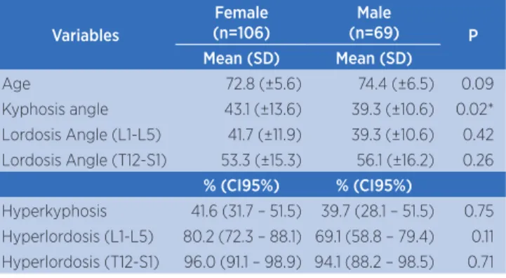
Table 4. Descriptive analysis of the average age and kyphosis and lordosis angles evaluated through two measuring strategies and proportion of individuals with hyperkyphosis and hyperlordosis calculated through the two measuring strategies
Variables
Female (n=106)
Male
(n=69) P
Mean (SD) Mean (SD)
Age 72.8 (±5.6) 74.4 (±6.5) 0.09
Kyphosis angle 43.1 (±13.6) 39.3 (±10.6) 0.02* Lordosis Angle (L1-L5) 41.7 (±11.9) 39.3 (±10.6) 0.42 Lordosis Angle (T12-S1) 53.3 (±15.3) 56.1 (±16.2) 0.26
% (CI95%) % (CI95%)
Hyperkyphosis 41.6 (31.7 – 51.5) 39.7 (28.1 – 51.5) 0.75 Hyperlordosis (L1-L5) 80.2 (72.3 – 88.1) 69.1 (58.8 – 79.4) 0.11 Hyperlordosis (T12-S1) 96.0 (91.1 – 98.9) 94.1 (88.2 – 98.5) 0.71 p*<0.05
SD: Standard Deviation; CI: Conidence interval
DISCUSSION
he changes in the curvature of the spine during the process of human aging are widely discussed, however, there is still disagreement among researchers concerning its measuring. he diversity of methods observed in the literature seems to demonstrate a free choice of the researcher, independently of the
relationship with the assessed structure1,2,6,9,10,14,16,17. In
the analyses in the sagittal plane, the position of the joints of the pelvic girdle interferes in the measures. hus, the measures that consider the angle of the lumbar region with inluence of the sacral position are frequent, such as the T12-S1 measure, also called
lumbosacral angle12,14,17,19-22. he present study was
based on two measures which are widely seen in researches with the elderly which are the lines on L1-L5 and T12-S1. Although the Cobb method is considered to be the standard, there is variability of
the starting point of the lines23,24.
Wang et al.6 discuss the measures of the thoracic and
lumbar spine in the Chinese population. he authors report the diiculty in identifying the best relationship between the thoracic and lumbar region, in the sagittal plane. hey highlight that there is inluence of one curvature over the other. Similarly, the works of Erkan
et al.3 and Quek4, which focus on the cervical spine,
also reveal uncertainty about the measures, pointing
to the need for standardization of the measures in the sagittal plane.
he use of measure L5-S1 is associated with changes in the position of the sacrum. Recent studies indicate that the pelvic position is important in the deinition of sagittal alignment, since it participates in the compensations in the lumbar region, as well as in the thoracic and cervical regions. he study by Russouly
& Pinheiro-Franco12 shows that the pelvis undergoes
retroversion as the reduction of lumbar lordosis occurs. his retroversion is increased when there is thoracic hyperkyphosis and generates other compensations, such as hip extension and knee lexion.
On the other hand, increased lumbar lordosis is
also evidenced in studies. he work by Abreu et al.24
found an important percentage of hyperlordosis after radiographic analysis in elderly people (using measures L1-L5). According to the researchers, the reduction of the muscular strength of the muscles that support the lumbar region, such as the rectus abdominis and abdominal oblique and the glutes, explain this angle change in the aging process.
Smith et al.18 discuss the radiographic measures and
mention the T12-S1 measure as the most common way to analyze the lumbar curve. However, they also report that, in the correlation of this line with the other curve of the sagittal plane (kyphosis), there was no signiicance. he same indings were observed in this study (Table 1), even with the separation by gender for obtaining greater homogeneity.
he study by Miyasaki et al.21 evaluated the lumbar
angle of elderly men and identiied associations with walking skills and the strength of the lower limbs. In their work, the deined measure was L1-L5. In the present study, there was a signiicant association of this measurement strategy with the thoracic curve (Tables 1 and 2). here was signiicance both for the group of elderly males as for the females (p=0.01 for men and p<0.01 for women).
he thoracic spine was used as a basis for comparison with the lumbar region. he angle of the thoracic spine also showed diferences concerning the cutof point for the elderly. Although recent studies use degrees between
40° and 50°, as shown in the research by Burke at al.5,
the studies by Katzman et al.7 have identiied that the
he measuring of hyperkyphosis was carried out for comparison with the changes in the lumbar spine because they share the same plane of movement, the sagittal plane. When testing the correlations between the values of the lumbar angles measured through L1-L5 and L1-S1, it was observed that only the irst was related to the thoracic curve. An odds ratio of 1.81 (p=0.02) was veriied for the L1-L5 measure with hyperkyphosis (Table 3). hese data indicate that the L1-L5 measure is most appropriate to assess lumbar lordosis.
CONCLUSION
he results of this research suggest a reassessment of the best strategy of measurement of the lumbar angle using the Cobb method. In this research, the line starting from L1-L5 was identiied as having a greater relationship with the thoracic curve. he other model analyzed, T12-S1, showed no relationship with the thoracic curve. It is important that further research is carried out with the same goal, to deepen the discussion on methods of measurement for deining the strategy that best represents the lumbar curve.
REFERENCES
1. Kado DM, Huang MH, Karlamangla AS, Cawthon P, Katzman W, Hillier TA, et al. Factors associated with kyphosis progression in older women: 15 years’ experience in the study of osteoporotic fractures. J Bone Mineral Res. 2013;28(1):179-87. doi: 10.1002/jbmr.1728.
2. Imagama S, Hasegawa Y, Matsuyama Y, Sakai Y, Ito Z, Hamajima N, et al. Inluence of sagittal balance and physical ability associated with exercise on quality of life in middle-aged and elderly people. Arch Osteoporos. 2011;6(1-2):13-20. doi: 10.1007/s11657-011-0052-1.
3. Erkan S, Yercan HS, Okcu G, Ozalp RT. The inluence of sagittal cervical proile, gender and age on the thoracic kyphosis. Acta Orthop Belg. 2010;76(5):675-80.
4. Quek J, Pua YH, Clark RA, Bryant AL. Efects of thoracic kyphosis and forward head posture on cervical range of motion in olders adults. Man Ther. 2013;18(1):65-71. doi: 10.1016/j.math.2012.07.005.
5. Burke TN, França FJ, Meneses SR, Cardoso VI, Pereira RM, Danilevicius CF, et al. Postural control among elderly women with and without osteoporosis: is there a diference? Sao Paulo Med J. 2010;128(4):219-24.
6. Wang HJ, Giambini H, Zhang WJ, Ye GH, Zhao C, An KN, et al. A modiied sagittal spine postural classiication and its relationship to deformities and spinal mobility in a Chinese
osteoporotic population. PLoS One. 2012;7:1-8. doi: 10.1371/ journal.pone.0038560.
7. Katzman WB, Vittinghof E, Kado DM. Age-related hyperkyphosis, independent of spinal osteoporosis, is associated with impaired mobility in older community-dwelling women. Osteoporos Int. 2011;22(1):85-90. doi: 10.1007/s00198-010-1265-7.
8. Katzman WB, Wanek L, Shepherd JA, Sellmeyer DE. Age-related hyperkyphosis: its causes, consequences, and management. J Orthop Sports Phys Ther. 2010;40(6):352-60. doi: 10.2519/jospt.2010.3099.
9. Polly DW Jr, Kilkelly FX, McHale KA, Asplund LM, Mulligan M, Chang AS. Measurement of lumbar lordosis: evaluation of intraobserver, interobserver, and technique variability. Spine (Phila PA 1976). 1996;21(13):1530-5.
10. Bruno AG, Anderson DE, D’Agostino J, Bouxsein ML. The efect of thoracic kyphosis and sagittal plane alignment on vertebral compressive loading. J Bone Miner Res. 2012;27(10):2144-51. doi: 10.1002/jbmr.1658.
11. Anderson DE, D’Agostino JM, Bruno AG, Manoharan RK, Bouxsein ML. Regressions for estimating muscle parameters in the thoracic and lumbar trunk for use in musculoskeletal modeling. J Biomech. 2012;45(1):66-75. doi: 10.1016/j. jbiomech.2011.10.004.
12. Roussouly P, Pinheiro-Franco JL. Biomechanical analysis of the spino-pelvic organization and adaptation in pathology. Eur Spine J. 2011;20(5):609-18. doi: 10.1007/s00586-011-1928-x. 13. Marras WS, King AI, Joynt RL. Measurements of loads on the
lumbar spine under isometric and isokinetic conditions. Spine. 1984;9(2):176-87. doi: 10.1097/00007632-198403000-00008. 14. Gonçalves GB, Pereira JS. Radiological assessment of the
angular values of back-lumbar and sacral-lumbar curvature in adolescents. Acta Fisiatr. 2008;15(2):92-5.
15. Russell BS, Muhlenkamp KA, Hoiriis KT, DeSimone CM. Measurement of lumbar lordosis in static standing posture with and without high-heeled shoes. J Chiropr Med. 2012;11(3):145-53. doi: 10.1016/j.jcm.2012.02.002.
16. Iyer S, Christiansen BA, Roberts BJ, Valentine MJ, Manoharan RK, Bouxsein ML. A biomechanical model for estimating loads on thoracic and lumbar vertebrae. Clin Biomech (Bristol, Avon). 2010;25(9):853-8. doi: 10.1016/j. clinbiomech.2010.06.010.
17. Cho IY, Park SY, Park JH, Kim TK, Jung TW, Lee HM. The efect of standing and diferent sitting positions on lumbar lordosis: radiographic study of 30 healthy volunteers. Asian Spine J. 2015;9(5):762-9. doi: 10.4184/asj.2015.9.5.762.
18. Smith JS, Shafrey CI, Fu KM, Scheer JK, Bess S, Lafage V, et al. Clinical and radiographic evaluation of the adult spinal deformity patient. Neurosurg Clin N Am. 2013;24(2):143-56. doi: 10.1016/j.nec.2012.12.009.
19. Henneman SA, Antoneli PHL, Oliveira GC. Incidencia pélvica: um parâmetro fundamental para deinição do equilíbrio sagital da coluna vertebral. Coluna/Columna. 2012;11(3):237-9. doi: http://dx.doi.org/10.1590/S1808-18512012000300011. 20. Araújo THP, Francisco LTP, Leite RF, Iunes DH. Posicionamento
21. Miyazaki J, Murata S, Horie J, Uematsu A, Hortobágyi T, Suzuki S. Lumbar lordosis angle (LLA) and leg strength predict walking ability in elderly males. Arch Gerontol Geriatr. 2013;56(1):141-7. doi: 10.1016/j.archger.2012.09.004.
22. Chen YL. Vertebral centroid measurement of lumbar lordosis compared with the Cobb technique. Spine (Phila Pa 1976). 1999;24(17):1786-90.
23. Chernukha KV, Dafner RH, Reigel DH. Lumbar lordosis measurement: a new method versus Cobb technique. Spine (Phila Pa 1976). 1998;23(1):74-9.