Acta Palaeontol. Pol. 61 (1): 97–117, 2016 http://dx.doi.org/10.4202/app.00126.2014
Morphology and histology of dorsal spines of
the xenacanthid shark
Orthacanthus platypternus
from the Lower Permian of Texas, USA:
Palaeobiological and palaeoenvironmental implications
KIMBERLY G. BECK, RODRIGO SOLER-GIJÓN,
JESSE R. CARLUCCI, and RAY E. WILLIS
Beck, K.G., Soler-Gijón,R., Carlucci, J.R., and Willis, R.E. 2016. Morphology and histology of dorsal spines of the xenacanthid shark Orthacanthus platypternus from the Lower Permian of Texas, USA: Palaeobiological and
palaeoen-vironmental implications. Acta Palaeontologica Polonica 61 (1): 97–117.
Detailed studies on Carboniferous species of the xenacanth Orthacanthus have shown that the xenacanth dorsal fin
spine can be used for skeletochronological analyses and provides valuable information about development, growth and environmental life conditions of those extinct sharks. We report here for the first time the histology and skeletochro-nology of Permian specimens, dorsal spines of Orthacanthus platypternus from the Craddock Bone Bed (lower Clear
Fork Formation; Early Permian, Leonardian age) of northern Baylor County (north-central Texas, USA).Twelve dorsal spines of O.platypternus preserve a highly vascularized wall mainly composed of centrifugally growing dentine in a
succession of dentine layers, probably deposited with an annual periodicity. As expected, spines of individuals with 1–2 dentine layers, presumably juveniles, present the smallest sizes. However, spines of individuals showing at least 3–4 dentine layers and interpreted to be subadults/young adults, are distributed in two spine-size clusters corresponding to females (probably the largest spines) and males, in agreement with the hypothesis of sexual size dimorphism proposed in a previous biometric analysis. Our comparative study of O. platypternus and the Stephanian species O. meridionalis
further suggests that spine denticulation can be useful for distinguishing between species of Orthacanthus and sexually
dimorphic forms (juvenile to adults) in each species. Total body length estimations of O. platypternus from the Craddock
Bone Bed point to relatively large juveniles and small subadults/young adults (less than 2 m in total length), living as opportunistic predators in the pond-channel coastal plain environments represented by the bone bed deposits. The com-parative analyses of the ontogenetic stages of the recorded specimens of O. platypternus and their distribution along
dif-ferent facies and localities indicate that this species was euryhaline, diadromous with a catadromous life-cycle which was strongly regulated by the semi-arid, seasonally dry tropical climate affecting western Pangaea during the Early Permian. Key words: Chondrichthyes, Xenacanthiformes, Orthacanthus, dorsal spine, diadromy, histology, Permian, Texas.
Kimberly G. Beck [kimberly.beck@mwsu.edu] and Ray E. Willis [raymond.willis@mwsu.edu], Department of Biology, Midwestern State University, 3410 Taft Boulevard, Wichita Falls, TX 76308, USA.
Rodrigo Soler-Gijón [rodrigo.solergijon@gmail.com], Museum für Naturkunde–Leibniz Institute for Evolution and Biodiversity Science, Invalidenstrasse 43, 10115, Berlin, Germany.
Jesse R. Carlucci [jesse.carlucci@mwsu.edu], Department of Geology, Midwestern State University, 3410 Taft Boule-vard, Wichita Falls, TX 76308, USA.
Received 30 September 2014, accepted 25 November 2014, available online 18 December 2014.
Copyright © 2016 K.G. Beck et al. This is an open-access article distributed under the terms of the Creative Commons Attribution License (for details please see http://creativecommons.org/licenses/by/4.0/), which permits unrestricted use, distribution, and reproduction in any medium, provided the original author and source are credited.
Introduction
The red beds of north-central Texas are well known for their preservation of Permian faunas in increasingly and season-ally xeric environments. These fossil beds have produced the fossil Seymouria baylorensis (Williston 1911) and have
provided the source material for numerous historically
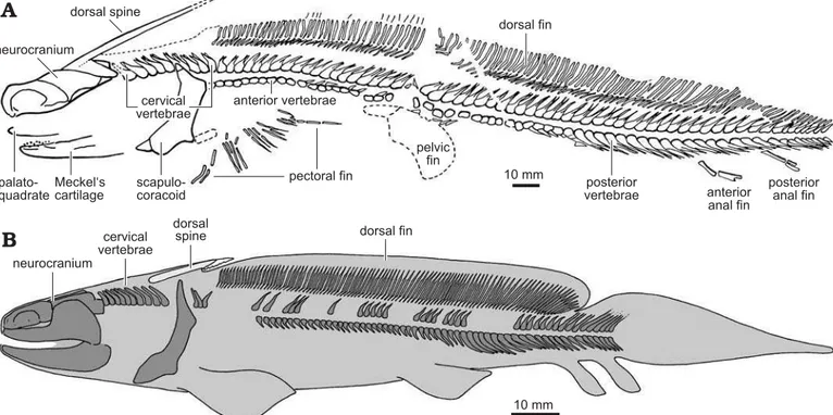
Johnson 1999, 2012). An articulated, nearly complete skele-ton of Orthacanthus platypternus is known from the Upper
Carboniferous of Hamilton, Kansas, USA (Fig. 2A). This fos-sil, representing a juvenile, is the most complete xenacanth recovered in the Permo-Carboniferous American localities (Zidek 1993a, b) and shows the typical eel-like morphol-ogy of the derived xenacanthiforms (family Xenacanthidae), well-known from many articulated specimens of European localities (see Schneider and Zajíc 1994; Heidtke 2003 and references therein). As indicated in Fig. 2A, juvenile O. platypternus had a dorsal spine already contacting the
occip-ital region of the neurocranium (but see the condition in early juvenile O. bohemicus; Fig. 2B), an elongated dorsal fin and
two anal fins. Dorsal spines of Orthacanthus, the subjects
of our study, are very common in the Craddock Bone Bed. Donelan and Johnson (1997) gave the first description and biometry referring the isolated spines to O. platypternus due
to the association with numerous teeth of that species. The xenacanth dorsal spine is a modified dorsal-fin spine, homologous to the fin-spine of fossil and modern phalacanthous sharks and structurally comparable with the fin spines of modern elasmobranchs lacking the ornamented mantle (e.g., Oxynotus; Soler-Gijón 2004). The xenacanth
spine was developmentally connected to the dorsal-fin mod-ule, which was probably under the positional regulation of the Hox and Tbx genes, as occurs in modern sharks (Maisey
2009). The occipital/cervical location of the spine in late juvenile-adults of derived xenacanthiforms (Orthacanthus, Xenacanthus, Triodus = Bohemiacanthus and Plicatodus)
is secondary, and a consequence of the forward differen-tial growth of the proximal end of the spine early in on-togeny (Soler-Gijón 2004). Articulated skeletons of juve-nile and adult primitive xenacanthiforms (Diplodoselache, Lebachacanthus) always exhibit the dorsal spine behind the
pectoral girdle, in front of the dorsal fin (Dick 1981; Heidtke 1982, 2007); a postcranial location is also suggested for
Dicentrodus and Reginaselache, until now only known by
disarticulated remains (Hampe 2003; Hampe et al. 2006; Turner and Burrow 2011).
The function of the dorsal spine is usually accepted as a defensive structure (Evans 1924; Hampe 1997; Johnson 1999: 253) but it has also been proposed that the spine re-duced lateral bending of the anterior body and therefore sta-bilized swimming (Soler-Gijón 2004). The xenacanth dor-sal spine was a non-replaced structure, in similar fashion to the dorsal-fin spines of extinct and modern chondrichthyans (Maisey 1978, 1979; Soler-Gijón 1999; Clarke et al. 2002; Ramos 2007; Tovar-Avila et al. 2008; Barnett et al. 2009). As such, dorsal spines have been the subject of histological and skeletochronological studies (Dick 1981; Soler-Gijón 1999; Soler-Gijón and Siebert 2001; Turner and Burrow 2011). Past studies have noted cyclical deposition in xenacanth dorsal spines and fin spines, allowing the study of growth rates through ontogeny, as well as interpretation of environmental conditions (see Soler-Gijón 1999). Dorsal spine histology and skeletochronology have only been studied in detail in Late Carboniferous xenacanths (Orthacanthus meridionalis from
the Puertollano coal basin, Spain and Orthacanthus sp. from
the Robinson locality, Kansas, USA) living in tidally influ-enced, marginal marine environments with a humid tropi-cal climate (Schultze 1995, 1998; Soler-Gijón and Moratalla 2001; Schultze and Soler-Gijón 2004). Permian Orthacanthus platypternus from the Craddock Bone Bed gives the
oppor-tunity to study xenacanth sharks that were growing, at least temporarily, in coastal plain environments under a semi-arid, seasonally dry tropical climate (Nelson et al. 2013).
Evidence for an increasingly dry and seasonal climate during the Permian in Baylor County and the surrounding area has been noted through the study of Permian floras (Chaney and DiMichele 2007; DiMichele et al. 2006) and palaeosols (Tabor and Montañez 2004). Evidence for cyclical growth has been found in the long bones of other vertebrates in the same bone bed (Beck 2014), and was expected in the hard structure of the xenacanth dorsal spine. The goals of this study were to (i) describe the morphology and histology of the dorsal spines of O. platypternus; (ii) determine if cyclical
growth was recorded in the dorsal spines of O. platypter-nus; (iii) investigate if cyclical growth would correspond to
spine size and answer previous questions about ontogeny and sexual size dimorphism in O. platypternus (Donelan
and Johnson 1997); (iv) compare dorsal spine histology of O. platypternus with an earlier xenacanth, O. meridionalis, to
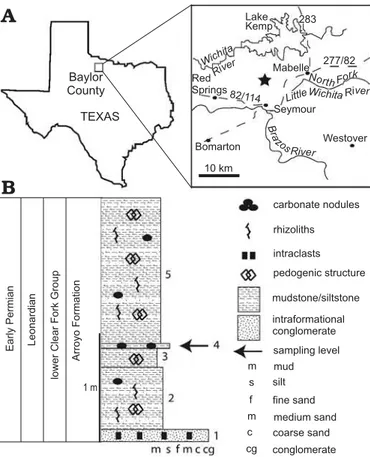
Fig. 1. Baylor County map (A) and stratigraphic section (B) at the
BECK ET AL.—DORSAL SPINES OF PERMIAN XENACANTHID SHARK 99
note any interspecific variability and test the hypothesis that spine denticle density corresponds to growth rates or is di-agnostic of species within thegenus; (v) discuss the possible diadromy and habitat of O. platypternus consistent with our
histological and skeletochronological results.
Institutional abbreviations.—HMNS, Houston Museum of
Natural Science, Houston, Texas, USA; KUVP, Museum of Natural History (Division of Vertebrate Paleontology), Lawrence, Kansas, USA; MCN, Museum of Natural Sciences, Madrid, Spain; PU-XE, Department of Paleontology (Vertebrates collection, Puertollano Basin), Universidad Complutense, Madrid, Spain; SMU, Southern Methodist University Dallas, Texas, USA.
Geological setting
The Early Permian of north-central Texas (Fig. 1) is char-acterized by a dynamic sequence of terrestrial red beds that grades into a coeval shallow marine shelf environment in the northern Midland basin. The Craddock Bone Bed (lower Clear Fork Formation; Nelson et al. 2013) exposes terrestrial dominated deposits that have previously attracted the at-tention of vertebrate specialists because of their articulated
Dimetrodon and temnospondyl specimens. The bone beds
are located approximately 12 km north of Seymour, TX on the Craddock Ranch. These Early Permian deposits have pre-viously been assumed to have accumulated under a strongly seasonal climate that was undergoing a transition from wet
conditions in the late Pennsylvanian to drier conditions in the Early Permian (Chaney and DiMichele 2007; Tabor and Montañez 2004). Tabor and Montañez (2004) noted that cli-mate changes leading up to the deposition of the Clear Fork Group (e.g., the Wichita Group) were consistent with atmo-spheric circulation over western equatorial Pangaea. More specifically, Permian deposits possibly record the onset of monsoonal atmospheric circulation across this region.
A vertical stratigraphic column (Fig. 1) through the col-lection sectors at the main quarry of the Craddock Ranch re-veals five distinct units. Unit 1 is an intraformational, poly-mict conglomerate that forms the quarry floor. The matrix is composed of clay and silt that surrounds mudstone and siltstone intraclasts, carbonate nodules, and other palaeosol debris. Secondary calcite forms the cement and coats many of the intraclasts. The conglomerate unit is interpreted as an event deposit, possibly deposited during large storm events that reworked underlying palaeosol deposits and concen-trated them in ephemeral alluvial fans. Units 2, 3, and 5 are primarily siliciclastic mudstone and siltstone that display pedogenic structures, carbonate nodules, rhizoliths, and subangular aggregates. They vary slightly in clay content or degree of induration, but all are consistent with vertisols based on their v-shaped cracks and pedogenic carbonate (analogous to the type G profiles of Tabor and Montañez 2004.) The lack of redoximorphic features (e.g., hematite nodules) indicates that units 2, 3 and 5 were relatively well drained in between periods of prolonged drying. Broadly, these units represent floodplain fines or oxbow (abandoned) channel deposits typical of the lower Permian. Chaney and
Fig. 2. Juvenile Orthacanthus platypternus, Stephanian B (Upper Carboniferous) of Hamilton, USA (A) compared to juvenile of O. bohemicus, Westphalian
D (Upper Carboniferous) of Bohemia, Czech Republic (B), showing the relative position of the dorsal spine. A, modified from Zidek (1993b: fig. 1);
DiMichele (2007) also noted that much of the “basal” and “lower” Clear Fork Group (both included now in the lower Clear Fork Formation; see Nelson et al. 2013: fig. 5) was consistent with vertisol and oxbow siltstone deposition. Unit 4 is a layer of slightly more indurated siltstone (minor clay and fine grained sand) with small carbonate nodules, con-taining the Orthacanthus spines, xenacanth calcified
car-tilage, scales of palaeonisciforms, and bones and teeth of
Trimerorhachis and Dimetrodon. Orthacanthus spines were
collected from two sectors of unit 4: the first consisting of a clay-rich mudstone and siltstone where many xenacanth spines and cartilage have been found among shed teeth of
Trimerorhachis and Dimetrodon, and the second sector
con-sisting of a carbonate-rich mudstone where the xenacanth spines are associated with large Dimetrodon bones (sectors
are named “Tuffy” and “Jonathan”, respectively, in Houston Museum of Natural Science records). The thin layer with
Orthacanthus was likely deposited during overbank
flood-ing events, or potentially in ephemeral streams or oxbow lakes formed during severe storms.
Material and methods
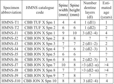
Twelve dorsal spine fragments of Orthacanthus platypter-nus, housed at the Houston Museum of Natural Science
(HMNS) Craddock Bone Bed collections, form the main data set for this histological study. Three additional spines (SMU 68799–801), collected by Donelan and Johnson and housed at Southern Methodist University, were also exam-ined in order to study the general morphology of spines of
O. platypternus (Fig. 3). Due to the destructive nature of
histological sampling, specimens selected for study were from non-articulated and incomplete spine fragments (many fragments were proximal to the denticulate region), and ranged in diameter from approximately 4 to 10 mm (see Table 1 for measurements and complete HMNS catalogue codes). Two specimens (HMNS-T1 and HMNS-T2) were collected in sector “Tuffy” of unit 4 (see Geological setting section above and Fig. 1). The rest of the studied specimens (HMNS-J1 through HMNS-J10) were collected in sector “Jonathan” of unit 4.
All spine fragments were photographed and measured prior to thin-sectioning. Measurements included length of fragments, width and height at their widest and narrowest points, and width at the site of thin-sectioning. Denticulate re-gions were present but poorly preserved on spines HMNS-T2 and HMNS-J3, so lengths of the denticulate regions, sizes of denticles, and distances between denticles could not be re-corded. The denticulate region of HMNS-T1 was present and complete, but was not histologically sampled.
To protect and stabilize spines during thin-sectioning, specimens were vacuum embedded in a 15 × 15 cm (6 × 6 inch) polypropylene container of Silmar-41 polyester resin (U.S. Composites, Inc.) with methyl ethyl ketone perox-ide catalyst at 0.7–1% by mass, according to the standard
thin-sectioning procedures described by Lamm (2013). The resin was allowed to cure in a vent hood for 24–72 hours. Embedded spines were cut into thick sections with a dia-mond-embedded steel blade. The mounting sides of thick sections were ground and polished on an Ingram thin-sec-tion silicon carbide abrasion wheel (Ward’s Natural Science Est., Inc.) with 220 grit per inch silicon carbide powder, fol-lowed by 400 grit per inch silicon carbide powder. Polished samples were mounted on frosted glass petrographic slides with clear 2-ton epoxy (Devcon) and allowed to cure for at least 24 hours. The backs of thick sections were cut off with a model 135 Ingram thin-section cut-off saw (Ward’s Natural Science Est., Inc.) and thin-sections were ground to approximately 0.1 mm in thickness with a model 400 Ingram thin-section grinder (Ward’s Natural Science Est., Inc.). Thin-sections were viewed and photographed through a Leica EZ4 D stereomicroscope (Leica Microsystems) with normal transmitted light, as well as through a Labophot-pol microscope (Nikon), using normal transmitted and polar-ized light. A small amount of water was added to each slide as it was viewed to enhance microstructure visibility.
All spines were sampled in cross-section, except for spine HMNS-T2, which was sampled as one proximal cross-sec-tion and a series of longitudinal seccross-sec-tions through the den-ticulate region. Only spines HMNS-T2 and HMNS-J3 were sampled through denticulate regions; all other thin-sections were made from fragments proximal to denticulated regions (see Table 1).
Table 1. Orthacanthus platypternus dorsal spines sampled.
Measure-ments are given for the most proximal point available and are round-ed to the nearest millimeter. The measurround-ed cross-sections are from non-denticulated regions, except for specimen HMNS-J3, cut at the level of the denticulated region. With the exception of HMNS-T1 and HMNS-J3, dentine layer 2 is assumed to be the older layer recorded in the cross-sections of the spines because dentine layer 1, represent-ing the juvenile stage SP1, becomes restricted to the distal part of the Orthacanthus spines during growth (cf. Soler-Gijón 1999) and
it is already absent in the non-denticulated region of the small spine HMNS-T2 (see longitudinal section in Fig. 4). Abbreviation: dl, den-tine layer of centrifugally growing denden-tine.
Specimen
abbreviation HMNS catalogue code Spine width (mm) Spine height (mm) Number dentine layers (dl) Esti-mated age (years)
HMNS-T1 CBB TUF X Spn 1 4 4 1 (dl1) 1
HMNS-T2 CBB TUF X Spn 2 6 6 1 (dl2) 2
HMNS-J1 CBB JON X Spn 1 9 10 3 (dl2–4) 4
HMNS-J2 CBB JON X Spn 2 8 8 ? ?
HMNS-J3 CBB JON X Spn 3 7 7 2 (dl1–2) 2
HMNS-J4 CBB JON X Spn 4 7 6 2 (dl2–3) 3
HMNS-J5 CBB JON X Spn 5 6 6 ? ?
HMNS-J6 CBB JON X Spn 6 8 6 2 (dl2–3) 3
HMNS-J7 CBB JON X Spn 7 10 8 >3 (dl2–n) >4 HMNS-J8 CBB JON X Spn 8 7 7 >3 (dl2–n) >4
HMNS-J9 CBB JON X Spn 9 7 8 ? ?
BECK ET AL.—DORSAL SPINES OF PERMIAN XENACANTHID SHARK 101
The terminology used here to describe the morphology and microstructure of the xenacanth spines basically follows that of Soler-Gijón (1999). In this study, “major growth line”
is used to describe a dark, hypomineralized line marking an unconformity in dentine deposition. “Minor growth lines” refer to finer lines within lamellar dentine usually arranged
Fig. 3. External morphology of dorsal spines of Orthacanthus platypternus (Cope, 1884), Lower Permian, Craddock Bone Bed, Texas, USA. A. HMNS-T1,
juvenile, lateral view. B. SMU 68799, juvenile, posterior view. C. SMU 68800, juvenile, posterior (C1) and postero-lateral (C2) views. D. SMU 68801, adult,
denticulated, postero-lateral view (D1) and non-denticulated, posterior view (D2) regions. Numbers 1 to 17 point to the positions of the denticles along the
in a close, but evenly spaced series. A dentine layer (dl) within the spine proper (SpPr) was identified as a band or zone of trabecular and lamellar dentine that was generally homogenous in appearance, especially in terms of colour and vascular structure, and usually separated from succes-sive layers by a major growth line. Successucces-sive dentine layers (dl1–n) preserved in the outer (centrifugally growing) part of the wall of each spine proper represent the successive on-togenetic stages (SP1–n). “Lamellar” is used to describe less vascular dentine, such as that located immediately around the pulp cavity or around the periphery of the spine wall.
Systematic palaeontology
Class Chondrichthyes Huxley, 1880
Order Xenacanthiformes Berg, 1940
[= Xenacanthida Glikman, 1964]
Family Xenacanthidae Fritsch, 1889
Genus
Orthacanthus
Agassiz, 1843
Type species: Orthacanthus cylindricus (Agassiz, 1843), Late
Carbon-iferous, Coal Measures, Manchester, England. See Hampe (2003) for additional information about this species and the Carboniferous bio-stratigraphy of the British localities.
Orthacanthus platypternus
(Cope, 1884)
Figs. 3–7, 8A, 9.Referred material.—15 isolated dorsal spines (HMNS-T1–2,
HMNS-J1–10, SMU 68799–68801) from the Craddock Bone Bed (northern Baylor County, Texas, USA), lower Clear Fork Formation, Early Permian (Leonardian age; see Nelson et al. 2013).
Description
General morphology.—The external morphology of a dorsal
spine of Orthacanthus platypternus is well represented by
the specimen SMU 68801 (Fig. 3D), 114 mm long, and lack-ing the most distal region. Two posterior rows of denticles covered about 48 mm of the complete length of the spine, indicating that the denticulation could be extended for the distal half of the complete spine. The cross-section was oval near the opening of the pulp cavity but became circular to subtriangular in the distal part of the non-denticulated re-gion and circular in the denticulated rere-gion. The spine was 8.4 mm wide at the proximal end of the denticulated region and reached a maximum width of 9.4 mm proximally, near the basal opening of the pulp cavity. The robustness index (maximum width to total length ratio) of the preserved spine is 1:12. The denticle sizes (base length) ranged 1.7–2.3 mm. A density of denticulation of 0.40 denticles/mm remained uniform through the spine SMU 68801 and in the much smaller fragmentary specimens SMU 68799 and 68800 (ju-veniles) that showed the most proximal denticles (Fig. 3B, C). The most distal denticulation (Fig. 3A) of a juvenile spine was preserved on the smallest spine sampled (HMNS-T1).
Thirteen denticles covered approximately 23.5 mm of the total spine fragment length (40 mm). These denticles in-creased in base length and denticle height proximally except for the most proximal denticle, which was approximately 1 mm shorter in length than the denticle before (distal to) it. Spacing between denticles on HMNS-T1 increased prox-imally until the 6th denticle, after which spacing between
denticles fluctuated. The denticle sizes ranged from 0.4 mm (first denticle, distally) to 2 mm (denticle 13). The density of denticulation dramatically increased from 1.1 denticles/mm in the distal part to 0.32 denticles/mm in the proximal part.
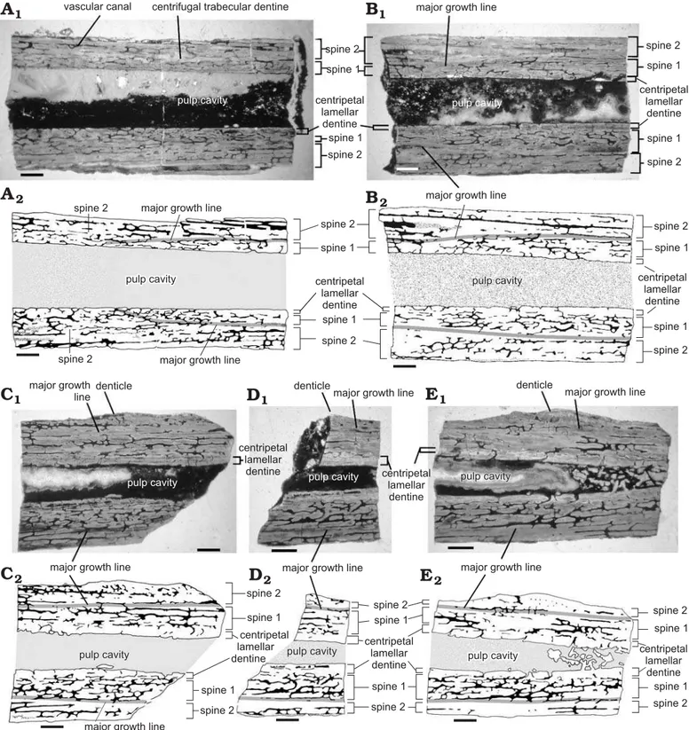
Histology.—The general histology of spines thin-sectioned
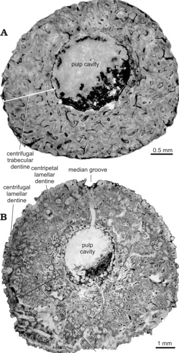
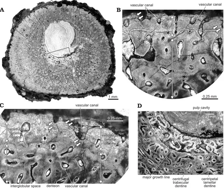
is shown in serial longitudinal sections (Fig. 4) and serial cross-sections (Fig. 5). The pulp cavity of many spines was filled with calcite and quartz, as well as opaque minerals such as iron oxides. The pulp cavity was located slightly off-center in the spine (as seen in cross-sections) at the proximal end of the denticulated region (Fig. 5), but became closer to the posterior side of the spine in the non-denticu-lated region (Figs. 6, 7). The wall of the spine was composed of two structural components—an outer layer of centrifu-gally growing dentine (growing outward from the periphery of the spine), and a layer of centripetally deposited dentine (growing inward) lining the pulp cavity.
The centrifugal dentine was composed primarily of highly vascular trabecular dentinal layers. Centrifugal lamellar dentine was in the most distal parts of the dentinal layers and at the periphery of large spines. Most spines had occasional enlarged vascular canals in the outer half of the spine wall (Fig. 6B, see also Fig. 5A, C). Dentinal tubules radiated out from the outermost lamellae of denteons, al-though some vascular canals were simple and did not have distinct lamellae. When a major growth line was present, denteons below the growth line were more developed than those outside of the line. Colour banding was apparent in most spines (Figs. 5A–D, 6A, 7).
The centripetal dentine mainly exhibited a lamellar structure with incremental deposition of hard tissue (Figs. 4, 5). Centripetal lamellar dentine thickened distally, and filled the pulp cavity of the most distal sections. Spines HMNS-T2 and HMNS-J3, shown in Figs. 4 and 5, respectively, each had a region of centripetal trabecular dentine in the pulp cavity near the distal end.
BECK ET AL.—DORSAL SPINES OF PERMIAN XENACANTHID SHARK 103
denticulate regions (see Fig. 5). Where both dentine layers were present, a major growth line separated them.
Major growth lines were most evident in spines HMNS-T2 and HMNS-J3 (Figs. 4, 5). A growth line was evident in four out of five cross-sections of specimen HMNS-J3 (all except for the most distal) and can be seen
most clearly in Fig. 5A. Vascular canals to the inside of the growth line were lined with lamellae, whereas vascular ca-nals to the outside of the ring were not. In serial longitudinal sections (Fig. 4), the major growth line gradually projected toward and eventually contacted the pulp cavity, proximally.
Some of the larger spines also had minor growth lines
in the lamellar dentine at the periphery of SP2 (Fig. 5, 7). In specimen HMNS-J3, three minor growth lines were fine, dark and evenly spaced (Fig. 5B). In specimen HMNS-J7, growth lines were arranged in at least three pairs (Fig. 7B), although the spine fragment was not long enough to deter-mine if these growth lines were major or minor growth lines. Growth lines, such as those seen in Fig. 5B, were also present in the lamellar dentine around the pulp cavity of four spines (specimens HMNS-J1, J3, J7, J8). In most specimens, colour-banding and growth lines usually circumvented vas-cular canals.
Cross-sections of specimen HMNS-J3 show the ortho-dentine (dark colour) forming the distal part of the denticles (see Fig. 5D). The inner region of denticles consisted of white dentine, with many long dentinal tubules radiating outward from at least one vascular canal at the base of or in the innermost region of the denticle. In serial longitudinal sections of HMNS-T2, the denticulate region did not extend to the entire length of SP1 and all denticles present on the available fragment were added to SP2 (Fig. 4).
Interglobular spaces were found in interdenteonal areas of all spines. These spaces were generally more numerous near the pulp cavity of all spines, and were especially dense in proximal sections, near the pulp cavity of larger spines.
In proximal specimen HMNS-J7, interglobular spaces and iron oxides were extremely dense in some regions near the pulp cavity and also markedly decreased in number and size at the border between the innermost layer of white centrip-etal dentine and the following, darker coloured trabecular dentine of the spine wall (Fig. 7D). Spine HMNS-J8 had a similar distinct decrease in interglobular spaces where the centripetal lamellar dentine lining the pulp cavity met the centrifugal trabecular dentine of the spine wall.
In several spines, calcospherites on the periphery of the spine wall formed a translucent border (Fig. 5B). Globular calcospherites were most clearly visible on the outer edge of specimen HMNS-J3, and were associated with numerous small interglobular spaces. Interglobular spaces represent poorly mineralized regions between calcospherites, which did not completely fuse during dentinogenesis (Currey 2006). In most cases, interglobular spaces were distributed sporadically through the spine wall, and in the highest num-bers and density near the pulp cavity of spines. The high density of interglobular spaces within the centripetal lamel-lar dentine of specimens HMNS-J7 and HMNS-J8 supports the hypothesis that younger dentine was hypomineralized and became more mineralized as the individual aged (as suggested by Soler-Gijón 1999). Hypomineralized dentine
Fig. 5. Serial cross-sections (A, C, D) of dorsal spine of Orthacanthus platypternus (Cope, 1884), Lower Permian, Craddock Bone Bed, Texas, USA;
specimen HMNS-J3, where A represents the most proximal section and D is the most distal. B. Detail of section in A showing three minor growth lines
BECK ET AL.—DORSAL SPINES OF PERMIAN XENACANTHID SHARK 105
(particularly that near the pulp cavity) was probably more susceptible to diagenesis and degradation following inva-sion of microorganisms, leading to the deposition of dark, opaque minerals (such as iron oxide) seen associated with interglobular spaces of the centripetal lamellar dentine of some spines (Fig. 7D) (Turner-Walker 2008). Altered den-tine and interglobular spaces filled by authigenic minerals are relatively common in fossil chondrichthyan and acan-thodian fin-spines (see discussion and references in Botella et al. 2012).
The colour-banding present in almost all spines was also noted by Maisey (1978) in finspines of hybodont sharks. The colour of fossil dentine can be related to diagenesis, where well-preserved dentine often appears reddish in colour, whereas layers of dentine exposed to the environment may be white-coloured. However, in some cases in this study, a major growth line separated two colour bands as well as two different stages in denteon development (such as in Fig. 5A), suggesting that colour banding can also be related to dentine deposition. The smallest spine sampled here (HMNS-T1) corresponded to a very young juvenile individual as indi-cated by the short denticulation, and lacked colour banding. The largest spine sampled (HMNS-J1) also showed weaker colour banding than most other spines, probably due to a very fast mineralization during dentinogenesis (see discus-sion below, in section “Histology and skeletochronology”).
Discussion
Growth and ontogeny.—The xenacanth spine appears to be structurally comparable to the dentinal trunk (stem) of the dorsal-fin spines of holocephalians and phalacanthous sharks (Soler-Gijón 2004; Maisey 2009). The comparative information concerning skeletochronology and growth pat-terns in Recent chondrichthyans facilitates a more objective interpretation of the fossil forms. In this respect, the recent publication on Heterodontusportusjacksoni by Tovar-Avila
et al. (2008) is especially relevant to our study. The authors found evidence of annual growth increment banding in both inner (centripetal) and outer (centrifugal) trunk dentine. Interestingly, the model of chondrichthyan dorsal-fin spine growth proposed by Tovar-Avila et al. (2008: fig. 6) is basi-cally similar to the pattern of deposition of dentinal layers described for xenacanths (compare with Soler-Gijón 1999: fig. 10 and Fig. 8 in this paper). In both Heterodontus and
xenacanths, the successively deposited dentinal layers of the outer trunk do not reach the apex of the spine but instead extend more proximally. The most distal part of the spine contains the first spine (termed SP1 for xenacanths, cf. Soler-Gijón 1999). Successive dentinal layers represent the suc-cessive ontogenetic stages (SP1–n). Fluorescent (oxytetracy-cline) marking in sections of Heterodontus spines suggests
the annual timing of dentine deposition and shows how the outer trunk dentine always surrounds the inner trunk dentine lining the pulp cavity, in a fashion similar to that observed in the sections of xenacanth spines. These comparative results suggest that the centripetal lamellar dentine of xenacanth spines can also be useful for age estimations. However, there is no correlation between the growth marks of centrifugal and centripetal dentine in the cross-sections of spines of xenacanths, in contrast to those described in Heterodontus
(Tovar-Avila et al. 2008: fig. 5; see also Ramos 2007: pl. 4.2). Centripetal lamellar dentine was not produced at the same rate or at the same time as centrifugal lamellar (and trabecular) dentine in the same cross-section (Soler-Gijón
Fig. 6. Cross-sections of dorsal spines (non-denticulated region) of Ortha-canthus platypternus (Cope, 1884), Lower Permian, Craddock Bone Bed,
Texas, USA. Comparison between the smallest, specimen HMNS-T1 (A),
and the largest Orthacanthus dorsal spine sampled, specimen HMNS-J1
1999), as observed by the difference in thickness and number of growth lines within both kinds of dentine on the same thin-section. This apparent asynchrony in the deposition of dentine in the xenacanths can be explained by the histological differences between outer (centrifugal) and inner (centripe-tal) dentines: the centrifugal trabecular dentine grew faster than the centripetal lamellar dentine. In Heterodontus, and
other neoselachians, the complete spine trunk is composed of lamellar dentine. For clarity, the succession of dentinal layers (SP) of the outer part of the spine (probably deposited with an annual periodicity) is chosen for skeletochronologi-cal and developmental studies in Orthacanthus platypternus
(cf. Soler-Gijón 1999), as well as to test the hypothesis of sexual size dimorphism by Donelan and Johnson (1997).
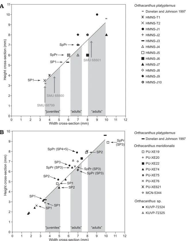
Donelan and Johnson (1997) presented the first biomet-ric analysis of the ontogenetic and sexual size variations in
the species Orthacanthus platypternus based on the
mea-surement (width × height of cross-sections) of a relatively large collection (65 specimens) of isolated dorsal spines. The obtained data, ranging from 3.5 × 3.2 mm to 9.9 × 9.6 mm (fitting the linear regression y = 0.91x + 0.15, where y and x represent height and width, respectively, of the spine cross-sections; see Fig. 9), are grouped in three clusters that are assigned by Donelan and Johnson (1997) to juveniles (38 smallest analyzed specimens), and sexually dimorphic adults (17 specimens of intermediate sizes and 9 specimens of the largest sizes).
It was expected that juvenile shark spines would dif-fer histologically from adult spines, and those difdif-ferences would correlate with morphological and biometric differ-ences proposed previously by Donelan and Johnson (1997) for dorsal spines from the same bone bed. Also, based
Fig. 7. Cross-section of dorsal spine of Orthacanthus platypternus (Cope, 1884), Lower Permian, Craddock Bone Bed, Texas, USA; specimen HMNS-J7
(non-denticulated region) showing color-banding in centrifugal trabecular dentine and growth lines in lamellar dentine. A. General view (posterior side
BECK ET AL.—DORSAL SPINES OF PERMIAN XENACANTHID SHARK 107
on several histological studies of fossil and extant shark spines, (such as those including xenacanthid dorsal spines (Soler-Gijón 1999), ctenacanth fin spines (Stamberg 2001), hybodont fin spines (Maisey 1978), and neoselachian fin spines (Tovar-Avila et al. 2008 and references therein), it was expected that juvenile and adult spines would show a cyclical growth pattern.
Our histological and skeletochronological analyses sup-port the conclusion of Donelan and Johnson (1997) con-cerning the smallest, juvenile remains. However, our data and the comparative analyses with ontogenetic data from other xenacanths and Recent sharks reveal a growth pat-tern in the subadults/adults of Orthacanthus platypternus
from the Craddock Bone Bed that is far more complex than that indicated by the biometric analyses of Donelan and Johnson (1997). We considered two different aspects in the discussion of the Craddock material: (i) comparative study of the histology and skeletochronology of the isolated spines in order to analyze the relationship between size (width × height of cross sections) and ontogenetic age, development and possible sexual dimorphism, and (ii) inference of the body size of the sharks for each ontogenetic stage in order to discuss the structure of the shark population, sexual size dimorphism, and ontogenetic habitat partitioning.
Histology and skeletochronology: We integrate the
skel-etochronological and biometric data from Orthacanthus
platypternus and two very well-known species O. meridio-nalis (Puertollano basin) and Orthacanthus sp. (Robinson
locality), including information of the ontogenetic trajecto-ries of several individuals as indicated by the measurements of histological sections (Fig. 9).
Many spine fragments sampled in this study were proxi-mal to their respective denticulate regions and therefore did not record juvenile growth stages. However, two large spines with denticulate regions thin-sectioned as serial cross-sec-tions and serial longitudinal seccross-sec-tions, as well as one small spine sampled, were supportive of the hypotheses proposed above. The smallest spine sampled (HMNS-T1) was compa-rable in diameter to the smallest spines collected by Donelan and Johnson (1997) (Fig. 9A), and was therefore assumed to be juvenile. This assumption was supported by the histology of the spine, in that no growth lines were present, the dentine structure (as well as colour) was homogeneous through the entirety of the spine wall, and the diameter of the spine was comparable to SP1 in larger spines sampled at a similar loca-tion (just proximal to the denticulate region). No centripetal lamellar dentine was present in HMNS-T1, in contrast to larger spines sampled at a corresponding location. Specimen HMNS-T1 then, represents the first dentine layer, or SP1, and the ontogenetic stage of a juvenile (Fig. 9A).
Spines HMNS-T2, HMNS-J3, and HMNS-J8 were com-parable in diameter to the smaller of two spine-size clusters
Fig. 8. A. Reconstruction of the dorsal spine (mainly the denticulated region) of Orthacanthus platypternus, based on specimens HMNS-T1 and HMNS-T2.
B, C. Reconstructions of the dorsal spine of Orthacanthus meridionalis (modified from Soler-Gijón 1999: fig. 10); PU-XE76 (B) PU-XE19 (C). Note that
Fig. 9. Bivariate plots of relationship between width and height in cross sections of Orthacanthus dorsal spines. A. Orthacanthus platypternus (Lower
Permian, Craddock Bone Bed, Texas, USA). B. Comparison of O. platypternus with O. meridionalis (Upper Carboniferous, Puertollano, Spain) and Orthacanthus sp. (Upper Carboniferous, Robinson, Kansas, USA). Note the linear regression (y = 0.91x + 0.15; n = 65, r2 = 0.98) calculated by Donelan
and Johnson (1997) for the isolated dorsal spines of O. platypternus. The maximum and minimal values are also included here for a comparative reference;
values of the rest of the specimens are not included for clarity. Grey shaded areas indicate approximate intervals of the three size clusters corresponding to juveniles and adults according the biometric analysis by Donelan and Johnson (size intervals are based on unpublished data presented in a poster at 57th Annual Meeting of the Society of Vertebrate Paleontology, Chicago, October 1997). Note the ontogenetic trajectories of several individuals of
O. platypternus (HMNS-T2 and HMNS-J3), O. meridionalis (PU-XE19, 20, 74 and 76) and Orthacanthus sp. (KUVP-72324) showing stages SP1–n
(spine proper). Data from O. meridionalis and Orthacanthus sp. after Soler-Gijón (1999: table 1 and figs. 4–8). Vertical grey arrows point to the position
BECK ET AL.—DORSAL SPINES OF PERMIAN XENACANTHID SHARK 109
attributed to adults by Donelan and Johnson (1997) (Fig. 9A). However, despite their similarity in sizes, the internal struc-tures of these spines correspond to two different ontogenetic stages (juveniles and subadult/adults). Specimen HMNS-J8 appears to fit in the model proposed by Donelan and Johnson as a spine belonging to a relatively small, but already adult individual (indicated by the number of preserved dentinal layers) (Table 1). Spines HMNS-T2 and HMNS-J3, which may be considered adults due to their sizes, actually corre-spond to young, juvenile individuals according to the histo-logical and skeletochronohisto-logical examinations. Both spines had one major growth line separating two dentine layers (see Figs. 4, 5). This growth line was not visible in the most distal sections of HMNS-J3, where only SP1 was present, or the most proximal part of HMNS-T2, where only SP2 was pres-ent. The second and more recent layer of dentine (SP2) thick-ened proximally and in some areas, contained minor growth lines within the centrifugal lamellar dentine (Fig. 5B).
A complete denticulate region is needed to associate ma-jor growth lines with growth rates, but the presence of one growth line indicates that both HMNS-J3 and HMNS-T2 are between one and two years old. The denticulate re-gion of HMNS-T2 does not extend the entire length of SP1 (Fig. 8), as has been described for Orthacanthus by
Soler-Gijón (1999) and so it is likely that HMNS-T2, the sim-ilarly sized HMNS-J3, and part of the middle spine-size cluster of Donelan and Johnson (1997) represent juveniles. Interestingly, the ontogenetic stage SP1 of HMNS-T2, as measured at the level of most distal part of the preserved spine proper, appears close to juvenile spine HMNS-T1 in Fig. 9A, though the two spines’ sizes place them in different size clusters of Donelan and Johnson (1997.)
Finally, spines HMNS-J1, HMNS-J7, and HMNS-J10 were comparable in diameter to the largest spine cluster reported previously (Donelan and Johnson 1997) (Fig. 9A). The three specimens show at least three dentinal layers pointing to larger and relatively older animals than those in the smaller clusters, in agreement with the biometric model. Spine HMNS-J7 corresponds to the oldest individual in the collection, showing several growth lines arranged in pairs in the centrifugal lamellar dentine, which suggests multiple dentine layers. In spite of its large size (almost of the same width as HMNS-J7), spine HMNS-J1 represents a relatively young individual recording only three dentinal layers and weakly developed growth lines. Such histology can be ex-plained by a very fast deposition of hard tissue.
Our results indicate that size does not always correlate with ontogenetic age and degree of development. The clus-ter of spines with inclus-termediate sizes includes relatively old and small “adult” individuals, as proposed by Donelan and Johnson (1997), but also younger individuals relatively large in size, which correspond to juveniles of the second group of “adults”. This conclusion for Orthacanthus platypternus
is also supported by the comparative study of Orthacanthus meridionalis from Puertollano (Spain) and Orthacanthus sp.
from Robinson (Kansas, USA) (see Fig. 9B). In similar
fash-ion to O. platypternus, there are two groups of spines of O. meridionalis belonging to old, adult individuals, which differ
in size and growth pattern. Spines PU-XE19 and PU-XE621 correspond with the large “adults” of O. platypternus whereas
the rest of the Spanish spines are located in or close to the interval of small “adults” of O. platypternus (cf. Donelan
and Johnson 1997). Specimens 72324 and KUVP-72325 of Orthacanthus sp. are located in the small-adults
cluster in Fig. 9B. The ontogenetic trajectories of spines of
O. meridionalis and Orthacanthus sp. (e.g., Fig. 9B:
speci-mens PU-XE19, PU-XE74, and KUVP-72324) show a typical decrease in growth rate, indicating the gradual ontogenetic change into sexual maturity. This ontogenetic trend is very well known from dorsal fin spines of Recent neoselachians (Guallart-Furió 1998; Ramos 2007; Irvine et al. 2006a, b). According to the record of complete articulated xenacanths (Schwind 1991; Schneider and Zajíc 1994; Heidtke 1999, 2007; Heidtke and Schwind 2004; Heidtke et al. 2004), there are no significant differences in the relative sizes of the spines between males and females; consequently the two “adult” groups of isolated spines can be correlated with two groups of individuals differing in total length because of sexual size dimorphism (see discussion below).
The denticulation of spines also provides ontogenetic information. As indicated by Soler-Gijón (1999), the den-ticles were added successively to the developing spines in the proximal direction (Fig. 8). Size and rate of denticle deposition are developmentally linked to the growth rate of the spine proper. Because spine histology just proximal to the denticulate region of HMNS-T1 does not show a second dentine layer, the thirteen denticles of HMNS-T1 can also be attributed to SP1, or the “first” spine. The most proximal denticle is shorter than the preceding denticle. In another study, a decrease in denticle size occurred near a major growth line and was associated with a decreased growth rate (see Soler-Gijón 1999). It is then possible that the most proximal denticle of HMNS-T1 could indicate a change in growth rate. Such timing in the first decrease in the growth rate (between in the twelfth and thirteenth den-ticle) is similar to a spine of Orthacanthus meridionalis, in
which the first decrease in denticle size and corresponding major growth line occur at the twelfth denticle (Fig. 8B).
In addition to the similarity in number of denticles depos-ited per unit of time, we have to point out a more important conclusion after the comparison of SP1 in O. platypternus,
as indicated by specimen HMNS-T1, and SP1 of O. me-ridionalis: the American species deposited larger denticles
and grew faster than the European species from the very early ontogenetic stage on (see Fig. 8 and Soler-Gijón 1999: figs. 22, 23). Consequently, in spite of the typical seasonal variations of the growth rate (e.g., Soler-Gijón 1999: fig. 9A for O. meridionalis), the two species differed in the speed
of Orthacanthus and sexually dimorphic forms (juvenile
to adults) in each species, after a careful examination of the ontogeny (as reported by the histology and skeletochro-nology). As shown in Soler-Gijón (1999: fig. 22; compare with Fig. 9B), two well-defined groups of spines have been reported for O. meridionalis, differing in the relative sizes
and density of denticles. Specimens PU-XE20, PU-XE74, and PU-XE76 of O. meridionalis present relatively small
denticles from the distal (juvenile spine) to the proximal parts (density: 0.55–0.60 denticles/mm), in clear contrast with PU-XE75, PU-XE22, and PU-XE621 which exhibit a larger general size and bigger denticles all along the spine (density: 0.38–0.41 denticles/mm). These two groups could correspond to males and females (or vice versa), but still are distinct from O. platypternus. The denticulated region
corre-sponding to stage SP1 in the largest spine of O. meridionalis
(PU-XE621) is less than 2 cm long, significantly shorter than in O. platypternus HMNS-T1 (2.3 cm). The denticle density
is 0.85 denticles/mm (12 denticles) for PU-XE621 but 0.55 denticles/mm for HMNS-T1. Donelan and Johnson (1997) even reported spines of O. platypternus with a density of
0.21 denticles/mm (for most proximal denticles) for large spines, far distant from the maximal values reached by O. meridionalis. Interestingly, the relationship between width
and height of the cross-sections of spines of O. meridionalis
group specimens together with very different patterns in the denticulation (PU-XE74, PU-XE75, PU-XE76, PU-XE20) and separate others with similar denticulations because of the size differences (Fig. 9B). In conclusion, our results sug-gest that the analysis of the denticulation can be adequate to distinguish between sexes in Orthacanthus species even
with a relatively small number of examined specimens.
Body sizes, sexual size dimorphism, and ontogenetic habi tat partitioning: The study of the ontogenetic variation
of the total animal length in Orthacanthus platypternus
is greatly constrained by the quality of the fossil record. In contrast to Lebachacanthus, Xenacanthus, and Triodus,
known by numerous nearly complete articulated skeletons,
Orthacanthus is represented mainly by isolated dorsal
spines and teeth. Rare articulated juvenile skeletons come from O. platypternus (Zidek 1993a, b; Fig. 2A) and O. bo-hemicus (Westphalian D of Bohemia; see Soler-Gijón 2004;
Fig. 2B). Semiarticulated partial skeletons or isolated cra-nial remains of subadult/adult individuals are recorded from
O. platypternus (Cope 1884; Broili 1904; Hotton 1952), O. bohemicus (Fritsch 1889; Heidtke 1998; Soler-Gijón 2000), O. buxieri (Lower Permian of French Massif Central; see
Heyler and Debriette 1986; Poplin and Heyler 1989) and
O. meridionalis (Soler-Gijón 1993, 1997b). Despite those
limitations, we can infer the complete sizes of juvenile and adult individuals in O. platypternus from the isolated dorsal
spines and neurocrania after the analysis of the allometric relationships between those dermal and endoskeletal ele-ments and the complete body in different xenacanth taxa.
The smallest spines found in the Craddock Bone Bed (e.g., HMNS-T1) and ontogenetic stage SP1 recorded in the
histo-logical structure of larger spines (Figs. 8A, 9A) correspond in dimension (about 60 mm long, 4 mm in maximum width) and development with the dorsal (occipital) spine of a nearly complete late juvenile individual of O. platypternus, 345 mm
in total length, from the Late Carboniferous of Hamilton (Fig. 2A; Zidek 1993a). There is no evidence of newborns in the Craddock Bone Bed. We expect much smaller spines (less than 2 cm long, less than 2 mm maximum width) with short or no denticulation for neonates or early juveniles less than 20 cm in total length, as suggested by the articulated material of O. bohemicus (see Fig. 2B; Soler-Gijón 2004).
Total body length estimation of larger (older) individuals of O. platypternus is problematic because of the absence
of complete subadult/adult specimens of any Orthacanthus
species for comparison. Soler-Gijón (1999) was incorrect in giving a total length of 75–90 cm to the largest individual of O. platypternus from the Craddock Bone Bed after the
application of the ratio 1:5–1:6 (dorsal spine length:total animal length ratio; cf. Zidek 1992) to the largest dorsal spine, 15 cm long. That ratio matches the data of juvenile
O. platypternus and the Xenacanthus species but
under-estimates the dimensions of subadult/adult individuals of
Triodus, Orthacanthus, and Lebachacanthus. Descriptions
of numerous articulated xenacanths, mainly from the Lower Permian of Saar-Nahe basin (Germany) reveal the large variation of the “dorsal spine length:total animal length” ratio in xenacanth sharks in correlation with the phyloge-netic position of the taxa (see Turner and Burrow 2011: fig. 9) and the morphological features of the spines (i.e., robust-ness and extent of denticulation; see Soler-Gijón 2004: table 1). The primitive Lebachacanthus has a relatively short,
robust spine and a ratio spine/animal of 1:13–1:22 (Probst 1986; Soler-Gijón 1997a; Heidtke 1999, 2007; Krätschmer 2004), whereas the derived Xenacanthus presents a slender
relatively long spine and a ratio spine/animal of 1:4–1:6 (Schwind 1991; Heidtke and Schwind 2004; Krätschmer 2005). Orthacanthus, as represented by fragmentary
spec-imens of O. meridionalis and O. buxieri, exhibits an
inter-mediate ratio spine/animal between Lebachacanthus and Xenacanthus. The most complete remains of O. meridiona-lis come from the bituminous layer B, above coal seam V in
the “Calvo Sotelo” quarry at the Puertollano basin, Spain (Soler-Gijón 1997b). An X-ray examination of one of the bi-tuminous slabs from Puertollano (unpublished material, col-lected by Alex Ritchie and currently housed in Australian Museum, Sydney) shows the postcranial articulated skele-ton of a mature male of O. meridionalis including a dorsal
spine about 11 cm long and 8 mm in maximum width. The preserved specimen is 90 cm long but lacks the cranial, anal, and caudal parts so that the living animal could be about 150 cm in total length, suggesting a ratio spine/animal of ca. 1:13. Interestingly, the holotype of O. buxieri showing a
BECK ET AL.—DORSAL SPINES OF PERMIAN XENACANTHID SHARK 111
(as estimated from the dimension of the neurocranium; cf. Zidek 1993a) pointing out a ratio spine/animal of 1:8–1:11, in agreement with the articulated O. meridionalis.
Surprisingly, the application of the ratios spine/animal of mature individuals of O. meridionalis and O. buxieri to the
large spines of O. platypternus from the Craddock (no more
than 15 cm long; e.g., SMU 68801; Figs. 3E, F, 9A) gives to-tal animal length values of less than 2 m, still far lower than the estimates of 2.5 to 3 m based in the sizes of large cranial remains found in other localities of the Arroyo Formation (Zidek 1993a). This result appears to indicate that the stream channels and ponds in the coastal plain were inhabited by relatively small individuals (juveniles to subadults/young adults.)
The detailed sampling by Donelan and Johnson (1997) with a relatively high number of collected spines (all belong-ing to different individuals) and the distribution of the sizes as indicated by the biometric analyses, strongly suggests that the absence of very small and large spines indicative of newborn/early juveniles and very old individuals, respec-tively, is not due to taphonomic biases or sampling artifacts but to biological reasons such as absence of very young and mature old individuals because of habitat partitioning (cf. Recent euryhaline sharks; Simpfendorfer et al. 2005; Phillips 2012; for examples with fossil forms, see Fischer et al. 2011 and references therein). Our conclusions are also supported by the recent biometric analyses of isolated teeth O. platypternus from the Geraldine bonebed (Nocona
Formation, Wichita Group, Sakmarian age) (Johnson 2013). The measured O. platypternus teeth from Geraldine range
from 0.84 to 5.39 mm in anteromedial-posterolateral (am-pl) dimension, denoting small animals in comparison with the large (very old) animals with teeth up to 14 mm in am-pl dimension described in other localities (Cope 1884; Broili 1904; Hotton 1952; Olson 1989). Ontogenetic habitat parti-tioning might also explain the absence of large O. compres-sus teeth in the Upper Carboniferous faunas of Nebraska
and Pennsylvania (Johnson 1999: 254, figs. 2, 3, tables 1, 2). A palaeobiological aspect closely related to the ontoge-netic habitat partitioning and reproduction concerns the size differences between sexes. The skeletochrological studies in Recent chondrichthyans show that females are usually larger than males, growing faster and/or for a longer time (Guallart-Furió 1998; Cailliet and Goldman 2004; Irvine et al. 2006a, b; Ramos 2007). Despite the fact that some Palaeozoic chon-drichthyans show the opposite pattern with smaller females (e.g., the holocephalian Echinochimaera meltoni; Grogan
and Lund 2004) the fossil record of articulated complete xe-nacanths indicates that the females attained the largest sizes. Importantly, this pattern in xenacanths is shown by prim-itive and derived taxa and by large and small species. For example, females of the primitive Lebachacanthus colosseus
could reach 3 m in total length whereas the males reached 2.5 m in maximum total length; females of smaller L. polli-chiae attained 1.75 m in total length, in contrast to the males
which reached 1.3 m in maximum total length (Heidtke
2007). Females and males belonging to the derived genera
Xenacanthus and Triodus were relatively small sharks, never
larger than 2 m in total length (Soler-Gijón 2004), but ex-hibited a pattern of sexual size dimorphism similar to that in Lebachacanthus species (Schwind 1991; Schneider and
Zajíc 1994; Heidtke and Schwind 2004; Heidtke et al. 2004). Given the phylogenetic position of Orthacanthus (more
de-rived than Lebachacanthus and sister-group of Xenacanthus
+ Triodus; see Soler-Gijón 1997a, 2000; Turner and Burrow
2011), it is most parsimonious to conclude that the females of Orthacanthus were also larger than the males in similar
fashion to the rest of xenacanths. Consequently, the clusters of Orthacanthus spines with largest sizes, in O. platypternus
and O. meridionalis (Fig. 9) appear to correspond to females.
Habitat of Orthacanthus platypternus.—The determination
of the life conditions (e.g., osmotic tolerance) of the aquatic vertebrate taxa located in floodplain pond bone beds is al-ways a complex work because (i) the bone beds are usually composed of remains of organisms from very diverse ecolog-ical contexts and (ii) the remains, usually disarticulated, are accumulated by very different biotic and abiotic processes. The Early Permian bone beds in southwestern USA were generally formed in fluvio-lacustrine areas on the coastal plain, close to an epicontinental sea (Midland Basin) (Fischer et al. 2014b: fig. 1A). These particular palaeogeographical locations (freshwater/brackish areas in spatial proximity of the sea; Parrish 1978) explain some of the features of the bone beds. For example, predation of Orthacanthus on Xenacanthus, which may have been restricted to a near-shore
marine habitat, could explain the presence of numerous dor-sal spines of Xenacanthus (but not teeth) associated with
teeth and spines of Orthacanthus in Archer City Bone Bed 3
and Conner Ranch Bonebed (Johnson 2012; see also Sander 1989 for a general description of these bone beds.)
The xenacanth sharks are considered as euryhaline or-ganisms (Olson 1989; Johnson 1999; Schultze 1985, 1995, 1998, 2009, 2013; Gijón 1993; Schultze and Soler-Gijón 2004; Soler-Soler-Gijón and Moratalla 2001; Hampe et al. 2006; Laurin and Soler-Gijón 2010; Carpenter et al. 2011; but see Fischer et al. 2013, 2014b for a different conclusion).
Orthacanthus platypternus was diadromous, as indicated
by its presence in the fresh-brackish alluvial channel fa-cies of the Clear Fork Group but also in the marine fafa-cies of the Arroyo Formation (Olson 1989; Johnson 1999). As discussed below, the palaeobiogeographical and palaeoenvi-ronmental distribution of the different ontogenetic stages of
O. platypternus indicate that the species was catadromous, a
conclusion already suggested by Johnson (1999: 253) based in the low palaeolatitude of the Texas Permian localities.
Though the depositional environment of the Craddock Bone Bed differs from that in previous histology studies of Orthacanthus, a cyclical, environmentally influenced
physical and biological factors: water temperature, food re-sources and breeding behavior (Cailliet and Radtke 1987; Soler-Gijón 1999; Ramos 2007; Tovar-Avila et al. 2008). The presence of major growth marks in immature spec-imens of O. platypternus (and the juveniles of other
xe-nacanth species; see Soler-Gijón 1999) indicates that the growth cyclicity as recorded in the hard tissues was related to water temperature and food availability rather than re-production. Water temperature as a primary factor affecting many physiological parameters, could directly influence the food consumption according the seasonal metabolic require-ments in a similar fashion to that observed in Recent trop-ical sharks (Luer et al. 1990). On the other hand, seasonal variations in accessibility and abundance of prey according to rainy-dry cycles was probably one of the main factors modulating the growth in O. platypternus, as occurs today
in fishes from non-marine and marginal marine tropical/ subtropical regions (e.g., Lowe-McConnell 1964; Lecomte et al. 1993; Robins et al. 2006). As opportunistic predators, the xenacanths could move between the diverse aquatic en-vironments of the coastal plain, intermittently connected by seasonal floodings, and exploit the high productivity of those freshwater/brackish habitats.
Increasing aridity during the deposition of the Early Permian bone beds is also pointed out by Fischer et al. (2014b) in their isotopic analyses of shark teeth from sev-eral localities of Texas and Oklahoma. Interestingly, oxygen and strontium isotope analysis of teeth of the xenacanths
Orthacanthus texensis (Waurika, southern Oklahoma; latest
Sakmarian) and Barbclabornia luedersensis (Spring Creek
B, northern Texas; Kungurian) indicate that during the time of dental mineralization, the sharks were living in shallow ponds of non-marine waters with significant evaporative enrichment of 18O, and quite close to the nearshore on the
coastal plain (see Fischer et al. 2014b: fig. 8).
There is no evidence supporting the idea that Orthacanthus
and Barbclabornia had an “obligate lifestyle in freshwater
without euryhaline behavior” as claimed by Fischer et al. (2014b: 721). These authors obtained 87Sr/86Sr ratios in the
shark teeth more radiogenic than 87Sr/86Sr of
contempora-neous seawater, correctly concluding non-marine conditions for the pond water visited by the sharks. However, the ob-tained values do not automatically exclude the possibility of brackish conditions (e.g., oligohaline to mesohaline) in the ponds according to Carpenter et al. (2011), who inter-preted the non-marine 87Sr/86Sr ratios from xenacanths of
the Upper Pennsylvanian Cohn Coal Member, Mattoon Fm., Illinois (USA) as indicative of brackish (estuarine) water. Dipnoans (e.g., Gnathorhiza) and aquatic tetrapods (e.g., Trimerorhachis), associated with the xenacanths in the bone
beds, also could tolerate brackish and even saline waters (Laurin and Soler-Gijón 2010; Schultze 2013) in a typical coastal plain ecosystem, in proximity to the sea and conse-quently affected by the dynamics of fluvial and marine (tidal) waters (Parrish 1978). Recently, Johnson (2013) reported for first time Orthacanthus platypternus and petalodonts
(prob-ably Janassa), a typical marine component, in the Geraldine
Bonebed (Nocona Formation, Wichita Group, Sakmarian age) in association of other xenacanths (Orthacanthus tex-ensis, Xenacanthus sp.), hybodont sharks, Helodus,
acantho-dians, actinopterygians, dipnoans (Sagenodus) and
numer-ous tetrapods including Trimerorhachis and Dimetrodon.
Johnson (2013) concluded that the depositional area of the Geraldine Bonebed was closer to the marine palaeoshore than suggested in previous studies (see Sander 1987).
The climatic conditions with a strong wet-dry season-ality (as indicated by the xenacanth teeth isotopes, palae-osols and palaeoflora) and the inferred small sizes of the depositional bone bed areas (“shallow ponds or backswamp areas generally smaller than one square mile” Fischer et al. 2014b: 721) suggest that the sharks, as euryhaline organ-isms, could move away from the ponds for reproduction and feeding (see Johnson 2012). This conclusion is also indi-cated by Zidek et al. (2004) who described the association of
Orthacanthusplatypternus with O. texensis, B. luedersensis
and other chondrichthyans (the holocephalian Helodus sp.,
and diverse hybodont sharks) in fluvio-lacustrine deposits of Northeast Frederick and Lake Frederick (Early Permian, upper Garber Sandstone, Tillman County, southwestern Oklahoma, USA). According to Zidek et al. (2004: 136–138) who cite the works of Simpson (1976, 1979), most of the Permian lakes of the coastal plain, as represented in the deposits of the Garber Sandstone, were intermittent, kill-ing their aquatic inhabitants in dry seasons. The authors concluded (Zidek et al. 2004:146) that the lakes represented by Northeast Frederick and Lake Frederick sites, covering about 160 acres each, were not large enough to sustain a breeding population of large predatory fishes.
Certainly the palaeobiogeographical distribution of
Orthacanthus platypternus suggests ontogenetic
habi-tat partitioning in this xenacanth. As indicated above, the dorsal spines and teeth of O. platypternus recorded in the
Craddock and Geraldine bone beds point to relatively small individuals, juveniles (but not newborn) to subadult/young adults, less than 2 m in total length, which probably inhab-ited shallow waters of small ponds and stream channels of the upper part of a lower coastal plain. Large dermal and cranial remains of larger individuals up to 3 m in total length are known from other Early Permian localities from Texas (Zidek 1993a). Medium size teeth of O. platypternus are
re-ported from marine limestone from the Arroyo Formation, representing estuarine-lagoonal near-shore conditions (Olson 1989). Most importantly, the articulated skeleton of a juvenile of O. platypternus (Fig. 2A) comes from
estua-rine facies of the Upper Carboniferous of Hamilton, Kansas (Zidek 1993a; Schultze 1995, 1998; Schultze et al. 1994). In summary, O. platypternus occurred in a wide range of
pres-BECK ET AL.—DORSAL SPINES OF PERMIAN XENACANTHID SHARK 113
ence of the giant Barbclabornia luedersensis which could
reach 4.5 to 5 m in total length, according the estimations based in cranial remains from Northeast Frederick locality (Zidek et al. 2004).
The distribution of O. platypternus also suggests the
migratory pattern of this diadromous xenacanth. Large, ma-ture individuals bred in a marine marginal environment. Neonates and early juveniles inhabited estuarine areas be-fore they moved into fresh-brackish habitats of the coastal plain. As a catadromous species, juveniles and subadult to young adults grew and developed in the ponds and channels of the coastal plain during several years before they re-turned to the sea for mating and spawning. We assume here that O. platypternus was an oviparous species as oviparity
appears to be the general reproductive strategy in xenacanth sharks (see Fischer et al. 2014a and references therein).
Early juveniles of O. platypternus probably grew very
fast in the estuarine waters, reaching more than 30 cm (total length) in less than one year, as indicated by the articu-lated material from Hamilton and the skeletochronological analyses from the isolated dorsal spines from Craddock (e.g., specimens HMNS-T1 and T2). Despite the risk of pre-dation and cannibalism (for evidences in xenacanths see Soler-Gijón 1995; Heidtke 2012), the young O. platypternus
spent a relatively long time in the marginal marine habi-tat before they migrated to the very low salinity areas of the coastal plain. With that behavior, the young xenacanths probably minimized the energetic costs of osmoregulation, using more energy on growth, in a fashion similar to that suggested for the neonates and early juveniles of the Recent euryhaline shark Carcharhinus leucas in the estuaries of
the southwest Florida, USA (Simpfendorfer et al. 2005). The selection of brackish waters (salinities between 7‰ and 17.5‰) by the young C. leucas in order to meet optimal
osmoregulation is logically connected to the fact that the smaller individuals have the highest surface-area-to-volume ratio and consequently, the highest energetic cost per unit weight for osmoregulating (Simpfendorfer et al. 2005: 83).
Two main factors probably triggered the migration of the juveniles of O. platypternus into the coastal plain habitats
and far inland areas: protection against predation and food resources. These factors control the distribution of young Recent sharks (e.g., selection of nursery areas; see review of the topic and fossil examples in Fischer et al. 2011) and the migratory pattern of fishes in general (see Tsukamoto et al. 2009 and references therein). Additionally, the distribution of juvenile and subadult-young xenacanths in the coastal plain can be explained by the combination of two circumstances: (i) the low topography and large geographical dimension of that region as indicated by palaeogeographical locations of the Early Permian localities around the Midland Basin (Nelson et al. 2013; Fischer et al. 2014b), and (ii) the par-ticular climatic conditions with marked seasonality in pre-cipitations. Relevant in this respect is the comparison with the life conditions of the modern sawfish Pristis microdon
in the Kimberley region of Western Australia, which
pres-ents an arid to semi-arid monsoonal climate, receiving the rains mainly during the wet season (Thorburn et al. 2007; Phillips 2012). While adults of P. microdon live and breed in
marginal marine environments, the juveniles penetrate into the Fitzroy River, move very fast through the fluvial system and reach freshwater areas far inland where they find food but few large predators (Thorburn et al. 2007). These recent studies on P. microdon also conclude that the juvenile and
subadult-young adult (small individuals less than 2.8 m in total length) stay several years (a maximum of four or five) in the freshwater rivers before they return to the marine regions for reproduction and growth (the species appears to reach up to 7 m in total length). The easy access to the Fitzroy River and wide distribution of immature P. microdon along the
non-marine environments are due to high tides, high run-off and large catchment of the river, and a lowland topography. However, because of extreme climatic conditions, the sea-sonal fluctuations in the water level of the rivers constrain the developmental time and maximum growth of P. microdon in
the fluvial system. Relatively small individuals appear to use pools as refuge habitats during the dry season (Martin 2005) but the large mature or nearly mature individuals move back to the sea. Orthacanthus platypternus and other xenacanth
sharks (e.g., O. texensis and B. luedersensis; Zidek et al.
2004: 146) which grew very fast and reached several meters in total length, could already present such adaptive behavior migrating from one habitat to another in order to respond to the unstable palaeoenvironmental conditions of a broad, low relief coastal plain under the influence of tropical semi-arid to sub-humid climate with seasonal rainfalls (DiMichele et al. 2006).
Conclusions
Dorsal spines of the xenacanth Orthacanthus platypternus
from the Permian Craddock Bone Bed preserve the typical histological structure of Orthacanthus spines, well known
from Carboniferous species, mainly O. meridionalis of
the Puertollano Basin (Spain) and Orthacanthus sp. of the
Robinson locality, Kansas (USA). The wall of the spine, highly vascularized, is mainly composed by centrifugally growing dentine, which appears as a succession of dentine layers, usually bounded by growth lines. In several spines of
O. platypternus, the growth lines are not clearly visible but
a distinctive colour-banding shows the cyclical centrifugal deposition of dentine layers and allows the skeletochrono-logical analyses. Dentine layers were probably deposited with an annual periodicity following the cyclical (seasonal) variations of two main environmental factors, the water temperature and food availability.
Despite the diagenetic alteration and fragmentation of the material, the dorsal spines of Orthacanthus platypter-nus provide valuable information about several ontogenetic