Panorama of Reconstruction of Skull Base
Defects: From Traditional Open to Endonasal
Endoscopic Approaches, from Free Grafts to
Microvascular Flaps
Camilo Reyes
1Eric Mason
1C. Arturo Solares
11Department of Otolaryngology, Georgia Regents University,
Augusta, Georgia, United States
Int Arch Otorhinolaryngol 2014;18:S179–S186.
Address for correspondence C. Arturo Solares, MD, Department of Head and Neck/Skull Base Surgery, Neurosurgery, Center for Skull Base Surgery, Georgia Regents University, 1120 15th Street BP 4109, Augusta, GA 30912, United States (e-mail: [email protected]).
Introduction
Skull base defects can derive from both traumatic and non-traumatic causes. In the non-traumatic group, nonsurgical trauma is the most common overall, and surgical (iatrogenic) damage is a minor cause. In the nontraumatic group, skull base erosion can be caused by high intracranial pressure due to tumor obstruction or from malignant neoplasms. Less
com-monly, skull base defects may be caused by radiotherapy or infections. Spontaneous cerebrospinalfluid (CSF) leaks (idio-pathic) can also occur.
As resections for skull base pathology become more complex, the resultant defects require more difficult and extensive reconstructions. This principle has become the pillar for most skull base reconstruction techniques, from traditional open to novel endoscopic endonasal or other
Keywords
►
skull base
►
cerebrospinal
fl
uid
leak
►
dura
►
reconstruction
►
endoscopic surgery
►
pedicled
fl
aps
Abstract
Introduction
A substantial body of literature has been devoted to the distinct
characteristics and surgical options to repair the skull base. However, the skull base
is an anatomically challenging location that requires a three-dimensional reconstruction
approach. Furthermore, advances in endoscopic skull base surgery encompass a wide
range of surgical pathology, from benign tumors to sinonasal cancer. This has resulted in
the creation of wide defects that yield a new challenge in skull base reconstruction.
Progress in technology and imaging has made this approach an internationally accepted
method to repair these defects.
Objectives
Discuss historical developments and
fl
aps available for skull base
reconstruction.
Data Synthesis
Free grafts in skull base reconstruction are a viable option in small
defects and low-
fl
ow leaks. Vascularized
fl
aps pose a distinct advantage in large defects
and high-
fl
ow leaks. When open techniques are used, free
fl
ap reconstruction
techni-ques are often necessary to repair large entry wound defects.
Conclusions
Reconstruction of skull base defects requires a thorough knowledge of
surgical anatomy, disease, and patient risk factors associated with high-
fl
ow
cerebro-spinal
fl
uid leaks. Various reconstruction techniques are available, from free tissue
grafting to vascularized
fl
aps. Possible complications that can befall after these
procedures need to be considered. Although endonasal techniques are being used
with increasing frequency, open techniques are still necessary in selected cases.
DOI http://dx.doi.org/ 10.1055/s-0034-1390016. ISSN 1809-9777.
Copyright © 2014 by Thieme Publicações Ltda, Rio de Janeiro, Brazil
Evolution of Reconstruction“from above”to Reconstruction“from below”
The presence of a wide variety of available surgical techniques poses the question if it is better to access skull base lesions via traditional transcranial routes or via minimally invasive expanded EEAs. Historically, transcranial resection has been considered the gold standard for surgical removal of numerous suprasellar lesions.1
In 1907, Herman Schloffer performed thefirst transsphe-noidal surgery.2In 1916, Cushing reported thefirst successful removal of a tuberculum sellae meningioma via a unilateral subfrontal approach. In 1950, Norman Dott introduced the lighted nasal speculum retractor. Continuing the trend, Ger-ard Guiot introduced the X-rayfilm intensifier andfl uoros-copy and pioneered image guidance surgery in 1956. In 1965, Jules Hardy introduced the use of the microscope in skull base surgeries, and in 1971, Donald Wilson introduced“keyhole surgery,”transitioning the trend to minimally invasive sur-gery.3Resection of skull base tumors has evolved to integrate modified skull base techniques such as supraorbital, orbito-zygomatic, and orbitopterional approaches.
Regardless of the surgical technique, the primary objec-tives of skull base tumor resection remain the same: gross total tumor resection with adequate decompression and preservation of surrounding structures, prevention of future recurrence, support of the brain and orbit, complete separa-tion of the cranial cavity from the sinonasal tract, eliminasepara-tion of dead space, and a watertight seal to avoid the consequences of CSF leaks, such as meningitis and pneumocephalus.
The disadvantages of the supraorbital keyhole approach include narrower viewing angles with limited maneuvering of instrumentation, especially in patients with optic canal involvement. Although the bilateral frontal and unilateral frontal approaches provide excellent views of critical struc-tures, the bilateral subfrontal approach has been noted to carry a greater risk of CSF leak, olfactory nerve damage, and postoperative brain edema.4,5 Additionally, with the pterional approach there can be significant frontal lobe retraction.
The introduction of the endoscope to skull base surgery eliminated many of the previous problems associated with microsurgical techniques. Casiano et al described the first pure EEA for resection of an esthesioneuroblastoma.6Since its introduction, this approach has been internationally utilized for the resection of a variety of skull base lesions.
understand the indications and limitations of each approach. Preferably, the same route used for tumor removal should be used to repair the skull base defect, thus avoiding the comorbidity of a second incisional approach. In specific scenarios, gross tumor removal and avoidance of skull base defects can be achieved using the endoscope.
Endoscopic closure of CSF leaks was first described by Wigand in 1981 using free mucosal grafts, and to date, it continues to be the preferred method of CSF leak closure because of its high success rate (90 to 97%).10Repairs can be achieved by mucosal grafting or a pedicled flap based on branches of the sphenopalatine, anterior ethmoidal, and facial arteries.11 Also, collagen matrix materials, fascia, or fat can be used as inlay grafts to help seal these defects.
Free Grafts and Pedicled Flaps
A free graft is a tissue cut from one site and transplanted to another site. A pedicledflap is tissue that is left attached to its donor site and transposed to a new location keeping its
“pedicle”intact. Prior to the routine use of vascularized tissue
flaps for skull base reconstruction, free grafts of biologic or synthetic material were used primarily in a multilayer ap-proach. Reconstructive allografts include DuraGen (Integra Lifescience Corporation, Plainsboro, New Jersey, United States) and Alloderm (Lifecell Corporation, Branchburg, New Jersey, United States), dural sealants such as DuraSeal (Confluent Surgical, Inc., Waltham, Massachusetts, United States), and fibrin glue. These materials are still part of the armamentarium for skull base reconstruction.
The middle turbinate is the most common donor site for the harvesting of intranasal free grafts. Other non-nasal free grafts, such as temporalis fascia, fascia lata, and palatal mucosa, can be used. After8 weeks, theseflaps are completely integrated
within the surrounding tissue. It is recommended that the free graft be 25% larger than the defect, because there is a 20% reduction in size.12Free grafts have the advantage of easy harvest with low donor site morbidity.
Among pedicled vascular flaps, the posteriorly pedicled nasoseptal flap or Hadad-Bassagaisteguy flap (NSF) is the
“tip of the spear”of most endoscopic skull base defect repairs (►Figs. 1 to3).13However, there are other intranasal pedicled flaps such as inferior turbinateflaps11,14and middle turbinate
skull base, from the posterior wall of the frontal sinus to the sella turcica, and from orbit to orbit. It should be used in conjunction with a multilayer reconstructive approach. The
flap should be separated from any nonabsorbable packing with nonadherent material so that the graft is not disrupted upon removal of the packing. Packing is typically left in place for 3 to 5 days. The size of the NSF can be compromised by lesions that involve the cropped area or septal spurs and in patients under 10 years of age where the septum is not fully developed.16
The NSF is limited in its ability to reach extremely anterior defects, such as those involving the posterior frontal table, frontal break, and anterior cribriform plate. Posteriorly17or anteriorly18pedicled inferior turbinateflaps or middle
turbi-nateflaps can be considered as a second option for smaller defects when an NSF is not available or not feasible. Terminal branches of the posterior lateral nasal septal artery supply both. Hadad et al described a modification of the anteriorly pedicle inferior turbinateflaps where dissection of the lateral nasal wall was added (Hadad-Bassagaisteguyflap 2 or HB2).11 The HB2 has the capacity to cover combined defects (tran-scribriform and transplanum). Patients in whom the NSF is not available are candidates for this reconstructive technique. It pedicle includes the territory of the facial (angular) artery and the anterior ethmoidal artery. Thisflap can be modified to have a posteriorly based pedicle to cover defects of the planum sphenoidale, sella, clivus, and nasopharynx.19These lateral nasal wall flaps require special surgical endoscopic
Fig. 1 (A) Unilateral anterior cranial base defect following the resection of a sinonasal malignancy. (B) Inlay placement of DuraGen (Integra Lifescience Corporation, Plainsboro, New Jersey, United States). (C) Positioning of the nasoseptalflap over the reconstruction. (D) Application of DuraSeal (Confluent Surgical, Inc., Waltham, Massachusetts, United States).
Fig. 3 Nasoseptalflap fully healed and integrated in the sphenoclival region.
skills, as they can be difficult to dissect. The success of these reconstructionflaps should be individualized to every patient needs. Significant crusting can occur, as with other nasal
flaps, which usually continues until total remucosalization occurs. Postoperative recommendations are the same as for other reconstructive techniques.
Pedicled extranasalflap options are also available. The use of theseflaps arises when the pedicled NSF is not available. Suchflaps are harvested from regional areas including the palatalflap, the pericranialflap, facial buccinatorsflap, and temporoparietal fascialflap.
The palatal flap has a vascular supply derived from the greater palatine artery, providing a 3-cm pedicle that could potentially reach any area of the skull base. This makes it, theoretically, very useful. Nevertheless, this technique has mainly been described in cadaveric studies, making it a resource of last choice.20,21
The pericranialflap was historically used as thefirst option before the use of endoscopic repairs.21Its pedicle is based upon the supraorbital and supratrochlear arteries, and it is transposed through a small nasionectomy into the endoscop-icfield. It can cover from the anterior cranial fossa as far as the sella, without reaching the posterior cranial base defects. In the past, thisflap was considered to have a negative impact on cosmetics; however, with the use of the endoscope, better visualization and minimally invasive surgery have avoided this problem.22
The facial buccinatorflap is pedicled upon the facial artery after it branches off the external carotid artery.23It can be a solely muscular or a combined myomucosalflap. Transposi-tion of the facial buccinator flap takes place through a maxillary window. It has a coverage area that fluctuates from 2 cm2 to 20.76 cm2, with an average of 15.90 cm2,
and it is able to reach the anterior skull base and planum sphenoidale.24Thisflap has only been described in cadavers, so the drawbacks are so far theoretical. These include the introduction of oral flora to the surgicalfield, vascular and nerve injuries, and the most important of them all, variability inflap extensions due to gravitational forces and retraction ability. However, it is important to acknowledge thisflap’s advantages, such as its extension, the axis of rotation, and the absence of external scars.
The temporoparietalflap is a good reconstructive option for defects of the sella, parasellar area, and clivus (►Fig. 4).
The superficial temporal artery supplies theflap. It requires a broad dissection, both endoscopically and externally. The pterygopalatine fossa is dissected, the vidian nerve is sec-tioned, and the anterior aspects of the pterygoid plates are drilled. Externally, a hemicoronal incision is carried down to the level of the hair follicles, a wide tunnel beneath the superficial layer of the deep temporalis fascia is created, and a lateral canthotomy incision is used to expose and separate the temporalis muscle from the lateral orbital wall and pterygomaxillary fissure, creating a tunnel that
fl fl
spine (black arrow) and the temporoparietal fasciaflap covering the parapharyngeal space and ICA (white arrow). Abbreviations: ICA, internal carotid artery; SS, sphenoid sinus.
communicates the temporal, infratemporal fossa, and endo-scopic transpterygoid approach. A portion of the lateral wall of the maxillary sinus is removed to open a wide communi-cation with the infratemporal fossa, thus establishing com-munication between theflap and the skull base.
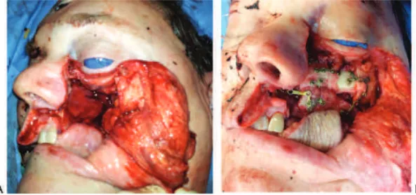
The temporalis muscle is a good reconstructive option for intratemporal fossa defects. It is helpful in separating the intracranial/extracranial spaces and provides sufficient bulk to obliterate the dead space created by the resection of infratemporal fossa/nasopharyngeal pathology. For those lateral skull base defects that require skin as well as soft tissue bulk, the pectoralis major myocutaneous flap still remains a viable option (►Fig. 5).
Success rates of pedicled flaps approximate 95%, which makes them a reliable reconstruction option for skull base defects.25However, when the size and location of the skull base defect to be repaired exceeds the excursion limits of the pedicledflap, freeflaps can be considered.
Free Flaps
The rectus abdominis myocutaneousflap has been commonly used in skull base reconstruction. Its blood supply is the inferior epigastric artery and vein. The insertion of the muscle
is detached and the harvest is completed. The major disad-vantage of thisflap is it bulkiness due to the width of the subcutaneous tissue of the abdominal wall. Also, there is risk of a ventral hernia owed to abdominal wall weakness.
The radial forearmflap is a fasciocutaneousflap supplied by the radial artery and vein. It is very pliable, which makes it versatile for skull base defect reconstruction (►Fig. 6). The flap has a very long pedicle, which allows for usage of neck vessels as recipient vessels. Although it is very rare, hand ischemia can occur because of radial artery sacrifice. In our experience, when the physical exam suggests poor hand perfusion during occlusion of the radial artery, Doppler ultrasonography is helpful in determining the patency of the palmar arch. The anterolateral thigh has become the preferred freeflap by many reconstructive surgeons; howev-er, in our practice we still favor the forearm freeflap due to pedicle length.
Osteocutaneous free flaps can be used in cases where significant orbital reconstruction is required. In our experi-ence, the stacked fibula free flap reconstruction is very versatile in orbitomaxillary reconstruction (►Fig. 7).26Other
osteocutaneous options include the scapula and hip bones with respective overlying skin.
Fig. 6 (A) Lateral skull base defect following the excision of a cutaneous malignancy with extension to the temporal bone and middle cranial fossa. (B) Dural reconstruction was performed with suturable DuraGen (not shown; Integra Lifescience Corporation, Plainsboro, New Jersey, United States). The residual craniotomy boneflap was repositioned and the soft tissue and skin defect were reconstructed with a radial forearm freeflap.
Also, complications can occur depending of the type offlap used. The nasoseptal flap can lead to displacement of the olfactory epithelium, persistent nasal crusting, and sphenoid sinus obstruction due to the pedicle’s orientation. Special areas of skull base reconstruction like the frontal sinus, the orbit, or major neurovascular structures must have special consideration. Complications from external incisions like alopecia, pain, hypesthesia, and/or infections can arise.27 Crusting is the most common symptom after skull base reconstruction (3 to 4 weeks of duration),28followed by nasal discharge, which is associated with more complex dissec-tions.29Resections of large portions of mucosa, such as the inferior turbinate, could lead to atrophic rhinitis. Improve-ment in nasal quality of life is usually achieved 4 months after surgery in most cases and in 6 months for more complex cases.30The use of endoscopic skull base techniques usually does not significantly contribute negatively to nasal quality of life scores; however, careful perioperative planning must be preformed to avoid such complications.
Early complications (<28 days postoperatively) of
microvascular free flaps include partial or total flap loss, CSF leak, infection, pneumocephalus, facial nerve lesion, blindness, seizures, and complications derived from long surgical procedures. Late complications (>28 days
postoper-atively) include palatalfistula, wound infection, meningitis, intracranial abscess, ectropion, enophthalmos, orbital dysto-pia, persistent diplodysto-pia, and facial cellulitis. Prior radiothera-py is a statistically significant predictor of wound complications, and the existence of medical comorbidities is the only independent risk factor for death.31The incidence of local and/or systemic postoperative complications follow-ing microvascular reconstruction of the skull base ranges between 30 and 40%.32–36Mortality rates are close to 4.7%
following craniofacial resection.31
Discussion: What Reconstruction Should Be
Used?
The reconstructive technique largely depends on the ap-proach used for resection and the nature of the resection. Both extradural or intradural tumor resection might be necessary. In the extradural surgery, the primary reconstruc-tive goal is coverage of the defect to facilitate healing. Intra-dural tumor surgery can be extra-arachnoidal (pituitary
surgery) or intra-arachnoidal surgery (where an
intra-operative CSF leak can always be expected). Intra-arachnoidal
systematic review,37the authors suggested that anterior fossa lesions can be repaired with inlay grafts, because the pressure from the brain could hold the material in position and avoid its migration; defects of the tuberculum sellae or the clivus are best reconstructed with pedicled flaps due to their proximity to the anterior brain cisterns and ventricles.
In general, low-flow CSF leaks or small defects (<1 cm) are
consistently repaired using multilayered free grafts, recon-structive allografts, and dural sealants with success rates
>90%.37–39Vascularized skull base reconstructions for large
dural defects (>3 cm) involving wide dural and arachnoid
dissection, high-flow CSF leaks, and poorly vascularized beds (defects) have a clear and significant advantage over free grafting in the prevention of postoperative CSF leaks.37,38,40 When open techniques are utilized, the extent of injury to the entry point needs to be assessed and reconstructed accordingly. With free flaps being more reliable in recent years, they should be considered and utilized whenever possible as they provide excellent functional and cosmetic results.
Final Comments
Skull base surgery has been revolutionized by the use of the endoscope. Endoscopic skull base surgery techniques have advanced considerably in recent years mainly be-cause of advances in instrumentation and imaging. Skull base reconstruction of endonasal defects has not been left behind; however, it still remains a challenge. The intro-duction of vascularizedflaps for endonasal reconstruction has improved the outcomes in several series, but it re-quires a technique that is able to recapitulate the mor-phology of the cranial base while simultaneously withstanding the forces exerted by the brain, brainstem, and CSF compartment. Although endonasal techniques are being used with increasing frequency, open techniques are still necessary in many cases and should remain an impor-tant part of the armamentarium. Microvascular recon-structive techniques are an excellent option in cases where extensive reconstruction is needed.
References
meningiomas. Neurosurgery 2007;61(5, Suppl 2):229–237, dis-cussion 237–238
2 Schmidt RF, Choudhry OJ, Takkellapati R, Eloy JA, Couldwell WT, Liu JK. Hermann Schloffer and the origin of transsphenoidal pituitary surgery. Neurosurg Focus 2012;33(2):E5
3 Liu JK, Das K, Weiss MH, Laws ER Jr, Couldwell WT. The history and evolution of transsphenoidal surgery. J Neurosurg 2001;95(6): 1083–1096
4 Mahmoud M, Nader R, Al-Mefty O. Optic canal involvement in tuberculum sellae meningiomas: influence on approach, recur-rence, and visual recovery. Neurosurgery 2010;67(3, Suppl Oper-ative):ons108–ons118, discussion ons118–ons119
5 Chokyu I, Goto T, Ishibashi K, Nagata T, Ohata K. Bilateral subfrontal approach for tuberculum sellae meningiomas in long-term post-operative visual outcome. J Neurosurg 2011;115(4):802–810
6 Casiano RR, Numa WA, Falquez AM. Endoscopic resection of esthesioneuroblastoma. Am J Rhinol 2001;15(4):271–279
7 Kassam A, Snyderman CH, Mintz A, Gardner P, Carrau RL. Expand-ed endonasal approach: the rostrocaudal axis. Part I. Crista galli to the sella turcica. Neurosurg Focus 2005;19(1):E3
8 Kassam A, Snyderman CH, Mintz A, Gardner P, Carrau RL. Expanded endonasal approach: the rostrocaudal axis. Part II. Posterior clinoids to the foramen magnum. Neurosurg Focus 2005;19(1):E4
9 Dehdashti AR, Ganna A, Witterick I, Gentili F. Expanded endoscop-ic endonasal approach for anterior cranial base and suprasellar lesions: indications and limitations. Neurosurgery 2009;64(4): 677–687, discussion 687–689
10 Prosser JD, Vender JR, Solares CA. Traumatic cerebrospinalfluid leaks. Otolaryngol Clin North Am 2011;44(4):857–873, vii
11 Hadad G, Rivera-Serrano CM, Bassagaisteguy LH, et al. Anterior pedicle lateral nasal wallflap: a novel technique for the recon-struction of anterior skull base defects. Laryngoscope 2011; 121(8):1606–1610
12 Hosemann W, Goede U, Sauer M. Wound healing of mucosal autografts for frontal cerebrospinalfluid leaks—clinical and ex-perimental investigations. Rhinology 1999;37(3):108–112
13 Hadad G, Bassagasteguy L, Carrau RL, et al. A novel reconstructive technique after endoscopic expanded endonasal approaches: vascular pedicle nasoseptal flap. Laryngoscope 2006;116(10): 1882–1886
14 Harvey RJ, Sheahan PO, Schlosser RJ. Inferior turbinate pedicleflap for endoscopic skull base defect repair. Am J Rhinol Allergy 2009; 23(5):522–526
15 Prevedello DM, Barges-Coll J, Fernandez-Miranda JC, et al. Middle turbinateflap for skull base reconstruction: cadaveric feasibility study. Laryngoscope 2009;119(11):2094–2098
16 Shah RN, Surowitz JB, Patel MR, et al. Endoscopic pedicled naso-septalflap reconstruction for pediatric skull base defects. Laryn-goscope 2009;119(6):1067–1075
17 Fortes FSG, Carrau RL, Snyderman CH, et al. The posterior pedicle inferior turbinateflap: a new vascularizedflap for skull base reconstruction. Laryngoscope 2007;117(8):1329–1332
18 Gil Z, Margalit N. Anteriorly based inferior turbinate flap for endoscopic skull base reconstruction. Otolaryngol Head Neck Surg 2012;146(5):842–847
19 Rivera-Serrano CM, Bassagaisteguy LH, Hadad G, et al. Posterior pedicle lateral nasal wallflap: new reconstructive technique for large defects of the skull base. Am J Rhinol Allergy 2011;25(6): e212–e216
20 Patel MR, Stadler ME, Snyderman CH, et al. How to choose? Endoscopic skull base reconstructive options and limitations. Skull Base 2010;20(6):397–404
21 Zanation AM, Thorp BD, Parmar P, Harvey RJ. Reconstructive options for endoscopic skull base surgery. Otolaryngol Clin North Am 2011;44(5):1201–1222
22 Zanation AM, Snyderman CH, Carrau RL, Kassam AB, Gardner PA, Prevedello DM. Minimally invasive endoscopic pericranialflap: a new method for endonasal skull base reconstruction. Laryngo-scope 2009;119(1):13–18
23 Rivera-Serrano CM, Oliver C, Prevedello D, et al. Pedicled facial buccinator (FAB)flap: a newflap for reconstruction of skull base defects. Laryngoscope 2010;120(Suppl 4):S234
24 Xie L, Lavigne F, Rahal A, Moubayed SP, Ayad T. Facial artery musculomucosalflap for reconstruction of skull base defects: a cadaveric study. Laryngoscope 2013;123(8):1854–1861
25 Herr MW, Lin DT. Microvascular freeflaps in skull base recon-struction. Adv Otorhinolaryngol 2013;74:81–91
26 Shipchandler TZ, Waters HH, Knott PD, Fritz MA. Orbitomaxillary reconstruction using the layeredfibula osteocutaneousflap. Arch Facial Plast Surg 2012;14(2):110–115
27 Chaaban MR, Woodworth BA. Complications of skull base recon-struction. Adv Otorhinolaryngol 2013;74:148–162
28 Suh JD, Chiu AG. Sphenopalatine-derived pedicled flaps. Adv Otorhinolaryngol 2013;74:56–63
29 de Almeida JR, Snyderman CH, Gardner PA, Carrau RL, Vescan AD. Nasal morbidity following endoscopic skull base surgery: a pro-spective cohort study. Head Neck 2011;33(4):547–551
30 Pant H, Bhatki AM, Snyderman CH, et al. Quality of life following endonasal skull base surgery. Skull Base 2010;20(1):35–40
31 Ganly I, Patel SG, Singh B, et al. Complications of craniofacial resection for malignant tumors of the skull base: report of an International Collaborative Study. Head Neck 2005;27(6):445–451
32 Patel SG, Singh B, Polluri A, et al. Craniofacial surgery for malignant skull base tumors: report of an international collaborative study. Cancer 2003;98(6):1179–1187
33 Disa JJ, Rodriguez VM, Cordeiro PG. Reconstruction of lateral skull base oncological defects: the role of free tissue transfer. Ann Plast Surg 1998;41(6):633–639
34 Teknos TN, Smith JC, Day TA, Netterville JL, Burkey BB. Microvas-cular free tissue transfer in reconstructing skull base defects: lessons learned. Laryngoscope 2002;112(10):1871–1876
35 Neligan PC, Mulholland S, Irish J, et al. Flap selection in cranial base reconstruction. Plast Reconstr Surg 1996;98(7):1159–1166, dis-cussion 1167–1168
36 Califano J, Cordeiro PG, Disa JJ, et al. Anterior cranial base recon-struction using free tissue transfer: changing trends. Head Neck 2003;25(2):89–96
37 Soudry E, Turner JH, Nayak JV, Hwang PH. Endoscopic reconstruc-tion of surgically created skull base defects: a systematic review. Otolaryngol Head Neck Surg 2014;150(5):730–738
38 Hegazy HM, Carrau RL, Snyderman CH, Kassam A, Zweig J. Trans-nasal endoscopic repair of cerebrospinalfluid rhinorrhea: a meta-analysis. Laryngoscope 2000;110(7):1166–1172
39 Briggs RJS, Wormald PJ. Endoscopic transnasal intradural repair of anterior skull base cerebrospinal fluid fistulae. J Clin Neurosci 2004;11(6):597–599