Evolution of Minimally Invasive Approaches to
the Sella and Parasellar Region
Robert G. Louis
1Amy Eisenberg
1Garni Barkhoudarian
1Chester Grif
fi
ths
1Daniel F. Kelly
11Brain Tumor Center & Pituitary Disorders Program, John Wayne Cancer Institute, Providence Saint John’s Health Center, Santa Monica, California, United States
Int Arch Otorhinolaryngol 2014;18:S136–S148.
Address for correspondence Daniel F. Kelly, MD, Brain Tumor Center, John Wayne Cancer Institute, 2200 Santa Monica Blvd., Santa Monica, CA 90404, United States (e-mail: [email protected]).
Introduction
Successful resection of sellar and parasellar tumors amidst the confines of the carotid arteries, optic nerves, hypothala-mus, normal pituitary gland, and cavernous sinuses can
present a significant technical challenge. Moreover, the sellar region is relatively centrally located within the head, meaning any approach requires not only precise surgical technique but also the use of relatively long instruments through a deep and Keywords
►
endoscopy
►
pituitary neoplasms
►
craniopharyngioma
►
chordoma
►
meningioma
►
skull base
Abstract
Introduction
Given advancements in endoscopic image quality, instrumentation,
surgical navigation, skull base closure techniques, and anatomical understanding, the
endonasal endoscopic approach has rapidly evolved into a widely utilized technique for
removal of sellar and parasellar tumors. Although pituitary adenomas and Rathke cleft
cysts constitute the majority of lesions removed via this route, craniopharyngiomas,
clival chordomas, parasellar meningiomas, and other lesions are increasingly removed
using this approach. Paralleling the evolution of the endonasal route to the parasellar
region, the supraorbital eyebrow craniotomy has also been increasingly used as an
alternative minimally invasive approach to reach this skull base region. Similar to the
endonasal route, the supraorbital route has been greatly facilitated by advances in
endoscopy, along with development of more re
fi
ned, low-pro
fi
le instrumentation and
surgical navigation technology.
Objectives
This review, encompassing both transcranial and transsphenoidal routes,
will recount the high points and advances that have made minimally invasive
approaches to the sellar region possible, the evolution of these approaches, and their
relative indications and technical nuances.
Data Synthesis
The literature is reviewed regarding the evolution of surgical
ap-proaches to the sellar region beginning with the earliest attempts and emphasizing
technological advances, which have allowed the evolution of the modern technique.
The surgical techniques for both endoscopic transsphenoidal and supraorbital
ap-proaches are described in detail. The relative indications for each approach are
highlighted using case illustrations.
Conclusions
Although tremendous advances have been made in transitioning toward
minimally invasive transcranial and transsphenoidal approaches to the sella, further
work remains to be done. Together, the endonasal endoscopic and the supraorbital
endoscope-assisted approaches are complementary minimally invasive routes to the
parasellar region.
received May 17, 2014 accepted July 3, 2014
DOI http://dx.doi.org/ 10.1055/s-0034-1390012. ISSN 1809-9777.
Copyright © 2014 by Thieme Publicações Ltda, Rio de Janeiro, Brazil
narrow corridor. Since the first attempts at resection of a sellar tumor in the early 1900s, stepwise advances in tech-nique and technology have paved the way for safer and more effective access to the sellar region. This review, encompass-ing both transcranial and transsphenoidal routes, will re-count the high points and advances that have made minimally invasive approaches to the sellar region possible in the modern era.
Review of the Literature
Earliest Attempts
Transcranial Approaches
Although he did not report on it until 1906, Sir Victor Horsley performed thefirst transcranial pituitary operation in 1889 but met with limited success using the approach because of what was later determined to be forceful retraction of the frontal lobe.1,2In 1905, Fedor Krause of Berlin used a frontal transcranial approach to reach the sella turcica in a living patient.3It was this initial work that provided the basis on which the majority of subsequent variations on transcranial approaches were developed. One variation by McArthur involved an extradural approach with resection of the supra-orbital ridge and the supra-orbital plate, allowing dissection to extend posteriorly toward the optic chiasm.4Further mod-ifications were made by several neurosurgical pioneers in the early part of the 20th century, including Dandy, Heuer, Frazier, and Cushing.3 Harvey Cushing advocated a trans-frontal craniotomy with a direct right subtrans-frontal midline approach.5As a result of Cushing’s commitment to perfecting intracranial approaches and his powerful influence on Amer-ican neurosurgery, the mainstream neurosurgical teaching during the 1930s and 1940s continued to focus on a trans-cranial approach to the pituitary gland.3
Transsphenoidal Approach
Based upon the work of Giordano, Hermann Schloffer of Austria reported thefirst successful resection of a pituitary tumor via a transsphenoidal approach in 1907. With local anesthesia provided by cocaine, Schloffer performed a three-stage procedure that appeared to represent a modification of contemporaneous approaches to treat sphenoid sinusitis.6 Although thefirst transsphenoidal operations by Schloffer, von Eiselsberg, and Kocher required external rhinotomy incisions, techniques quickly developed that decreased the invasiveness of this approach.7In 1910, Hirsch introduced the endonasal approach by reporting two cases.8Nearly simulta-neously, Halstead pioneered the sublabial approach whereby he was able to preserve the cartilaginous septum, thus obtaining more pleasing postoperative aesthetic outcomes.9 Although these approaches both required some degree of turbinectomy or ethmoidectomy, they represent the earliest versions of the two most common transsphenoidal ap-proaches to the sella used today.
By 1914, Cushing described the successful use of the sublabial transseptal approach and used the transsphenoidal approach between 1910 and 1925 to operate on 231 pituitary
tumors, with a mortality rate of only 5.6%.10Despite being recognized as less invasive and providing better visualization, Cushing abruptly abandoned its use from 1929 to 1932 in favor of the transcranial route.7 Cushing returned to the transcranial approach for sellar tumors largely due to poor visualization and likely because he considered the extent of resection and intraoperative complications to be more easily evaluated and treated from above.
By 1956, one of Cushing’s pupils, Norman Dott of Edin-burgh, who recognized the importance of the transsphenoidal operation, had performed 80 consecutive transsphenoidal operations with no deaths.11He is also credited with develop-ing a lighted speculum retractor that improved illumination of the surgical site. Dott then introduced his method to Gerard Guiot, who began to perform the transsphenoidal approach in 1957 and subsequently accrued a series of more than 1,000 cases of pituitary adenomas. These few pioneers, by preserving and improving the transsphenoidal approach, paved the way for the modern era of neurosurgery. Although leaving no visible cosmetic defects, these early transsphenoidal opera-tions could hardly be considered minimally invasive. Still, over the ensuing decades, advances in technique and technology allowed future neurosurgeons to build upon the principles set forth by Cushing, Halstead, Hirsch, Dott, and Guiot.
Technological Advances: Paving the Way for Modern Approaches to the Sella
Beginning in the 1950s, a series of technical and technological innovations would set the stage for the transition toward 21st-century approaches to the sella. With the increased use of antibiotics and the introduction of hydrocortisone replace-ment, the mortality and morbidity associated with pituitary surgery continued to decrease. Subsequently, two innova-tions contributed to a renewed interest in the transsphenoi-dal approach.
Intraoperative Fluoroscopy
Soon after performing his first transnasal resection, Guiot introduced intraoperativefluoroscopy, allowing the surgeon to visualize the depth and positioning of surgical instruments in real time. This real-time visualization revolutionized the technical aspects of pituitary surgery and can be considered thefirst step toward intraoperative neuronavigation. Fluo-roscopy allowed for safer, more extensive resection of sellar, parasellar, and suprasellar lesions and was soon associated with improved surgical outcomes.
Jules Hardy—The Operative Microscope and Selective Adenomectomy
for metastatic breast cancer. The microscope improved illu-mination, added magnification, and provided stereoscopic visualization, allowing Hardy to develop the technique of selective adenomectomy with pituitary gland preservation.3 These benefits quickly became widely recognized, and the microscope was soon adopted as an essential component of the transsphenoidal approach.
Endoscope
Many argue that the major limitation of the microscope in the transsphenoidal approach is the restricted visualization lim-ited to a corridor confined within the nasal speculum. In contrast, the modern rod-lens endoscope provides a more panoramic view unobtainable with the microscope. This limited“tunnel vision”microscopic view coupled with tech-nical advances and a growing experience in sinonasal endos-copy fueled the revolution in endoscopic transsphenoidal surgery that began in the 1990s.13
Neuronavigation, Doppler Probe, and Electrophysiologic Monitoring
Prior to the development of modern neuroimaging techni-ques, tumor localization was typically based on surface or internal landmarks. Currently, with the ability to apply frameless image guided navigation, surgical planning and intraoperative maneuvering can be more precise and thus reduce the risk of collateral damage to the normal brain, cranial nerves, and cerebral vasculature. The application of image-guided navigation in both transcranial and transsphe-noidal surgery has allowed us to maximize surgical resection while minimizing risk. The enhanced ability afforded by neuronavigation to localize tumors has allowed surgeons to minimize the use of large craniotomyflaps in favor of more precise “keyhole” approaches.14 These minimally invasive approaches allow the surgeon to identify both the tumor and key anatomical structures while minimizing the risk of injury and the risks and discomforts of larger exposures and approaches. As a result, neuronavigation based on computed tomography (CT) or magnetic resonance imaging (MRI) has become a standard adjunct for sellar tumor resection.
Evolution of the Transsphenoidal Technique—Progress toward Minimally Invasive Neurosurgery
Fueled by advances in both technology and progressively more detailed understanding of microsurgical anatomy, the end of the 20th century saw a relatively rapid evolution in surgical techniques for access to the sellar region. Thanks to the increasing availability of information through online publication of medical literature, this evolution was led by a few pioneers but has spread rapidly to become widely accepted. In the late 1980s and 1990s, there was a transition for many neurosurgeons, including our group, away from the traditional sublabial transsphenoidal approach toward the direct endonasal transsphenoidal approach initially de-scribed by Griffith and Veerapen.15,16Although these techni-ques were performed primarily under microscopic visualization, they represent advances along the stepwise progression leading to our current technique.
As neurosurgeons gained more experience with the rod-lens endoscope, increasing collaboration with otolaryngolo-gists resulted in the elimination of the nasal speculum and microscope in transsphenoidal procedures. In 1997, Jho and Carrau published the first large series (50 patients) with predominantly pituitary adenomas treated via a fully endo-scopic transsphenoidal approach.17Subsequently, during the late 20th and early 21st centuries, many neurosurgeons began to transition initially from a traditional microscopic to an endoscope-assisted and eventually to a fully endoscopic approach. Cappabianca et al further refined the procedure by developing unique endoscopic instrumentation and identify-ing areas for technical improvement.18 In addition, by pro-viding a panoramic view, the endoscope has been increasingly utilized for lesions beyond the sella. Contribu-tions made by Kassam, Carrau, Snyderman, Gardner, Preve-dello, and others facilitated a further reach of extended approaches to the midline skull base, which were originally described using the microscope by Weiss, Oldfield, and Laws.13These endoscopic approaches include the transcribri-form and transplanum approaches to the anterior cranial fossa, ethmoid-pterygoid-sphenoid or direct transsphenoidal approach to the cavernous sinus, and the transclival approach for infrasellar skull base and prepontine lesions.
Discussion
Advantages and Limitations of Pure Endoscopic Transsphenoidal Approach
The advantages of the endoscope are not without cost. The endoscopic approach introduces an entirely new system for neurosurgeons, who are usually more accustomed to the operating microscope. The importance of surgeon familiarity with instruments and camera systems cannot be overstated, and there is a significant learning curve when attempting endonasal endoscopy. This fact is underscored by the reality that operator experience is associated with better outcomes and lower complication rates.19Besides the new technology, the presence of the endoscope within the operative field introduces a unique challenge, representing a physical obstacle that must be accommodated with wide exposure and instru-ment adjustinstru-ment. The endoscope itself limits maneuverability of other surgical instruments, which is exacerbated by its limited zoom capacity. Because the scope must often be advanced deep into the operativefield to achieve the optimal view, collision and conflict (“sword-fighting”) with other instruments is a frequent challenge that must be minimized. Because of the presence of multiple simultaneous instruments, the nasal mucosa is at greater risk of being injured and care must be taken to sufficiently lateralize the middle turbinates to minimize damage. Finally, and perhaps most importantly, to perform two-handed microneurosurgery, the endoscope must be held or driven by another surgeon who can provide an optimal view of the surgicalfield while minimizing conflict with the other surgical instruments. This cosurgeon is increas-ingly an otolaryngologist who is skilled in sinonasal endosco-py, as originally described by Jho and Carrau in 1997.17Static instrument holders have been used as an alternative to the two-physician strategy, though this comes with added equip-ment cost, cumbersome setup, and the need for manual adjustment of view by the primary surgeon.
Another limitation of the endoscope is that it provides only a two-dimensional image as compared with the three-di-mensional microscopic visualization. Although this is poten-tially a problem particularly for less experienced surgeons, the dynamic movement of the endoscope within the sino-nasal skull base space allows the surgeon to progressively gain a three-dimensional anatomical understanding. Whether newer three-dimensional endoscopes that are being progres-sively being improved will ultimately be proven superior over current two-dimensional endoscopes in terms of tumor removal rates and complications remains to be proven.20
Transsphenoidal Surgery in the 21st Century—Surgical Technique
Currently at our institution and many others around the world, endonasal endoscopic surgery for sellar lesions utilizes a binostril technique with a neurosurgeon and otolaryngolo-gist working together throughout the majority of the proce-dure. The operation is begun with a 4-mm 0-degree rod-lens endoscope with 30- and 45-degree endoscopes available for use later in the procedure.
Patient Positioning
The patient’s head is placed either in a horseshoe head-holder for standard transsphenoidal cases or in a three-point
May-fieldfixation for more extended endonasal procedures that
are anticipated to last over 6 hours. The head is tilted toward the left shoulder and turned 20 to 30 degrees toward the right, the endotracheal tube is positioned at the left side of the mouth, and the anesthesiologist and anesthesia equipment are positioned on the patient’s left side. This setup allows for both surgeons to have a comfortable operative position on the patient’s right. For sellar lesions, we position the head in a neutral plane (0 degrees) relative to thefloor; when removing lesions primarily in the suprasellar region, 10 to 15 degrees of neck extension is applied; for infrasellar and clival lesions, 10 to 15 degrees of neckflexion is used. Following positioning of the head, the surgical navigation mask (Stryker Navigation, Stryker Corp, Kalamazoo, MI) is placed on the patient’s face, and the system is registered to the preoperative MRI and/or CT angiogram. Finally two high-definition video monitors displaying the endoscopic picture are positioned at almost 90-degree angles to one another, allowing for each to be directed at one of the two operating surgeons. A third monitor for neuronavigation is placed between the two high-defi ni-tion monitors (►Fig. 1).
Sellar Exposure
Once the sphenoid sinus has been entered, any bony septations limiting access to the sella are carefully removed using a rongeur or high-speed drill with a 4-mm course diamond bit. Special attention is paid to lateral septations as they often lead directly to the petrous and cavernous carotid arteries. Aggressive removal or torquing of these septations can result in carotid artery laceration. The mucosa over the sella is then removed but the remaining sphenoid sinus mucosa is left intact to preserve as much normal sinus architecture and functional tissue as possi-ble. The sellar face is then opened to expose the sellar dura. Depending on the size of the tumor and the degree of surround-ing invasion, bone is typically removed laterally from cavernous sinus to cavernous sinus, superiorly to the tuberculum sella, and inferiorly to the sellarfloor. When possible, thefloor is preserved to facilitate fat graft placement. The opening is typically started with the drill and then completed with a 2-mm Kerrison rongeur. For large macroadenomas, there is often extensive bony erosion or thinning and dural invasion, which may extend out to and over the cavernous carotid arteries. If this is found to be the case, it should be assumed that the bone over the carotid arteries may also be eroded by tumor and extreme caution should be exercised before definitive localization of both arteries has occurred.
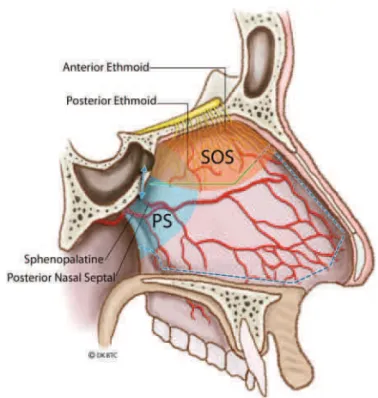
Fig. 2 Artist representation of the nasal septal anatomy, vasculature, and location of olfactoryfibers. Proper placement of the rescueflap incision allows elevation and preservations of the septal olfactory strip. Abbreviations: PS, posterior septum; SOS, septal olfactory strip.
Dural Opening
Prior to sellar dural opening, the location of the cavernous carotid arteries is precisely determined with a micro-Doppler probe (10-MHz ES-100X MiniDop with NRP-10H bayonet probe; Koven, St. Louis, Missouri, United States) and surgical navigation (►Fig. 3).22 The locations of the adenoma and pituitary gland should be anticipated based on the preopera-tive MRI. Ideally, the dura is opened without entering into the gland or transgressing the adenoma pseudocapsule. In most instances of macroadenomas, the pituitary gland will be compressed laterally and/or superiorly, but occasionally, a portion of the gland may be draped anteriorly over the tumor. A wide dural opening is performed inU-shaped fashion with a
standard straight microblade (Mizuho America Inc., Union City, CA). Next, angled microdissectors are used to carefully separate and elevate the dura from the underlying tumor and pituitary gland. The dural opening can then be extended more laterally as needed with a right-angle microhook blade or curved microscissors. Laterally, the opening will normally extend to within 1 to 2 mm of the medial wall of the cavernous sinus. If cavernous sinus bleeding is encountered, this low-pressure venous bleeding is generally easily con-trolled with Surgifoam (Ethicon Inc., Johnson & Johnson Co., Piscataway, New Jersey, United States) or Gelfoam (Pfizer Inc., New York, New York, United States) and gentle direct pres-sure. In patients with microadenomas and a low-lying dia-phragm, care must be taken to not extend the dural opening too far superiorly as this may often result in an early and anterior cerebrospinalfluid (CSF) leak.
Tumor Removal
Complete tumor resection with preservation or improvement in pituitary gland function is the goal for all patients under-going endonasal resection of a pituitary adenoma. Oldfield and Vortmeyer were thefirst to describe the technique of adenoma removal utilizing the tumor pseudocapsule in
Cushing disease as a means of achieving complete tumor resection.23This technique is particularly useful for micro-adenomas but can also be applied in macromicro-adenomas. For microadenomas, in which the tumor is behind a small rim of anterior gland, an incision can be made in the gland at its thinnest point to reach the pseudocapsule, which is a thin rim of compressed normal gland. A plane is then established between the adenoma and normal gland using micro-dis-sectors, irrigation, and gentle traction. The tumor is carefully separated from the compressed normal gland and gently removed with the surrounding pseudocapsule intact.
For larger macroadenomas with suprasellar extension, it is often best tofirst inferiorly and centrally debulk the tumor with ring curettes and suction. This initial decompression allows the more superior portion of the tumor with its pseudocapsule intact to be separated from compressed normal gland and the diaphragma sella and is often removed in one remaining large rind. With this technique, one avoids pulling the tumor down from the diaphragm sella without direct visualization, thus decreasing the likelihood of CSF leak.
Once all the visualized tumor has been removed with the 0-degree endoscope, the 30- or 45-0-degree angled lenses are utilized to obtain a clear view of regions not in direct line of sight.24This angled view is especially helpful for tumors with extensive suprasellar or cavernous sinus extension. The 45- and 90-degree up-angled ringed curettes may be used along with angled suctions to probe the folds of the diaphragma to dislodge residual tumor. In addition, a Valsalva maneuver or bilateral jugular vein compression can be administered to encourage downward descent of any suprasellar tumor that remains attached to the diaphragma. Ultimately, full inversion of the diaphragma into the enlarged sella should be seen if complete tumor removal has been accomplished. In cases of large macro-adenomas, the redundant and collapsed diaphragma sella often falls fully into the sella and obscures visualization of the sellar recesses. In such cases, it is extremely helpful to elevate this tissue with a spatula dissector and/or a cottonoid to facilitate an unobstructed view of these hidden regions to avoid missing residual adenoma. In all tumors with suspected cavernous sinus invasion, inspection of the medial cavernous sinus wall is performed with a 30- or 45-degree angled-lens endoscope. If present, tumor within the medial cavernous sinus may be safely removed using angled ring curettes and gentle suction. Venous bleeding from the cavernous sinus is once again controlled with Surgifoam or Gelfoam. In contrast to tumor within the medial cavernous sinus compartment, tumor that has extended along or lateral to the internal carotid artery (ICA) is difficult to access safely, and removal is associated with a higher risk of neuro-vascular injury. Monitoring and direct stimulation of cranial nerves III and VI is helpful for this lateral, posterior cavernous sinus dissection.
Skull Base Reconstruction and Cerebrospinal Fluid Leak Repair
Once tumor removal is complete, the sellar resection area is irrigated with full-strength hydrogen peroxide for approxi-mately 1 minute and hemostasis is achieved with Surgifoam. If there is significant diaphragmatic defect, irrigation with Fig. 3 Artist representation of micro-Doppler usage for localization of
hydrogen peroxide should not be performed. The type of skull base reconstruction performed depends primarily on wheth-er or not a CSF leak is present. Although the grading system for categorizing CSF leaks we described in 2007 is still quite useful, the technical details of repair have evolved in the endoscopic era.25 All repairs involved the use of collagen sponge (Helistat Integra, Hudson NH) as part of the recon-struction. In patients found to have no evidence of a CSF leak following a Valsalva maneuver (grade 0), a single layer of collagen sponge placed over the exposed dura is utilized as the only repair material. This is sealed in place usingfibrin glue. Exceptions include very large“dead space” defects or translucent diaphragm sellae that may have potential for hemorrhage or leakage. In such cases, the dead space isfilled with an abdominal fat graft. In cases where a small amount of CSF is detected following a Valsalva maneuver but no obvious diaphragmatic defect is visualized (grade 1), a layer of colla-gen sponge is initially placed under the dural edges. An intrasellar extradural buttress consisting of either the pa-tient’s previously harvested bony septum or a Medpore polyethylene plate (Stryker, Kalamazoo, Michigan, United States) is then placed over the collagen sponge followed by a second outer layer of collagen. The repair is bolstered with tissue glue (DuraSeal, Integra US, Hudson NH; or Tisseel, Baxter Healthcare Corp., Deerfield, IL). Medium-sized CSF leaks (grade 2) or grade 1 leaks with a large amount of intrasellar dead space require the placement of an abdominal fat graft. After harvesting from the lower abdomen, the fat graft is initially placed within the intrasellar space, taking care not to re-create too much mass effect on the suprasellar neurovascular structures. Again, this is followed by an intra-dural layer of collagen sponge and in some instances an intrasellar extradural rigid buttress. A second layer of fat and collagen is typically placed over this construct and bolstered with tissue glue. To assess the adequacy of the repair, prior to placing tissue glue, the anesthesiologist is asked to perform a Valsalva maneuver to raise the patient’s intracranial pressure. In some instances of grade 1 and 2 leaks in which a buttress is needed but no lateral bone edges are available to safely wedge the buttress in place, a temporary buttress with a Merocel sponge (Medtronic Inc, Minneapolis, MN) is placed in one or both nostrils for up to 5 days (continuous antibiotics while the packs are in place are necessary to prevent toxic shock syndrome). Large defects (grade 3) are typically seen only in extended suprasellar approaches for tumors such as craniopharyngiomas or tuberc-ulum meningiomas. The repair for such grade 3 leaks consists of virtually the same construct utilized to treat a grade 2 leak with the addition of a nasoseptal flap that is held in place with bilateral Merocel nasal packs, left in place for 5 days.26 Any patient with an intraoperative CSF leak is placed on acetazol-amide for 48 to 72 hours after surgery to decrease CSF produc-tion. CSF diversion with lumbar drainage is rarely employed.
Closure
Following the sellar reconstruction, blood is suctioned from the sphenoid sinus, nasal cavity, and nasopharynx, and good nasal mucosal hemostasis is obtained to minimize the
amount of blood that is swallowed in the immediate postop-erative period. If a nasoseptalflap is necessary to complete our repair, one of the two rescueflaps may be extended in the standard fashion. If not, then the rescueflaps are carefully elevated back along the remaining portion of the vomer and inferior nasal septum to their original location. The middle turbinates are then repositioned anatomically. Nasal packing is typically not utilized unless a buttress is needed as de-scribed above to help hold the repair in position. To minimize chances of a postoperative CSF leak, nasal epistaxis, or intra-sellar bleeding, excessive coughing or “bucking” should be avoided during extubation and blood pressure carefully monitored and controlled in the early postoperative period. The pharyngeal and gastric contents are typically aspirated of blood products to reduce postoperative nausea/vomiting. Throat packs are not routinely used as they are abrasive to the oropharynx, frequently resulting in patient discomfort.
Indications and Case Illustrations
The endoscopic endonasal transsphenoidal approach and its superior, inferior, and lateral extensions is ideal for a wide variety of sellar and parasellar lesions and is now the most common route for access to the pituitary gland. Lesions accessible through this route include pituitary adenomas, Rathke cleft cysts, clival chordomas, craniopharyngiomas, and sellar arachnoid cysts as well as tuberculum sellae, cavernous sinus, and petroclival meningiomas. The following cases are illustrative examples of lesions that are best suited for this technique.
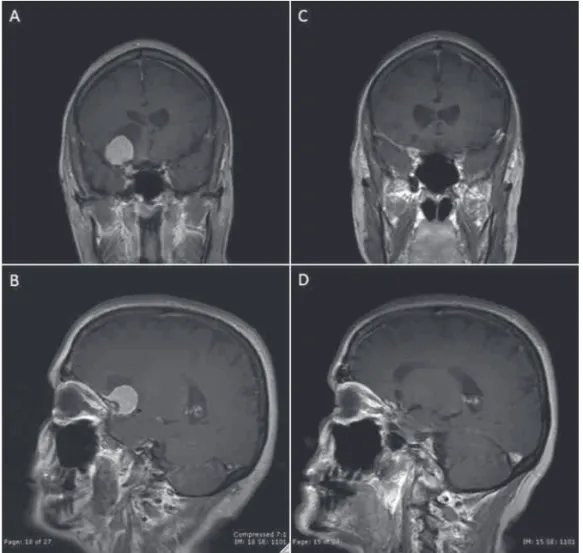
Case 1—Nonfunctioning Macroadenoma
The patient was a 39-year-old man with headaches and rapid visual loss in the left temporalfield. Ophthalmologic exami-nation demonstrated a bitemporal hemianopsia, which was more pronounced on the left. The patient was also found to have a left afferent pupillary defect as well as decreased visual acuity (20/100) in the left eye. The remainder of his neuro-logic exam was unremarkable. An MRI of the brain demon-strated a 252919-mm sellar mass with suprasellar extension, causing severe chiasmal compression (►Fig. 4). Pituitary hormonal testing revealed low T4 (3.5 μg/dL), inappropriately normal thyroid-stimulating hormone (1.95 μIU/mL), low free (23.5 pg/mL) and total (72ng/dL) testosterone, normal adrenocorticotropic hormone (58 pg/ mL) and cortisol (10.6μg/dL), elevated IGF-1 (388 ng/mL) and prolactin (1,253 ng/mL). The patient did not exhibit any of the usual stigmata of acromegaly or hyperprolactinemia. Due to the acute nature of the patient’s visual loss and the concern for a combined growth hormone/prolactin cosecreting tu-mor, surgical intervention was performed. The patient un-derwent an endoscopic endonasal transsphenoidal tumor resection with gross total removal. Pathology was consistent with a typical pituitary adenoma with immunohistochemical staining positively for both growth hormone and prolactin and an elevated Ki-67 proliferative index of 5 to 7%.
The sellarfloor was repaired in the standard fashion using collagen sponge, abdominal fat graft, nasal septal bone, and
observed, the decision to reinforce the sellar repair with an abdominal fat graft was made due to the large nature of the tumor and to prevent excessive diaphragmatic and optic chiasmal herniation. The patient initially tolerated the pro-cedure well without complications. His postoperative day 2 prolactin level decreased to 53.7 ng/mL. He noticed an immediate improvement in his visual function postopera-tively and was discharged home on postoperative day 2. At 3-month follow-up his visual acuity is 20/20 OU and his visual
fields are full to confrontation. His follow-up 3-month post-operative MRI demonstrated no obvious residual tumor with a small questionable nodule in the right posterior cavernous sinus region (►Fig. 4).
Case 2—Tuberculum Sella Meningioma
A 65-year-old woman presented with transient blurry vision. On further questioning, she reported gradual visual deterio-ration over several years. Visual acuity testing revealed 20/25 OU. Formal visualfield testing revealed bilateral optic nerve dysfunction but without respecting the vertical plane; oph-thalmoscopy confirmed bilateral optic atrophy. Sellar MRI demonstrated a 252423-mm contrast-enhancing suprasellar mass arising from the tuberculum sellae causing severe elevation of the optic apparatus, worse on the right, consistent with a tuberculum sellae meningioma that par-tially encased the left A2 segment. She had an extended
endoscopic transsphenoidal, transplanum tumor removal. At surgery, the tumor wasfibrous and significantly adherent to the right optic nerve and chiasm. Due to the adherent nature of the tumor, a small, 1- to 2-mm fragment of tumor was left along the inferior aspect of the right optic nerve and along the left A2 segment. A grade 3 CSF leak was repaired with collagen sponge, abdominal fat graft, nasoseptalflap, andfibrin glue. Two Merocel nasal packs were left in place as a temporary buttress to hold the repair. She tolerated the procedure well without complications and was discharged home on postoperative day 3. She had transient worsening of her right temporal visualfield, which improved by discharge. She had transient diabetes insipidus, requiring one dose of desmopressin (DDAVP), which then resolved and cortisol levels remained normal after surgery. Pathology confirmed a World Health Organization grade 1 meningothelial menin-gioma. Her MRI on thefirst postoperative day demonstrated no obvious evidence of residual tumor, despite the known small residual at the time of surgery. She will have another MRI at 3 months postsurgery (►Fig. 5).
Lessons Learned—Endoscopic Technique and Technology Applied to Intracranial Surgery: The Supraorbital Eyebrow Craniotomy
preserving the integrity of as much normal tissue as possible.” (p. 106)27 The supraorbital eyebrow craniotomy is a well-described, minimally invasive keyhole technique through a small anterolateral craniotomy that provides access to a wide range of anterior and midline skull base pathologies including the anterior fossa floor, the parasellar region, proximal sylvianfissure, circle of Willis, basal frontal lobe, and ventral brainstem.14,28As we described several years ago, this ap-proach is now routinely used as an alternative or comple-mentary approach to the endonasal endoscopic approach for parasellar tumors.29
Determining Which Approach for Which Parasellar Tumors
The most commonly encountered surgical lesions amenable to the supraorbital approach are tuberculum sellae, planum and anterior clinoid meningiomas, some olfactory groove meningiomas, craniopharyngiomas, and intra-axial tumors of the orbitofrontal region, frontal pole, and medial temporal lobe.14,28–31 We have employed the supraorbital approach most commonly for select tuberculum sella meningiomas that are over 3 cm in maximal diameter and have vascular encasement or extend well lateral to the supraclinoid carotid arteries or optic nerves. The supraorbital route is used for craniopharyngiomas that are not predominantly in the retro-chiasmatic space and instead extend lateral to the
supra-clinoid carotid arteries or into the anterior cranial fossa, as well as some recurrent craniopharyngiomas.
endoscope-assisted microsurgical techniques were used in 135 cases, giving the advantage of higher light intensity, a clear depiction of details in close-up positions, and an ex-tended viewing angle.32With the use of angled endoscopes, “blind”corners of the surgicalfield can be safely controlled without additional extension of the craniotomy. These areas would have been otherwise inaccessible through the keyhole approach using microscopic visualization.
Although the supraorbital approach offers access to most of the anterior fossafloor, parasellar area, and medial aspect of the middle fossa, there are four specific areas that can be difficult to reach and adequately visualize (►Fig. 6). These include the anterior aspect of the olfactory groove, the sellar
floor, the region under the ipsilateral optic nerve, and the anterior aspect of the middle fossa under the sphenoid ridge. The midline depression of the olfactory groove along the anterior skull base typically lies below the line of sight provided by the operating microscope. Likewise, a surgical trajectory along the orbital roof will not provide a direct line of sight into the sella itself. Similarly, the area directly under the ipsilateral optic nerve cannot be visualized with micro-scope without undue optic nerve manipulation. However, reaching these three relative blind spots is possible with the use of an angled endoscope and angled instrumentation. The fourth anatomic limit of the supraorbital approach is the lesser wing of the sphenoid. Lesions with significant exten-sion below the lesser sphenoid wing into the far anterior aspect of the middle cranial fossa may not be accessible from a supraorbital craniotomy. In such cases, a traditional pterional or minipterional craniotomy may be required to achieve access inferior and anterior to the sphenoid ridge.14
Surgical Technique—Supraorbital“Eyebrow”
Craniotomy
The operation is performed under general anesthesia with placement of a Foley catheter and arterial line in most cases. The patient’s head is fixed in a Mayfield three-pin head holder, angled 30 degrees away from the operative side
and extended such that malar eminence is prominent, similar to traditional pterional positioning. The Stryker navigation system is attached to the patient’s head and is registered to the surgical navigation MRI. The ipsilateral upper quadrant of the abdomen is marked and prepped in case an abdominal fat graft should become necessary. The eyebrow incision is marked and prepped and draped in usual sterile fashion. The eyebrow incision is made, starting from just medial to the supraorbital notch and extending laterally in the midst of the eyebrow to its termination with 5-mm extension in that trajectory. Sharp dissection is taken down to the pericranium, and a subgaleal plane is created in the supraorbital region. The dissection extends laterally to the superior part of the temporalis fascia and just lateral to the superior temporal line. The supraorbital nerve is identified medially and preserved. Multiplefishhooks are placed on the superior aspect of the incision to expose the supraorbital area. A pericranial cuff is then cut, extending from immediately lateral to the supraor-bital nerve, coursing superiorly in the arc of the planned craniotomy, over the superior temporal line and into the temporalis fascia and muscle. This pericranial cuff is elevated with subperiosteal dissection and retracted over the brow with a stitch.
A single bur hole is placed with a “matchstick” bit just inferior and lateral to the superior temporal line and behind the frontozygomatic process. The underlying dura is then elevated from the overlying bone. The craniotomy extends from the bur hole, medially along the supraorbital ridge. Although effort should be made to avoid entering the frontal sinus, this should not be at the expense of limiting the extent of the craniotomy, as maximizing the inferior extent of the craniotomy is critical both for visualization and for maneuver-ing of surgical instruments. The standard supraorbital craniot-omy is 2 to 2.5 cm wide and 1.5 to 2 cm tall (►Fig. 7). Again, proper positioning of the craniotomy as low as possible on the anterior fossafloor is important to obtain an optimal surgical trajectory. If the frontal sinus is entered, temporary occlusion accomplished with an iodine-soaked Gelfoam, and definitive repair is completed with an abdominal fat graft during closure. To facilitate exposure, the inner surface of the calvarium is drilled inferiorly along thefloor of the frontal fossa including
Fig. 6 Artist representation of the surgical access provided by the supraorbital craniotomy.
any protuberances of the orbital roof. This maneuver is essen-tial to optimize theflat surgical trajectory along the frontal
floor, as even small boney ridges may significantly impair the line of sight to the sellar region. The dura is then opened in aU
-shaped fashion and reflected inferiorly over the orbital rim. The duralflap is kept moist and under tension throughout the case to prevent shrinking and allow for a watertight closure. Immediately after dural opening, a small arachnoid incision into the opticocarotid cistern is made to allow for CSF egress as the microscope is brought into position. The remainder of the procedure is completed with a combination of microscopic and endoscopic visualization. Brain retractors are used infrequently.
Under microscopic visualization, the subfrontal corridor is dissected. The frontal lobe is protected with a strip of Telfa (Integra US, Hudson, NH), and the ipsilateral optic nerve and supraclinoid carotid artery are identified. The arachnoid of the opticocarotid and optico-oculomotor cisterns is opened widely to allow further CSF egress. The release of CSF is essential to achieve adequate brain relaxation and avoid frontal lobe retraction. Additional dissection of arachnoid at the base of the frontal lobe and within the proximal sylvian
fissure will free the frontal lobe from the basal cisterns and temporal lobe and allow it to fall away with gravity.
Following these maneuvers, afield of view including the ipsilateral proximal sylvianfissure, the ipsilateral third nerve,
both optic nerves, the optic chiasm, the lamina terminalis, both supraclinoid carotid arteries, both A1 segments, the anterior communicating artery, both A2 segments, and the pituitary stalk is possible. Additional dissection through the opticocarotid or optico-oculomotor windows will expose the ventral brainstem, basilar artery, and other posterior circula-tion vessels and perforators. Standard microsurgical dissec-tion and tumor removal then proceeds with care taken to preserve arachnoid membranes and key neurovascular struc-tures such as the superior hypophyseal arteries. Tumors densely adherent to the optic apparatus or circle of Willis vessels, which is common with craniopharyngiomas, are often best left behind to avoid new neurologic deficits.
With the use of a 30- or 45-degree angled endoscope, a view over the tuberculum sellae into the pituitary fossa, over sphenoid ridge into the middle fossa, over the dorsum sellae into the prepontine cistern, and over the orbital roof to the cribriform recess is possible.14When necessary, the supraorbital approach can be used to reach as far posteriorly and inferiorly to the ventral brainstem and superior one-third of the clivus.
The dural closure involves reapproximation and primary closure (when possible) of dural edges, followed by place-ment of a large piece of collagen sponge. Fat grafts are utilized to seal large defects of the frontal sinus. These are reinforced and secured with Tisseel. The boneflap is reapproximated with a lateral bur hole cover and a medial straight plate with
4-mm titanium screws. The boneflap is placed to eliminate any gap superiorly, and the gap inferiorly is filled with collagen matrix. The incision is irrigated with antibiotic-containing saline. The pericranial flap is repositioned with multiple 3–0 Vicryl stitches (Johnson & Johnson, New Bruns-wick, NJ). The scalp is closed with interrupted inverted 3–0 Vicryl sutures followed by a running subcuticular 5–0 Mono-cryl stitches (Johnson & Johnson, New Brunswick, NJ) Antibi-otic ointment, sterile dressing, and gentle head wrap compressive dressing is applied to help avoid a hematoma. Care is taken to avoid excess pressure with the head wrap to prevent pressure urticaria or skin necrosis.
Supraorbital Craniotomy—Case Illustration
Case 3—Clinoidal Meningioma
A 67-year-old woman suffered a ground-level fall and was found to have an incidental right paraclinoidal meningioma on CT. An MRI further characterized the lesion as a homo-geneously enhancing 262222-mm mass arising from the right anterior clinoid and medial sphenoid wing with an associated arachnoid cyst. Moderate surrounding vasogenic edema and 6 mm of right-to-left subfalcine herniation were noted. The imaging characteristics were consistent with an anterior clinoidal meningioma. Her neurologic examination was nonfocal. Due to the tumor extending lateral to the optic nerves and supraclinoid carotid artery along the sphenoid wing, a right supraorbital craniotomy was selected for resec-tion. The 30-degree angled endoscope was invaluable in visualizing and resecting tumor along the lateral wall of the cavernous sinus and extending into the sylvianfissure. At the time of surgery, it was felt that gross total resection was achieved. The patient tolerated the procedure well and there were no complications. Postoperative follow-up MRI demon-strates a small amount of persistent enhancement along the clinoidal dura and medial sphenoid wing. Two years after surgery, this has been stable and has not demonstrated any evidence of tumor progression or recurrence (►Fig. 8).
Final Comments
Although tremendous advances have been made in tran-sitioning toward minimally invasive transcranial and trans-sphenoidal approaches to the sella, further work remains to be done. Rather than representing afinished product, the current state of the art represents just another step in the continual march toward improved outcomes and decreased morbidity. When considering any approach to a pathologic entity, it is important to understand the advantages and disadvantages of a given procedure. Familiarity with the techniques and the indications and surgical anatomy for each approach will allow the surgeon to tailor the operation to the approach best suited for each patient.
References
1 Horsley V. On techniques of the operations of the central nervous system. BMJ 1906;2:411–423
2 Gandhi CD, Christiano LD, Eloy JA, Prestigiacomo CJ, Post KD. The historical evolution of transsphenoidal surgery: facilitation by technological advances. Neurosurg Focus 2009;27(3):E8 3 Liu JK, Das K, Weiss MH, Laws ER Jr, Couldwell WT. The history and
evolution of transsphenoidal surgery. J Neurosurg 2001;95(6): 1083–1096
4 Mcarthur L. An aseptic surgical access to the pituitary body and its neighborhood. JAMA 1912;58:2009–2011
5 Cushing H. The Pituitary Body and Its Disorders: Clinical States Produced by Disorders of the Hypophysis Cerebri. Philadelphia, PA: J.B. Lippincott; 1912:296–305
6 Kanter AS, Dumont AS, Asthagiri AR, Oskouian RJ, Jane JA Jr, Laws ER Jr. The transsphenoidal approach. A historical perspective. Neurosurg Focus 2005;18(4):e6
7 Jane JA Jr, Han J, Prevedello DM, Jagannathan J, Dumont AS, Laws ER Jr. Perspectives on endoscopic transsphenoidal surgery. Neuro-surg Focus 2005;19(6):E2
8 Hirsch O. Endonasal method of removal of hypophyseal tumors. With a report of two successful cases. JAMA 1910;5:772–774 9 Halstead A. Remarks on the operative treatment of tumors of the
hypophysis. With the report of two cases operated on by an oronasal method. Trans Am Surg Assoc 1910;28:73–93
10 Henderson WR. The pituitary adenomata. A follow-up study of the surgical results in 338 cases (Dr. Harvey Cushing’s series). Br J Surg 1939;26(104):811–921
11 Horwitz NH. Library: historical perspective. Norman M. Dott (1897–1973). Neurosurgery 1999;45(4):944–948
12 Landolt AM. History of pituitary surgery from the technical aspect. Neurosurg Clin N Am 2001;12(1):37–44, vii–viii
13 Cutler AR, Barkhoudarian G, Griffiths CF, Kelly DF. Transsphenoidal endoscopic skull base surgery: state of the art and future perspec-tive. Innovative Neurosurgery 2013;1:15–35
14 Wilson DA, Duong H, Teo C, Kelly DF. The supraorbital endoscopic approach for tumors. World Neurosurg 2013;(e-pub ahead of print)
15 Griffith HB, Veerapen R. A direct transnasal approach to the sphenoid sinus. Technical note. J Neurosurg 1987;66(1):140–142 16 Zada G, Kelly DF, Cohan P, Wang C, Swerdloff R. Endonasal trans-sphenoidal approach for pituitary adenomas and other sellar lesions: an assessment of efficacy, safety, and patient impressions. J Neurosurg 2003;98(2):350–358
17 Jho HD, Carrau RL. Endoscopic endonasal transsphenoidal surgery: experience with 50 patients. J Neurosurg 1997;87(1):44–51 18 Doglietto F, Prevedello DM, Jane JA Jr, Han J, Laws ER Jr. Brief history
of endoscopic transsphenoidal surgery—from Philipp Bozzini to the First World Congress of Endoscopic Skull Base Surgery. Neuro-surg Focus 2005;19(6):E3
19 Barahona MJ, Sojo L, Wägner AM, et al. Determinants of neurosur-gical outcome in pituitary tumors. J Endocrinol Invest 2005;28(9): 787–794
20 Barkhoudarian G, Del Carmen Becerra Romero A, Laws ER. Evalu-ation of the 3-dimensional endoscope in transsphenoidal surgery. Neurosurgery 2013;73(1, Suppl Operative):ons74–ons78, discus-sion ons78–ons79
21 Griffiths CF, Cutler AR, Duong HT, et al. Avoidance of postoperative epistaxis and anosmia in endonasal endoscopic skull base surgery: a technical note. Acta Neurochir (Wien) 2014;156(7):1393–1401 22 Dusick JR, Esposito F, Malkasian D, Kelly DF. Avoidance of carotid artery injuries in transsphenoidal surgery with the Doppler probe and micro-hook blades. Neurosurgery 2007;60(4, Suppl 2):322–328, discussion 328–329
23 Oldfield EH, Vortmeyer AO. Development of a histological pseu-docapsule and its use as a surgical capsule in the excision of pituitary tumors. J Neurosurg 2006;104(1):7–19
25 Esposito F, Dusick JR, Fatemi N, Kelly DF. Graded repair of cranial base defects and cerebrospinal fluid leaks in transsphenoidal surgery. Neurosurgery 2007;60(4, Suppl 2):295–303, discussion 303–304
26 Hadad G, Bassagasteguy L, Carrau RL, et al. A novel reconstructive technique after endoscopic expanded endonasal approaches: vascu-lar pedicle nasoseptalflap. Laryngoscope 2006;116(10):1882–1886 27 Wilson DH. Limited exposure in cerebral surgery. Technical note. J
Neurosurg 1971;34(1):102–106
28 Reisch R, Perneczky A, Filippi R. Surgical technique of the supra-orbital key-hole craniotomy. Surg Neurol 2003;59(3):223–227 29 Fatemi N, Dusick JR, de Paiva Neto MA, Malkasian D, Kelly DF.
Endonasal versus supraorbital keyhole removal of
craniopharyng-iomas and tuberculum sellae meningcraniopharyng-iomas. Neurosurgery 2009; 64(5, Suppl 2):269–284, discussion 284–286
30 Reisch R, Stadie A, Kockro R, Gawish I, Schwandt E, Hopf N. The minimally invasive supraorbital subfrontal key-hole approach for surgical treatment of temporomesial lesions of the dominant hemisphere. Minim Invasive Neurosurg 2009;52(4):163–169 31 McLaughlin N, Ditzel Filho LF, Shahlaie K, Solari D, Kassam AB,
Kelly DF. The supraorbital approach for recurrent or residual suprasellar tumors. Minim Invasive Neurosurg 2011;54(4): 155–161