Advances in Computed Tomography Evaluation
of Skull Base Diseases
Luciano M. Prevedello
11Department of Radiology, Ohio State Wexner Medical Center,
Columbus, Ohio, United States
Int Arch Otorhinolaryngol 2014;18:S123–S126.
Address for correspondence Luciano M. Prevedello, MD, MPH, Department of Radiology, Ohio State Wexner Medical Center, 395 W 12th Ave, 4th Floor, Columbus, OH 43210, United States
(e-mail: [email protected]).
Introduction
Due to its complex anatomy and close relationship with osseous structures, skull base lesions are often evaluated with both computed tomography (CT) and magnetic resonance imaging (MRI) for diagnostic and preoperative planning purposes. CT can delineate the osseous anatomy with increased precision in relation to MRI. Additionally, CT can provide important tips to diagnosis by identifying different patterns of bone involvement and periosteal reaction or by simply detecting the presence of ossification or calcification within a lesion.
Review of Literature and Discussion
Slow-growing lesions that do not infiltrate the bone tend to demonstrate smooth cortical expansion and bone remod-eling with preservation the bony cortex, whereas aggres-sive tumors or infection typically infiltrates the bone,
destroying the adjacent bony cortex in a permeative pat-tern.1This concept is often used to differentiate between schwannomas and paragangliomas in the skull base (►Fig. 1). Well-recognized exceptions to the rule are inva-sive pituitary adenomas and meningiomas. Bone invasion in pituitary adenomas does not typically represent a malignant feature and can be seen in 35% of the cases
of benign adenomas.2Clival invasion is more commonly seen in women, large tumors, and null-cell-type tumors. These infiltrative adenomas, however, have increased recurrence rates and complications and therefore their preoperative identification is important for surgical plan-ning and prognostic purposes.3
A subset of grade I meningiomas can also invade the bone.4 These cases tend to be clinically challenging as complete tumor resection becomes more difficult and therefore the risk of recurrence increases.5 It has been suggested that
Keywords
►
skull base
►
computed
tomography
►
dual energy
Abstract
Introduction
Computed tomography (CT) is a key component in the evaluation of
skull base diseases. With its ability to clearly delineate the osseous anatomy, CT can
provide not only important tips to diagnosis but also key information for surgical
planning.
Objectives
The purpose of this article is to describe some of the main CT imaging
features that contribute to the diagnosis of skull base tumors, review recent knowledge
related to bony manifestations of these conditions, and summarize recent technological
advances in CT that contribute to image quality and improved diagnosis.
Data Synthesis
Recent advances in CT technology allow
fi
ne-detailed evaluation of the
bony anatomy using submillimetric sections. Dual-energy CT material decomposition
capabilities allow clear separation between contrast material, bone, and soft tissues
with many clinical applications in the skull base. Dual-energy technology has also the
ability to decrease image degradation from metallic hardwares using some techniques
that can result in similar or even decreased radiation to patients.
Conclusions
CT is very useful in the evaluation of skull base diseases, and recent
technological advances can increase disease conspicuity resulting in improved
diagnos-tic capabilities and enhanced surgical planning.
DOI http://dx.doi.org/ 10.1055/s-0034-1390017. ISSN 1809-9777.
Copyright © 2014 by Thieme Publicações Ltda, Rio de Janeiro, Brazil
THIEME
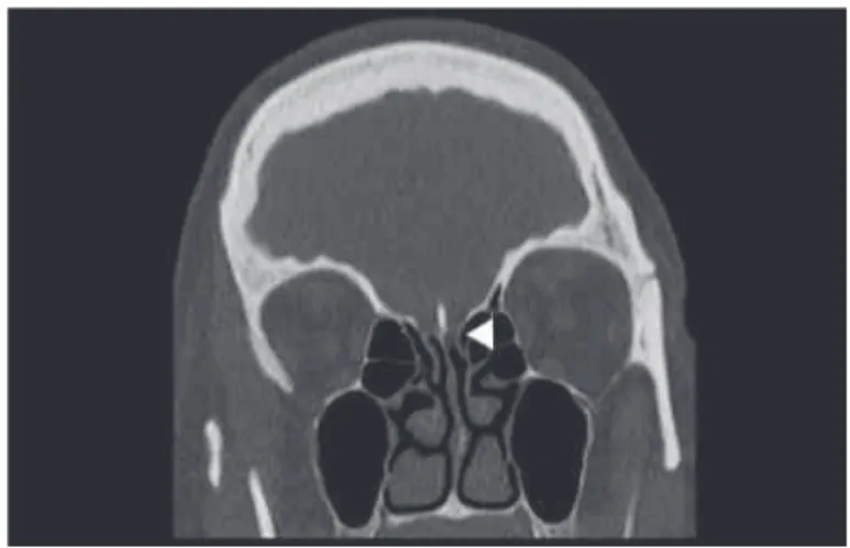
the molecular regulators of bone tropism in meningiomas may depend on their anatomical location as meningiomas of the anterior skull base show a distinct protein expression pattern compared with spheno-orbital meningiomas.5 The most typical bonyfinding in meningiomas, however, is bone hyperostosis (►Fig. 2). The cause of hyperostosis in meningi-omas is controversial. One theory favors focal vascular dis-turbances generated by the tumor,6–8 another suggests
osteoblastic stimulation by tumor secreting factors,9,10and another proposes bone production by the tumor itself.6,8It is important to recognize, however, that a significant number of patients with radiologic hyperostosis have been demonstrat-ed to have tumor invasion of the bone.11,12
Hemangiopericytoma is an important diagnostic consid-eration when bone invasion is identified in a dural-based mass. These tumors are typically multilobulated, extra-axial masses, with associated bone erosion. Unlike with meningio-mas, however, hyperostosis and intratumoral calcification are not typically present.13 Metastasis can also have a similar appearance to hemangiopericytomas and should also be considered in the appropriate clinical setting.
Another bony change related to meningiomas is the pres-ence of pneumosinus dilatans, which consists of abnormal expansion of one or more paranasal sinus. This can be another
helpful sign to indicate the presence of a meningioma in the anterior skull base.14
CT is also very helpful in identifying patterns of calcifi ca-tion or ossification to assist in the diagnosis. It is known that
90% of craniopharyngiomas calcify, making this an
impor-tant diagnostic feature.15Additionally, the distinct pattern of chondroid calcification (arc or ringlike calcifications) in chondrosarcomas can also be useful in pointing to this entity during diagnostic workup. Chordoma is one of the main differential considerations once chondrosarcomas are sus-pected, and it is important to differentiate between arc-whorl intralesional calcifications seen in chondrosarcomas with fragmented destroyed bone more often seen in chordomas.16 Another use of CT in the skull base is in the identification of bony defects in the evaluation of cerebral spinalfluid (CSF) leaks. Recent multislice CT scanners can acquire images with slice thickness as thin as 0.5 to 0.6 mm and can perform multiplanar reconstructions, providing greater ability to evaluate submillimetric defects. Interactive multiplanar eval-uation (axial, coronal, sagittal, and oblique planes) is impor-tant to identify and correctly describe osseous defects in the evaluation of CSF leaks (►Fig. 3).17The identification of bony defects in these cases is highly sensitive but not definitive for CSF leak. Stone et al observed that all patients in their 42-patient cohort with confirmed CSF leak demonstrated bony defects on high-resolution CT. Ten patients with bony defects demonstrated on CT, however, were not confirmed to have CSF leak.18
In addition to providing important characterization of the bony structures, CT can also provide invaluable infor-mation about the relationship of a lesion with the adjacent vascular structures through computed tomography angiog-raphy (CTA).
One major challenge related to vascular imaging in the skull base has been the evaluation of the cavernous internal carotid artery. This is particular true because the high-density contrast material within the vessels becomes less conspicuous when surrounded by bone. Several computer-ized bone subtraction algorithms have been proposed in the past in an attempt to overcome this issue. One technique utilizes two imaging acquisitions (pre- and postcontrast) to
Fig. 2 Hyperostosis (white arrowheads) in a cavernous sinus menin-gioma (black arrowheads).
Fig. 3 Bony defect along the left cribriform plate (arrowhead) in a patient with suspected cerebrospinalfluid leak.
Fig. 1 Demonstration of two distinctive patterns of bone ment. Note the permeative and destructive pattern of bone involve-ment typically seen in aggressive lesions such as paragangliomas (A) in relation to a smoothly marginated expansile lesion such as a schwan-noma (B) in the right jugular foramen in these two different patients.
International Archives of Otorhinolaryngology Vol. 18 No. S2/2014
subtract the background bone. The major disadvantage of this technique is the patient’s increased radiation expo-sure.19 In addition, motion between the two acquisitions can also impact the quality of the bone subtraction in this technique. Another bone subtraction method uses comput-erized imaging processing techniques to differentiate the vessel anatomy from the adjacent bone by segmenting out only the structure containing the contrast material. This technique relies on the variation between the densities of different structures as well as few anatomical landmarks to distinguish between bone and vessel. Unfortunately, overlap exists between the density of these structures, and the bone subtraction obtained with this methodology is not consis-tently reliable. Recent dual-energy technology can differen-tiate between contrast material and bone with high precision. This is possible because the density of the calcium and iodine varies, which causes them to behave differently depending on the energy applied to X-ray beams (different peak kilovoltages). Computer algorithms are then applied to the acquired images, allowing decomposition of few ele-ments and subtraction of the calcium (►Fig. 4).
The evaluation of the petrous, cavernous, and supraclinoid internal carotid artery is very important in the preoperative planning for tumors that invade the cavernous sinus. Cavern-ous sinus meningiomas, for instance, can cause significant narrowing of the cavernous internal carotid artery. Although this is not easily recognized with standard multiplanar re-constructions, techniques such as dual-energy bone subtrac-tion or some postprocessing tools such as curved reformats can be of great assistance. Even with postprocessing techni-ques, however, it may be difficult to appreciate smaller vessels such as posterior communicating arteries that may have been compressed by the tumor. In these situations, it is often helpful to use thin-section T2-weighted images to troubleshoot.
Another use of dual-energy technology in the evaluation of skull base pathology relates to its ability to decrease artifacts that are known to negatively impact image quality, particu-larly in the posterior fossa. The petrosal ridge of the temporal bone is the hardest bone in the human body and is responsible for significant artifact in the cerebellopontine region. Based on the two polychromatic X-ray beams available in
dual-energy CT, sophisticated reconstruction algorithms can be applied to estimate what a scan performed with a single monochromatic X-ray beam might have shown. With such approach, beam-hardening and streak artifacts can be signif-icantly reduced, although often at the expense of lower signal-to-noise ratio.20 The same technique can be applied to reduce artifact from external sources or metallic hardware in the craniocervical junction (►Fig. 5).21,22
Dual-energy CT can also be used to generate a virtual noncontrasted CT from a contrast enhanced study by sub-tracting the iodine material from the image using its material decomposition capabilities. Yet, there is strong evidence that dual-energy CT acquired via dual-source technology does not result in increased radiation to patients.23In fact, the radia-tion from dual-energy CTs measured by volume computed tomography dose index (CTDIvol) have been found to be 12% lower than single-energy CTs.24
Additionally, having the ability to generate two scans (contrast-enhanced and virtual noncontrast) from a single postcontrast acquisition may have further radiation exposure savings when these two scans are clinically needed. By decomposing the iodine component from the image, dual-energy CT can also provide maps on which iodine distribution is color-coded and superimposed on the virtual noncontrast CT, which is thought to increase visual detection of lesions in the head and neck.25,26
Final Comments
CT is an invaluable tool in the evaluation of skull base disease. In addition to providing important tips to diagnosis, it can also depict important landmarks for surgical plan-ning. Recent advances in CT technology allowfine-detailed evaluation of the bony anatomy with submillimetric imag-ing sections with increased overall image quality and similar or even decreased radiation exposure to patients.
References
1 Raut AA, Naphade PS, Chawla A. Imaging of skull base: pictorial essay. Indian J Radiol Imaging 2012;22(4):305–316
Fig. 4 Bone subtraction using dual-energy technique with clear separation between iodine and calcium.
Fig. 5 Dual-energy acquisition with two different monoenergetic selections (A: 50 keV; B: 100 keV). The 100-keV monoenergetic imaging shows decreased streak artifact from the suboccipital metallic hardware.
2 Hansen TM, Batra S, Lim M, et al. Invasive adenoma and pituitary carcinoma: a SEER database analysis. Neurosurg Rev 2014;37(2): 279–285, discussion 285–286
3 Chen X, Dai J, Ai L, et al. Clival invasion on multi-detector CT in 390 pituitary macroadenomas: correlation with sex, subtype and rates of operative complication and recurrence. AJNR Am J Neuroradiol 2011;32(4):785–789
4 Bikmaz K, Mrak R, Al-Mefty O. Management of bone-invasive, hyperostotic sphenoid wing meningiomas. J Neurosurg 2007; 107(5):905–912
5 Salehi F, Jalali S, Alkins R, et al. Proteins involved in regulating bone invasion in skull base meningiomas. Acta Neurochir (Wien) 2013; 155(3):421–427
6 Freedman H, Forster FM. Bone formation and destruction in hyperostoses associated with meningiomas. J Neuropathol Exp Neurol 1948;7(1):69–80
7 Bonnal J, Thibaut A, Brotchi J, Born J. Invading meningiomas of the sphenoid ridge. J Neurosurg 1980;53(5):587–599
8 Heick A, Mosdal C, Jørgensen K, Klinken L. Localized cranial hyperostosis of meningiomas: a result of neoplastic enzymatic activity? Acta Neurol Scand 1993;87(3):243–247
9 Pompili A, Derome PJ, Visot A, Guiot G. Hyperostosing meningio-mas of the sphenoid ridge—clinical features, surgical therapy, and long-term observations: review of 49 cases. Surg Neurol 1982; 17(6):411–416
10 Maroon JC, Kennerdell JS, Vidovich DV, Abla A, Sternau L. Recurrent spheno-orbital meningioma. J Neurosurg 1994;80(2):202–208
11 Goyal N, Kakkar A, Sarkar C, Agrawal D. Does bony hyperostosis in intracranial meningioma signify tumor invasion? A radio-patho-logic study. Neurol India 2012;60(1):50–54
12 Pieper DR, Al-Mefty O, Hanada Y, Buechner D. Hyperostosis associated with meningioma of the cranial base: secondary changes or tumor invasion. Neurosurgery 1999;44(4):742–746, discussion 746–747
13 Chiechi MV, Smirniotopoulos JG, Mena H. Intracranial hemangio-pericytomas: MR and CT features. AJNR Am J Neuroradiol 1996; 17(7):1365–1371
14 Parizel PM, Carpentier K, Van Marck V, et al. Pneumosinus dilatans in anterior skull base meningiomas. Neuroradiology 2013;55(3): 307–311
15 Eldevik OP, Blaivas M, Gabrielsen TO, Hald JK, Chandler WF. Craniopharyngioma: radiologic and histologicfindings and recur-rence. AJNR Am J Neuroradiol 1996;17(8):1427–1439
16 Cho YH, Kim JH, Khang SK, Lee J-K, Kim CJ. Chordomas and chondrosarcomas of the skull base: comparative analysis of clinical results in 30 patients. Neurosurg Rev 2008;31(1):35–43, discus-sion 43
17 La Fata V, McLean N, Wise SK, DelGaudio JM, Hudgins PA. CSF leaks: correlation of high-resolution CT and multiplanar reformations with intraoperative endoscopicfindings. AJNR Am J Neuroradiol 2008;29(3):536–541
18 Stone JA, Castillo M, Neelon B, Mukherji SK. Evaluation of CSF leaks: high-resolution CT compared with contrast-enhanced CT and radionuclide cisternography. AJNR Am J Neuroradiol 1999; 20(4):706–712
19 Seo H, Choi DS, Shin HS, Cho JM, Koh EH, Son S. Bone subtraction 3D CT venography for the evaluation of cerebral veins and venous sinuses: imaging techniques, normal variations, and pathologic findings. AJR Am J Roentgenol 2014;202(2):W169–W175
20 Pomerantz SR, Kamalian S, Zhang D, et al. Virtual monochromatic reconstruction of dual-energy unenhanced head CT at 65–75 keV maximizes image quality compared with conventional polychro-matic CT. Radiology 2013;266(1):318–325
21 Guggenberger R, Winklhofer S, Osterhoff G, et al. Metallic artefact reduction with monoenergetic dual-energy CT: systematic ex vivo evaluation of posterior spinal fusion implants from various ven-dors and different spine levels. Eur Radiol 2012;22(11):2357–2364
22 Lewis M, Reid K, Toms AP. Reducing the effects of metal artefact using high keV monoenergetic reconstruction of dual energy CT (DECT) in hip replacements. Skeletal Radiol 2013;42(2):275–282
23 Henzler T, Fink C, Schoenberg SO, Schoepf UJ. Dual-energy CT: radiation dose aspects. AJR Am J Roentgenol 2012;199(5, Suppl): S16–S25
24 Tawfik AM, Kerl JM, Razek AA, et al. Image quality and radiation dose of dual-energy CT of the head and neck compared with a standard 120-kVp acquisition. AJNR Am J Neuroradiol 2011; 32(11):1994–1999
25 Vogl TJ, Schulz B, Bauer RW, Stöver T, Sader R, Tawfik AM. Dual-energy CT applications in head and neck imaging. AJR Am J Roentgenol 2012;199(5, Suppl):S34–S39
26 Wichmann JL, Nöske E-M, Kraft J, et al. Virtual monoenergetic dual-energy computed tomography: optimization of kiloelectron volt settings in head and neck cancer. Invest Radiol 2014; [Epub ahead of print]
International Archives of Otorhinolaryngology Vol. 18 No. S2/2014