Case report
The first clinical case was that of a 40-year-old Caucasian female who sought emergency room treatment in May of 2003 presenting with a clin-ical profile characterized by dyspnea, irritating cough, night sweats, asthenia, myalgia, nausea and subfebrile temperatures (37.5-38.0°C) for 3 weeks.
In the pathological history, varicose veins of the lower limbs were worthy of note, as were an episode of renal colic in 1998 and an episode of biliary colic due to gallstones in 2000.
It is worthy of note that the patient was not a user of alcohol, tobacco or illicit drugs.
The patient had been using oral contracep-tives for several years.
In the epidemiological context, the fact that the patient lived in an urban area, with good hygienic and sanitary conditions, and had
Introduction
Although the etiology of ectopic thyroid is not well known, mutations in the thyroid tran-scription factor,(1-3) PAX8 genes and the thyroid stimulating hormone receptor gene have been implicated in a minority of patients with thyroid dysgenesis.(4-6)
The occurrence of ectopic thyroid tissue is a rare embryological aberration.(7,8) Among patients with hypothyroidism, the incidence of ectopic thyroid is estimated to range from 1/4,000 to 1/8,000.(9) When ectopic thyroid occurs, it is located along the path of descent of the devel-oping embryonic thyroid, from the foramen cecum(5) to its destined pretracheal location.(8)
The development of ectopic thyroid tissue can occur at any point during the migration of the thyroid, resulting in lingual, sublingual, prela-ryngeal and substernal (mediastinal) ectopia.(7,9)
Ectopic thyroid in the anterior mediastinum*
Tireoide ectópica no mediastino anterior
Maria José Araújo da Cunha Guimarães, Carla Manuela Silva Valente, Lèlita Santos, Manuel Fontes Baganha
Abstract
Ectopic thyroid is a rare condition, and its location in the anterior mediastinum is even rarer, there having been only 5 reported cases in the past 30 years. Here, we describe 2 clinical cases and present a review of the literature regarding the etiology, embryology and clinical manifestations of ectopic thyroid.
Keywords: Thyroid dysgenesis; Mediastinum; Goiter.
Resumo
A ectopia de tireoide é rara, e a sua localização no mediastino anterior é excepcional, estando descritos apenas 5 casos nos últimos 30 anos. Os autores apresentam 2 casos clínicos, além de uma revisão da literatura abordando a etiologia, a embriologia e manifestações clínicas de ectopia de tireoide.
Descritores: Disgenesia da tireoide; Mediastino; Bócio.
* Study carried out in the Department of Pulmonology and Allergology of the Hospitals of the University of Coimbra, Coimbra, Portugal.
Correspondence to: Maria José Araújo da Cunha Guimarães. Departamento de Ciências Pneumológicas e Alergológicas, Hospitais da Universidade de Coimbra, Praceta Mota Pinto, 2º Piso, 3000-075, Coimbra, Portugal.
Financial support: None.
The abdomen was soft, depressible and painless on palpation, there being no palpable masses or organomegalies.
The lower limbs presented varicose veins in the area of the great saphenous vein, and the results of the summary neurological tests were normal.
In view of the clinical profile, we considered the following working diagnoses: community-acquired pneumonia, namely bacterial (not excluding that caused by a specific germ), due to the fever and the alterations found during the physical examination; and neoplastic disease, namely lymphoma or lung neoplasia, due to the systemic component of the clinical evaluation associated with the presence of a mass in the chest during the physical examination.
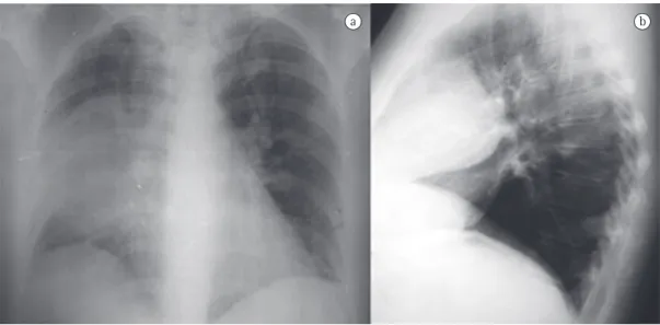
Complementary tests performed in the emer-gency room revealed normocytic normochromic anemia (hemoglobin, 11.3 mg/dL; mean glob-ular volume, 86.8 fl; and mean corpuscglob-ular hemoglobin, 33.7 mg/L), normal total leuko-cyte counts (7 g/L), hypokalemia (3.3 mmol/L) and hypoalbuminemia (3 g/dL), as well as an increase in the levels of aspartate aminotrans-ferase (60 U/L), alanine aminotransaminotrans-ferase (79 U/L), lactate dehydrogenase (196 U/L), alkaline phosphatase (272 U/L) and C-reactive protein (19.2 mg/dL). The patient also presented hypoxemia on room air (PaO2: 79.5 mmHg) and mild hypocapnia (33 mmHg). A chest X-ray revealed opacity in the middle third of the right lung field (Figure 1).
no contact with animals is worthy of note. In addition, she was not engaging in risky sexual behavior.
The patient had a history of allergy to penicillin.
Regarding the gynecologic and obstetric history, the patients reported a full-term (40-week) pregnancy and normal delivery in 1989.
In the family history, it was worthy of note that her father died at the age of 59 years and her mother died at the age of 70 years, both due to cerebrovascular accident, one brother had epilepsy, and two brothers had diabetes.
Upon physical examination, the patient (weight: 78 kg; height: 1.53 m; and body mass index: 32 kg/m2) was conscious, alert, coopera-tive, with good color and well hydrated, without cyanosis and without exanthema or adenopathy. She was subfebrile (37.5°C), presenting an arte-rial pressure of 120/70 mmHg, a pulse with a heart rate (HR) of 96 bpm and a respiratory rate of 32 breaths/min.
Although the patient had dyspnea, there was good chest expansion. Palpation revealed that the transmission of vocal vibration was main-tained, there being a parenchymal sound upon percussion of the entire lung field. Pulmonary auscultation revealed reduced breath sounds in the right lung field, from the third intercostal space to the lung base, without adventitious sounds. Heart auscultation was rhythmic, without heart murmurs and with a HR of 96 bpm.
Figure 1 - Anteroposterior and right lateral chest X-ray revealing opacification of the middle zone of the right lung field, with round borders and in an anterior location, in profile.
and the findings confirmed those of the CT images.
In view of these new data, we raised the hypothesis that we were confronted with a neoplastic disease.
In this context, we thought it could be a germ cell tumor, a neurogenic tumor, a lymphoma, a thymic tumor, a mesenchymal tumor or, although a more remote possibility, an ectopic mass in a gland, namely the thyroid.
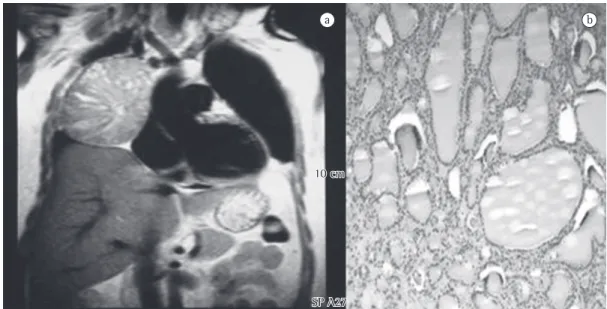
The patient was then referred for surgery, which allowed the excision of an anterior medi-astinal mass (measuring 12 × 9 cm and weighing 400 g), well delineated by a capsule, and whose histology corresponded to ectopic thyroid tissue, the morphology overlapping with that of nodular thyroid hyperplasia but differing from it by presenting microfollicles containing colloid as well as an adjacent inflammatory reaction (Figure 2).
The levels of thyroid-stimulating hormone and free thyroid hormone were normal. An ultra-sound of the thyroid revealed a heterogeneous gland of enlarged proportions, with various solid echogenic nodules in the left lobe (the largest one measuring 1.1 cm) and in the right lobe (the largest one measuring 2.8 cm), there being no cervical adenopathy.
In addition, the patient underwent techne-tium (99mTc) scintigraphy to determine whether there were other ectopic glands, as well as to allow the functional and morphological study of Bacterial pneumonia was considered as
the main working diagnosis. The patient was admitted, and treatment with clarithromycin, cefazolin and oxygen was initiated. Clinical and analytical improvement was obtained on the second day of treatment, although the patient still presented mild hypoxemia and increased globular sedimentation rate (90 mm/h).
At admission, blood and urine cultures were performed, as was serology for Chlamydia, Mycoplasma, Brucella, Salmonella, Rickettsia, Legionella and Coxiella, as well as for cytome-galovirus, Epstein-Barr virus, hepatitis A virus, hepatitis B virus, and hepatitis C virus. All results were negative. The urine test results revealed no alterations, and lymphocytic phenotyping was normal. The electrophoretic protein profile anal-ysis revealed no alterations, and an abdominal ultrasound revealed gallstones.
Another chest X-ray, performed on postad-mission day 8, showed that the opacity described above persisted. Subsequently, a CT scan of the chest, performed to clarify this issue, revealed a solid formation in the anterior mediastinum. The formation measured 10 cm in its largest diam-eter and maintained cleavage planes with the heart and the lung, as well as insinuating itself between the lung lobes and compressing them. In addition, a magnetic resonance imaging scan was performed to determine the true extent of the lesion, especially in terms of the cleavage plane with the mediastinal structures (Figure 2),
10 cm
SP A27
a b
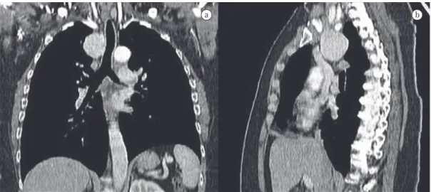
A CT scan of the chest revealed, in the upper mediastinum, a round, extensive forma-tion (4.5 × 4.3 cm), without signs of infiltraforma-tion and with well-defined borders, located laterally between the superior vena cava and the right contour of the trachea (Figure 3).
An ultrasound of the thyroid identified only two solid nodules, with high echogenicity, in the left lobe.
Fiberoptic bronchoscopy revealed tracheal deviation to the right and moderate signs of inflammation in the bronchial tree.
Thyroid hormone levels were normal, and there were no antimicrosomal or antithy-roglobulin antibodies. Tumor marker tests were negative.
Thyroid scintigraphy with 99mTc revealed that the thyroid was reduced to its left lobe.
The patients underwent excision of the medi-astinal tumor (7 × 5 × 4 cm), and the microscopic report of the surgical sample was consistent with intramediastinal thyroid with a macrofollicular pattern and no cellular atypia.
The patient was referred to an endocrinol-ogist, and levothyroxine (0.1 mg/day) was maintained.
Discussion
The prevalence of ectopic thyroid tissue ranges from 7% to 10%.(8)
Ectopia usually occurs along the midline, and locations lateral to that are rare,(10) with slightly more than half a dozen cases being known. the thyroid, which proved to present functional
integrity.
She was treated with levothyroxine in an attempt to reduce the substernal goiter and subsequently submitted to total thyroidectomy.
At this writing, the patient was being treated with levothyroxine (100 µg/day) and was asymptomatic.
The second clinical case was that of a 57-year-old Caucasian female nonsmoker who was admitted for investigation of an upper mediastinal lesion.
The patient presented with a clinical profile of severe asthenia, dyspnea upon moderate exer-tion, orthopnea and dysphonia for 4 months.
She had been treated with antibiotics at an outpatient clinic, due to suspected pharyngitis, and had shown no improvement.
The patient had a history of partial right thyroidectomy due to nodular hyperplasia 9 years prior. She was chronically treated with levothyroxine (0.1 mg/day).
Upon physical examination, the patient was conscious, alert and apyretic, as well as presenting normal respiration. Pulmonary auscultation revealed reduced breath sounds at the vertex of the right hemithorax, without adventitious sounds. Thyroid palpation revealed left lobe hypertrophy, without a palpable mass on the right.
Analytically, the patient presented no alterations.
An anteroposterior and lateral right chest X-ray revealed enlargement of the right upper mediastinum.
Figure 3 - Chest CT scan showing a round tumor formation (4.5 × 4.3 cm) in the upper mediastinum, with well-defined borders and without signs of infiltration of adjacent structures.
3. Kambe F, Seo H. Thyroid-specific transcription factors. Endocr J. 1997;44(6):775-84.
4. Macchia PE, Lapi P, Krude H, Pirro MT, Missero C, Chiovato L, et al. PAX8 mutations associated with congenital hypothyroidism caused by thyroid dysgenesis. Nat Genet. 1998;19(1):83-6.
5. Hazarika P, Siddiqui SA, Pujary K, Shah P, Nayak DR, Balakrishnan R. Dual ectopic thyroid: a report of two cases. J Laryngol Otol. 1998;112(4):393-5.
6. Macchiarini P, Ostertag H. Uncommon primary mediastinal tumours. Lancet Oncol. 2004;5(2):107-18. 7. Cherif L, Lakhoua Y, Khiari K, Hadj-Ali I, Rajhi H, Kaffel
N, et al. Ectopic thyroid: two cases [Article in French]. Ann Endocrinol (Paris). 2004;65(3):233-7.
8. Basaria S, Cooper DS. Graves’ disease and recurrent ectopic thyroid tissue. Thyroid. 1999;9(12):1261-4. 9. Baik SH, Choi JH, Lee HM. Dual ectopic thyroid. Eur
Arch Otorhinolaryngol. 2002;259(2):105-7.
10. Kumar R, Sharma S, Marwah A, Moorthy D, Dhanwal D, Malhotra A. Ectopic goiter masquerading as submandibular gland swelling: a case report and review of the literature. Clin Nucl Med. 2001;26(4):306-9. 11. Byrd MC, Thompson LD, Wieneke JA. Intratracheal
ectopic thyroid tissue: a case report and literature review. Ear Nose Throat J. 2003;82(7):514-8.
12. Specker R, Curti G, Müller W, Stulz P. Intrathoracic goiter--a rare mediastinal tumor [Article in German]. Swiss Surg. 2001;7(3):134-8.
13. Sand J, Pehkonen E, Mattila J, Seppănen S, Salmi J. Pulsating mass at the sternum: a primary carcinoma of ectopic mediastinal thyroid. J Thorac Cardiovasc Surg. 1996;112(3):833-5.
14. Sakorafas GH, Vlachos A, Tolumis G, Kassaras GA, Anagnostopoulos GK, Gorgogiannis D. Ectopic intrathoracic thyroid: case report. Mt Sinai J Med. 2004;71(2):131-3.
15. Bremerich J, Pippert H. Ectopic thyroid tissue: an unusual differential diagnosis of space-occupying mediastinal lesions [Article in German]. Schweiz Med Wochenschr. 1997;127(7):266-70.
16. Tojo K. Lingual thyroid presenting as acquired hypothyroidism in the adulthood. Intern Med. 1998;37(4):381-4.
17. Güngör B, Kebat T, Ozaslan C, Akilli S. Intra-abdominal ectopic thyroid presenting with hyperthyroidism: report of a case. Surg Today. 2002;32(2):148-50.
18. Yamauchi M, Inoue D, Sato H, Ashida C, Hiraumi H, Shan L, et al. A case of ectopic thyroid in lateral neck associated with Graves’ disease. Endocr J. 1999;46(5):731-4. Regarding intrathoracic (mediastinal)
loca-tion, we found only 7 cases in the literature. In 5 of those cases, the ectopia was located in the anterior mediastinum,(8,11-13) as in the cases reported here, and, in 2,(12,14) it was located posteriorly. Therefore, although entirely intrathoracic ectopic goiters are rare, they have to be considered in the differential diagnosis of all mediastinal masses.(15)
Most patients with ectopic thyroid are euthy-roid and asymptomatic. However, obstructive or compression-related symptoms, especially of the upper aerodigestive tract,(8) can appear, as can hypothyroidism(16) and, much more rarely, hyper-thyroidism.(8,17,18)
Other less common manifestations result from rarer locations. Cases of patients who only became symptomatic during pregnancy have been described, there having been regression after delivery.(16)
In conclusion, ectopic thyroid is a rare condition, and its location in the anterior medi-astinum is even rarer (only 5 cases having been reported in the past 30 years). We intend to draw attention to the importance of considering ectopic thyroid in the differential diagnosis of mediastinal masses, since, after surgery, there is a risk of hypothyroidism if the ectopic gland is not functioning. It should be emphasized that the diagnosis is made essentially through anato-mopathological examination and, in most cases, only after surgery.
References
1. Gillam MP, Kopp P. Genetic regulation of thyroid development. Curr Opin Pediatr. 2001;13(4):358-63. 2. Van Vliet G. Development of the thyroid gland: lessons
from congenitally hypothyroid mice and men. Clin Genet. 2003;63(6):445-55.
About the authors
Maria José Araújo da Cunha Guimarães
Intern. Complementary Internship Program in Pulmonology, Hospitals of the University of Coimbra, Coimbra, Portugal.
Carla Manuela Silva Valente
Intern. Complementary Internship Program in Pulmonology, Hospitals of the University of Coimbra, Coimbra, Portugal.
Lèlita Santos
Attending Internal Medicine Physician. Hospitals of the University of Coimbra, Coimbra, Portugal.
Manuel Fontes Baganha