Evolution of exogenous lipoid pneumonia in
children: clinical aspects, radiological aspects
and the role of bronchoalveolar lavage*
Evolução da pneumonia lipoide exógena em crianças: aspectosclínicos e radiológicos e o papel da lavagem broncoalveolar
Selma Maria de Azevedo Sias, Angela Santos Ferreira, Pedro Augusto Daltro, Regina Lúcia Caetano, José da Silva Moreira, Thereza Quirico-Santos
Abstract
Objective: To present aspects of the evolution of lipoid pneumonia in children, based on clinical, radiological and bronchoalveolar lavage fluid findings, emphasizing the importance of bronchoalveolar lavage for the diagnosis and treatment. Methods: We included 28 children, with a mean age of 20 months (range, 1-108 months), diagnosed with chronic pneumonia refractory to antimicrobial therapy, with TB or with a combination of the two. Most of the children had at least one risk factor for aspiration, and all of them had a history of mineral oil ingestion for intestinal constipation (23/28) or complicated ascaridiasis (5/28). Clinical evaluations, tomographic evaluations and analyses of bronchoalveolar lavage fluid were carried out at the beginning of treatment and throughout a follow-up period of 24 months. Results: Tachypnea and cough were the most common symptoms. The most common radiological alterations were areas of consolidation (23/28), perihilar infiltrates (13/28) and hyperinflation (11/28). Chest CT scans showed areas of consolidation with air bronchogram (24/28), decreased attenuation in the areas of consolidation (16/28), ground-glass opacities (3/28) and crazy-paving pattern (1/28). In the analysis of the bronchoalveolar lavage fluid, Sudan staining revealed foamy macrophages, confirming the diagnosis of lipoid pneumonia. After treatment with multiple bronchoalveolar lavages (mean = 9.6), 20 children became asymptomatic, 18 of those presenting normal tomographic images. Conclusions: A diagnosis of lipoid pneumonia should be considered in patients with chronic refractory pneumonia or TB, especially if there is a history of mineral oil ingestion. Bronchoscopy with multiple bronchoalveolar lavages was an efficient treatment for the clearance of mineral oil from the lung parenchyma and the prevention of fibrosis. This strategy contributed to reducing the morbidity of lipoid pneumonia, which remains a rare diagnosis.
Keywords: Pneumonia, lipid; Bronchoalveolar lavage; Treatment outcome.
Resumo
Objetivo: Descrever os aspectos da evolução da pneumonia lipoide em crianças, com base em aspectos clínicos, radiológicos e de achados no lavado broncoalveolar, enfatizando a importância diagnóstica e terapêutica da lavagem broncoalveolar. Métodos: Foram incluídas 28 crianças, com idade média de 20 meses (1-108 meses) e diagnóstico de pneumonia crônica refratária a antimicrobianos e/ou TB. A maioria apresentava um fator de risco para aspiração, e todas apresentavam história de ingestão de óleo mineral para o tratamento de constipação intestinal (23/28) ou de ascaridíase complicada (5/28). A avaliação clínica e tomográfica e análises do lavado bron-coalveolar foram realizadas no início do tratamento e em até 24 meses. Resultados: Os sintomas mais frequentes foram taquipneia e tosse. As principais alterações radiológicas foram consolidações (23/28), infiltrado peri-hilar (13/28) e hiperinsuflação (11/28). A TC de tórax mostrou consolidações com broncograma aéreo (24/28), dimi-nuição de atenuação nas áreas de consolidação (16/28), opacidade em vidro fosco (3/28) e padrão em mosaico (1/28). O estudo do lavado broncoalveolar apresentava macrófagos espumosos corados por Sudan, confirmando o diagnóstico da pneumonia lipoide. Após tratamento com múltiplas lavagens broncoalveolares (média = 9,6), 20 crianças tornaram-se assintomáticas, havendo normalização tomográfica em 18. Conclusões: O diagnóstico de pneumonia lipoide deve ser considerado na pneumonia crônica ou TB refratárias ao tratamento, especialmente se houver história de ingestão de óleo mineral. A broncoscopia com múltiplas lavagens broncoalveolares mostrou-se eficiente para a depuração do óleo aspirado do parênquima pulmonar e a prevenção da fibrose, contribuindo para a redução da morbidade dessa doença, que ainda é pouco diagnosticada.
Descritores: Pneumonia lipoide; Lavagem broncoalveolar; Resultado de tratamento.
* Study carried out in the Department of Respiratory Endoscopy and Pediatric Pulmonology of the Antonio Pedro University Hospital, Fluminense Federal University, Niterói, Brazil.
Correspondence to: Selma M. A. Sias. Departamento Materno Infantil, Hospital Universitário Antonio Pedro, Universidade Federal Fluminense, Rua Marques do Paraná, 303, Centro, CEP 24030-210, Niterói, RJ, Brasil.
Tel 55 21 2629-9015. Fax 55 21 2629-9017. E-mail: selma_sias@vm.uff.br
Financial support: This study received financial support from the Coordenação de Aperfeiçoamento de Pessoal de Nível Superior (CAPES, Coordination of the Advancement of Higher Education) and the Fundação Euclides da Cunha da Universidade Federal Fluminense (FEC-UFF, Euclides da Cunha Foundation of the Fluminense Federal University).
Methods
The present study was carried out at the Department of Respiratory Endoscopy of the Antonio Pedro University Hospital, Fluminense Federal University, Niterói, Brazil, between June of 2005 and June of 2008. We investigated children aged 13 or younger, diagnosed with chronic pneumonia refractory to medication and referred for diagnostic bronchoscopy. The inclu-sion criterion was history of mineral oil ingestion, and the exclusion criterion was the presence of purulent or hemorrhagic BAL fluid. A total of 28 children were included, and the diagnostic data from the BAL indicated LP in all cases. The children were monitored for 24 months. After the diagnosis of LP was confirmed, the parents were given instructions regarding the new treat-ment, which aimed at curing the disease. After agreeing with what had been proposed, the parents signed a written informed consent. The protocol of the present study included demo-graphic data, as well as clinical, laboratory, tomographic and BAL findings. Most of the
chil-Introduction
Lipoid pneumonia (LP) is not easily diag-nosed, since its clinical and radiological findings are similar to those of bacterial pneumonia and TB. Caused by the aspiration of fat substances from endogenous or exogenous sources, LP results in a proliferative, chronic interstitial inflammation of the lung parenchyma. It was first described by Laughlen in 1925,(1) when the author observed the presence of oil in the lungs of 3 autopsied children and of 1 autopsied adult. The most common cause of LP is the aspi-ration of mineral oil, which is often used in the treatment of intestinal constipation.(2) The high viscosity of mineral oil can inhibit the cough reflex, facilitating the aspiration of oil to the lower airway, even in the absence of risk factors, especially in suckling infants, because of the way in which they are positioned during feedings. Other known risk factors for LP are structural and functional disturbances of the gastrointes-tinal tract, neurological and muscular diseases that affect respiration and swallowing, comma and anesthesia, as well as accidental and forced oil ingestion.(3-5) The cultural practice, in certain countries (Mexico, Arabia, India and Guatemala), of using oily substances for the body hygiene of suckling infants and for nasal clearance might also be associated with the development of LP.(4,6-8) In Brazil, mineral oil is still often used for the treatment of intestinal constipation and partial bowel obstruction caused by severe ascariasis.(9)
The clinical manifestations of LP range from asymptomatic cases to severe pulmonary involvement, with respiratory failure and death, according to the quantity, quality and duration of the aspiration. The radiological alterations are nonspecific, ranging from perihilar infiltrate to extensive areas of consolidation, with air bronchogram predominantly in the lower and posterior regions of the lungs. The bronchoalve-olar lavage (BAL) fluid is opalescent, with a halo of fat, suggestive of alveolitis, and numerous foamy macrophages with intracytoplasmic and extracellular droplets of fat. The objective of the present study was to describe the aspects of the evolution of LP in children, based on clinical, radiological and BAL findings of children moni-tored for 24 months at the Service of Pediatric Pulmonology of the Antonio Pedro University Hospital, Niterói, Brazil.
Table 1 - Demographic and clinical characteristics of the children with lipoid pneumonia investigated.
Characteristic n %
Age, months
1-24 22 79
25-72 4 14
> 72 2 7
Indication of mineral oil
Intestinal constipation 23 82
Ascariasis 5 18
Risk factors
Age (suckling infant) 22 79
Gastroesophageal reflux 5 18
Swallowing disorder 5 18
None 1 4
Clinical manifestations
Tachypnea 27 96
Cough 24 86
Fever 23 82
Dyspnea 15 54
Lack of weight gain 9 32
Moaning 8 29
Recurrent respiratory infection 5 18
Rales 14 50
Wheezing 1 4
therapeutic BAL was performed weekly with five aliquots of 1 mL/kg of body weight of sterile saline solution warmed to 37°C, until the BAL fluid was nearly transparent with cellularity within the normal limits.(10)
Results
A total of 28 children were evaluated for 24 months (15 females and 13 males). Age ranged from 1 to 108 months (mean = 20 months; median = 5.5 months). All of the children had used mineral oil as treatment for intestinal constipation (23/28) or partial bowel obstruc-tion by Ascaris lumbricoides (5/28), for a mean period of 32 days (median = 15 days). Nearly all children (27/28) presented respiratory and other symptoms, including tachypnea, cough, fever, dyspnea, lack of weight gain, moaning and recurrent respiratory infection (Table 1). The most common laboratory alterations were neutrophilia (85%), leukocytosis (73%), eosinophilia (11%) and elevated erythrocyte sedimentation rate (58%). The blood workup was normal in 2 children. The mean time between the onset of symptoms and the diag-nostic bronchoscopy was 38 days. Regardless of the treatments for bacterial pneumonia or TB, the chest X-rays remained unaltered until the bronchoscopy was performed.
The chest X-rays revealed the following: extensive areas of consolidation, predominantly in the right lung (n = 23); perihilar infiltrate and consolidation (n = 9); bilateral perihilar infiltrate (n = 2); unilateral perihilar infiltrate (n = 2); and hyperinflation (n = 11). The chest CT scans revealed consolidation with air bronchogram (24/28), as well as a few cases of ground-glass opacities (3/28) and crazy-paving pattern (1/28). dren (n = 20) were submitted to treatment with
therapeutic BAL; 2 were treated with corticos-teroids and BAL; 6 abandoned treatment. The present study was previously approved by the Research Ethics Committee of the Fluminense Federal University School of Medicine (CEP/ CMM/HUAP nº 031/06).
Chest CT scan
The HRCT scan of the chest was performed, without sedation, at the time of admission and immediately after the treatment, in a four-channel equipment (Siemens, Erlanger, Germany), using a protocol of low-dose radiation (120 kV and 25-50 mA), and 1.0 mm thick axial slices with high spatial resolution algorithm.
Bronchoscopy and bronchoalveolar
lavage
Bronchoscopy was performed using a flex-ible bronchoscope (FB Olympus 3C-40, Tokyo, Japan), with intravenous sedation (midazolam) and the placement of an oxygen catheter; SpO2 and heart rate were monitored. After inspection of the tracheobronchial tree, the bronchoscope was introduced into the affected segment or lobe, which had been identified in the HRCT scan. The lavage consisted in introducing three aliquots of sterile saline solution (each 1 mL/kg of body weight) warmed to 37°C, followed by immediate aspiration. The first aliquot was set aside for microbiological study and the remaining aliquots were collected in a single vial for cell analysis. Cellularity was determined using a Neubauer chamber. The cell suspension was centrifuged for 5 min at 200 × g (Cytopro; Wescor, Logan, UT, USA) for staining with Sudan, periodic acid-Schiff and May-Grünwald-Giemsa. The
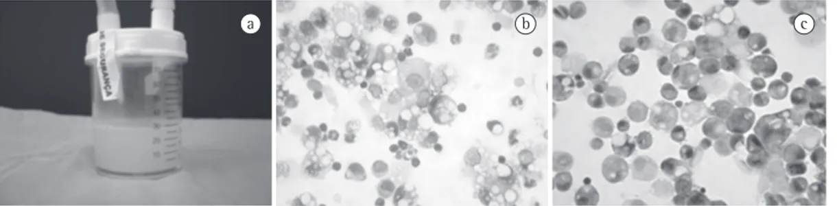
a b c
showed numerous macrophages with intracyto-plasmic vacuoles of various sizes, stained orange with Sudan staining (Figure 1). The BAL fluid cellularity ranged from 500 to 7,680 cells/mm3 (mean = 1,723 cells/mm3). The microbiological study was negative in all of the cases.
Of the 28 children investigated, 20 were treated with multiple BALs (mean = 9.35; vari-ation: 4-22) and became asymptomatic after the treatment; 18 children had normal CT scan results immediately after the treatment, 1 child presented a cystic image in the right lung and 1 child had a discrete area of segmented atel-ectasis 12 months after the treatment. Treatment abandonment occurred in 6 cases.
Discussion
The condition known as LP, lipid pneu-monia, oil pneupneu-monia, oil aspiration pneupneu-monia, lipoid cell pneumonia, pulmonary steatosis, pulmonary lipidosis or paraffin pneumonia is a chronic inflammation of the lung parenchyma with interstitial involvement due to the accu-mulation of oily material in the alveoli. This condition can be classified as exogenous LP, endogenous LP or idiopathic LP.(11) Exogenous LP—the most commonly described form of LP—is associated with the aspiration or inhalation of, principally, mineral oil, a substance used as a laxative in cases of intestinal constipation or as adjuvant treatment for partial bowel obstruc-tion by A. lumbricoides. Medications in which the oil acts as a vehicle—nasal drops, ointment, enema, contrast medium and industrialized lubricants—are also associated with LP.(9,12,13) Endogenous LP, which is less frequent, occurs due to the distal obstruction of the airways by malignant lesions, suppurative processes, oblit-erating bronchiolitis and diseases causing lipid deposition.(11) Idiopathic LP is rare, being associ-ated with smoking in healthy individuals.(14) In children, exogenous LP is the most common form of the disease, due to the aspiration of mineral oil, as shown in the present study.
Various pulmonary diseases, such as bacterial pneumonia, TB, cystic fibrosis, bronchiectasis and tumors, can be mimicked by LP.(11,15) None of the cases investigated in the present study were initially diagnosed as LP. All of the chil-dren we investigated had been treated with various antimicrobial treatment regimens; 9 of them had also initiated treatment for TB, which The lobes most affected were, in decreasing
order of frequency, the right upper lobe, the right lower lobe, the left lower lobe, the middle lobe and the left upper lobe. The lingula was affected in 5 cases. In 5 cases, the two lungs had been equally affected. In 16 cases, the density measurements within the consolidation
areas ranged between −11 and −118 HU. In spite
of the great improvement observed in the CT scan right after treatment, tomographic altera-tions were still observed in 6 children. These children were reevaluated after 6 months; 2 of them were once again submitted to HRCT after 12 months.
In all of the cases, the BAL fluid was opales-cent with a supernatant halo of fat. Cell analysis Figure 2 - HRCT scan of the chest, revealing extensive areas of consolidation with air bronchogram in a two-year-old child with lipoid pneumonia. This image was obtained before the treatment with multiple bronchoalveolar lavages.
rocyte sedimentation rate.(6,18) These alterations were observed in most of the children investi-gated in the present study, which probably led to the incorrect diagnosis of bacterial pneumonia.
In some cases, there was discordance between the clinical and radiographic findings. Children in whom the pulmonary auscultation findings were normal but chest X-rays showed extensive alterations were incorrectly diagnosed with pulmonary TB. In children, the radiological and tomographic alterations caused by TB vary greatly; such alterations can be similar to those caused by LP, and definitive diagnosis should be established before initiating a specific treatment for TB.(21) However, in cases of LP, there is no epidemiological history for TB, Koch’s bacillus cannot be identified in pulmonary granulomas, pulmonary involvement predominates in the lower lobes (preferential zones of aspiration), density measurements in the consolidation areas are negative and BAL fluid cytology reveals foamy alveolar macrophages with lipid vacuoles stained with Sudan.(5,16)
The radiological alterations of LP are nonspecific, generally involving multiple lobes and predominating in the posterior and infe-rior regions of the lungs.(5) In the present study, there was a predominance of consolidation, perihilar infiltrate (especially in the right lung) and hyperinflation. The principal tomographic alterations in LP are multifocal consolidation; linear and ground-glass interstitial opacities; thickening of intralobular septa; crazy-paving pattern; poorly-defined nodules; pleural effu-sion; and masses.(17,22) In the present study, the principal findings were consolidations with air bronchogram and few cases of ground-glass opacity. The crazy-paving pattern was seen only in 1 child. Such alterations were bilateral and multifocal, involving from three to five lobes and predominantly in the posterior regions of the right lung. Negative density values (between
−30 and −150 HU) within the consolidation area,
although nonspecific, can indicate the presence of fat and are frequently associated with LP, as shown in the present study.(23-25)
Currently, analysis of the BAL fluid is consid-ered the diagnostic method of choice for suspected cases of LP.(5) An opalescent macro-scopic aspect of the BAL fluid, with a halo of supernatant fat, is a strong indication of LP. However, only the cytochemical examination generates costs, increases the risk for the
devel-opment of resistant bacterial strains and delays diagnosis, as well as increasing morbidity and the risk of complications. In the present study, the diagnosis of LP was established, on average, 38 days after the onset of symptoms. The major risk factors observed were age (79% were suck-ling infants), gastroesophageal reflux (18%) and swallowing disorder (18%).
Mineral oil is an inert substance that is not metabolized by pulmonary enzymes when aspirated. Instead, it is emulsified and phago-cytosed by alveolar macrophages, returning to the alveolar space after cell death. The release of inflammatory cytokines by the activated macrophages probably leads to fever and to the presence of infection markers, which causes the misdiagnosis of LP as bacterial pneumonia.(16) The type and volume of oil aspirated, the time of permanence of such substance in the alveoli and individual defense mechanisms in the pulmo-nary microenvironment determine the various body responses. Initially, there is the develop-ment of a foreign body inflammatory response. Subsequently, with the permanence of the oil, there is the development of chronic interstitial inflammation, which can evolve to pulmonary fibrosis. In some cases, the coalescence of oil drops is slowly surrounded by fibrous tissue and giant cells, taking on a nodular or mass-like aspect, known as paraffinoma, more commonly observed in the adult population.(13,17) The radio-logical presentation of LP in the initial stage of alveolar involvement is consolidation; followed by the observation of interstitial infiltrate or mixed infiltrate (interstitial and alveolar); in cases of paraffinoma, a tumor lesion is observed.(17,18)
2. Meltzer E, Guranda L, Vassilenko L, Krupsky M, Steinlauf S, Sidi Y. Lipoid pneumonia: a preventable complication. Isr Med Assoc J. 2006;8(1):33-5.
3. Bandla HP, Davis SH, Hopkins NE. Lipoid pneumonia: a silent complication of mineral oil aspiration. Pediatrics. 1999;103(2):E19.
4. Annobil SH, el Tahir M, Kameswaran M, Morad N. Olive oil aspiration pneumonia (lipoid) in children. Trop Med Int Health. 1997;2(4):383-8.
5. Furuya ME, Martínez I, Zúñiga-Vásquez G, Hernández-Contreras I. Lipoid pneumonia in children: clinical and imagenological manifestations. Arch Med Res. 2000;31(1):42-7.
6. Castañeda-Ramos SA, Ramos-Solano F. Neumonia lipoídica exógena. Bol Med Hosp Infant Mex. 1989;46(9):597-602.
7. Riff EJ, Moore C, Tufenkeji H, Harfi H. Infantile lipoid pneumonia. Ann Saudi Med. 1990;10(4):378-82. 8. Hoffman LR, Yen EH, Kanne JP, Effmann EL, Gibson
RL, Van Niel CW. Lipoid pneumonia due to Mexican folk remedies: cultural barriers to diagnosis. Arch Pediatr Adolesc Med. 2005;159(11):1043-8.
9. de Oliveira GA, Del Caro SR, Bender Lamego CM, Merçon de Vargas PR, Vervloet VE. Radiographic plain film and CT findings in lipoid pneumonia in infants following aspiration of mineral oil used in the treatment of partial small bowel obstruction by Ascaris lumbricoides. Pediatr Radiol. 1985;15(3):157-60.
10. Tessier V, Chadelat K, Baculard A, Housset B, Clement A. BAL in children: a controlled study of differential cytology and cytokine expression profiles by alveolar cells in pediatric sarcoidosis. Chest. 1996;109(6):1430-8. 11. Spickard A 3rd, Hirschmann JV. Exogenous lipoid
pneumonia. Arch Intern Med. 1994;154(6):686-92. 12. Pujol JL, Barnéon G, Bousquet J, Michel FB, Godard P.
Interstitial pulmonary disease induced by occupational exposure to paraffin. Chest. 1990;97(1):234-6. 13. Aboudara M, Yun J. A case of fire-eater’s pneumonia in
an active-duty soldier. MedGenMed. 2006;8(2):67. 14. Sharma A, Ohri S, Bambery P, Singh S. Idiopathic
endogenous lipoid pneumonia. Indian J Chest Dis Allied Sci. 2006;48(2):143-5.
15. Balakrishnan S. Lipoid pneumonia in infants and children in South India. Br Med J. 1973;4(5888):329-31. 16. Mylonaki E, Voutsas V, Antoniou D, Papakosta D,
Kontakiotis T, Skordalaki A, et al. Hydrocarbon pneumonitis following liquid paraffin aspiration during a fire-eating performance: a case report. J Med Case Reports. 2008;2(1):214.
17. Lipinski JK, Weisbrod GL, Sanders DE. Exogenous lipoid pneumonitis: pulmonary patterns. AJR Am J Roentgenol. 1981;136(5):931-4.
18. Reyes de la Rocha S, Cunningham JC, Fox E. Lipoid pneumonia secondary to baby oil aspiration: a case report and review of the literature. Pediatr Emerg Care. 1985;1(2):74-80.
19. Weinstein M. Index of suspicion. Case 3. Lipoid pneumonia. Pediatr Rev. 2000;21(5):173, 176-7. 20. Simmons A, Rouf E, Whittle J. Not your typical
pneumonia: a case of exogenous lipoid pneumonia. J Gen Intern Med. 2007;22(11):1613-6.
21. Hugosson CO, Riff EJ, Moore CC, Akhtar M, Tufenkeji HT. Lipoid pneumonia in infants: a radiological-pathological study. Pediatr Radiol. 1991;21(3):193-7.
using Sudan staining, which stains orange the fat present in the extracellular medium and in the cytoplasmic vacuoles of macrophages, can confirm the diagnosis. All of the children included in the present study were diagnosed as having LP based on the BAL findings.
Although there is still no consensus regarding the treatment for LP, the interruption in the use of mineral oil leads to clinical improve-ment. The use of corticosteroids is controversial, since they are indicated principally in the most severe cases and in cases in which radiological and functional abnormalities persist in spite of a clinical improvement.(4,26,27) Depuration of the inhaled oil is a slow process, and the perma-nence of this substance in the lung parenchyma leads to inflammation and fibrosis.(11,17,28) Therefore, the best therapeutic strategy would be to remove the oil as early as possible through bronchoscopy with multiple BALs, especially in the segments most severely affected, therefore reducing the need for corticosteroids and their side effects.(25) In the present study, 2 children were treated with corticosteroids and multiple BALs. The result was clinical and tomographic normalization after 12 months of monitoring (Figures 2 and 3). A recent study involving chil-dren with LP caused by aspiration of mineral oil demonstrated the efficacy of the treatment with multiple BALs, with clinical and tomographic resolution.(25)
Cases of chronic pneumonia refractory to antimicrobial therapy, combined with a history of ingestion of mineral oil and the presence of condensation in lower and posterior regions of the lungs, with negative density measure-ments, should raise the clinical suspicion of LP. However, the diagnosis should be established based on the findings of the BAL, which can also be used as a therapeutic measure. Because health professionals are not aware of the risks of using mineral oil, especially for the very young and the elderly, and because of the indiscrimi-nate use of mineral oil, which is commercialized without directions for use and without the need for a medical prescription, LP continues to be underdiagnosed in Brazil.
References
of lipoid pneumonia with multiple bronchoalveolar lavages. Pediatr Pulmonol. 2009;44(4):309-15. 26. Fan LL, Graham LM. Radiological cases of the month.
Lipoid pneumonia from mineral oil aspiration. Arch Pediatr Adolesc Med. 1994;148(2):205-6.
27. Ciravegna B, Sacco O, Moroni C, Silvestri M, Pallecchi A, Loy A, et al. Mineral oil lipoid pneumonia in a child with anoxic encephalopathy: treatment by whole lung lavage. Pediatr Pulmonol. 1997;23(3):233-7.
28. Midulla F, Strappini PM, Ascoli V, Villa MP, Indinnimeo L, Falasca C, et al. Bronchoalveolar lavage cell analysis in a child with chronic lipid pneumonia. Eur Respir J. 1998;11(1):239-42.
22. Lee KH, Kim WS, Cheon JE, Seo JB, Kim IO, Yeon KM. Squalene aspiration pneumonia in children: radiographic and CT findings as the first clue to diagnosis. Pediatr Radiol. 2005;35(6):619-23.
23. Agarwal R. Low-attenuation consolidation - the most characteristic finding in lipoid pneumonia. Eur J Intern Med. 2006;17(4):307.
24. Zanetti G, Marchiori E, Gasparetto TD, Escuissato DL, Soares Souza A Jr. Lipoid pneumonia in children following aspiration of mineral oil used in the treatment of constipation: high-resolution CT findings in 17 patients. Pediatr Radiol. 2007;37(11):1135-9.
25. Sias SM, Daltro PA, Marchiori E, Ferreira AS, Caetano RL, Silva CS, et al. Clinic and radiological improvement
About the authors
Selma Maria de Azevedo Sias
Adjunct Professor of Pediatrics. Fluminense Federal University, Niterói, Brazil.
Angela Santos Ferreira
Professor of Pulmonology. Occupational Pulmonary Diseases Outpatient Clinic of the Antônio Pedro University Hospital, Fluminense Federal University, Niterói, Brazil.
Pedro Augusto Daltro
Assistant Professor. Department of Education, Fernandes Figueira Institute, Fundação Oswaldo Cruz – FIOCRUZ, Oswaldo Cruz Foundation – Rio de Janeiro, Brazil.
Regina Lúcia Caetano
Pharmacist. Department of Cellular and Molecular Biology, Institute of Biology, Fluminense Federal University, Niterói, Brazil.
José da Silva Moreira
Associate Professor. Postgraduate Program in Pulmonology, Federal University of Rio Grande do Sul, Porto Alegre, Brazil.
Thereza Quirico-Santos