ULTRASOUND FINDINGS IN THYROIDITIS*
Ilka Yamashiro1, Osmar de Cássio Saito2, Maria Cristina Chammas3, Giovanni Guido Cerri4
OBJECTIVE: The aim of this study was to evaluate sonographic features of thyroid gland in patients with thyroiditis. MATERIALS AND METHODS: During a nine-month period, 38 patients previously diagnosed with thyroiditis by the Ultrasound Unit of “Instituto de Radiologia da Faculdade de Medicina da Universidade de São Paulo”, São Paulo, SP, Brazil, were studied. Thirteen-six of these patients were women in the age range between 17 and 78 years. The following variables were observed and studied: thyroid volume, echogenicity, echotexture, and the presence of level VI chain lymph nodes (pre-tracheal, the preferential site of thyroid drainage), besides the gland dimensions. RESULTS: Thirteen patients had goiter; 37 of 38 thyroid glands presented heterogeneous echotexture and diffuse hypoechogenicity. Thyroiditis was diagnosed by labora-tory tests in all patients. Level VI chain (pre-tracheal) lymph nodes were observed in 28 cases, all of them with reactional features. Of these cases, ten patients were submitted to fine needle aspiration biopsy, and the cytological result indicated inflammatory reaction. No case of nodule-simulating focal thyroiditis was found. CONCLUSION: The thyroid gland sonographic hypoechogenicity and heterogeneity, in association with the presence of cervical chain lymph nodes, constitute findings of great importance in the diagnosis of thyroiditis when correlated with clinical and laboratory tests findings.
Keywords: Ultrasound; Thyroid; Thyroiditis.
Achados ultra-sonográficos na tireoidite.
OBJETIVO: Avaliar os aspectos ultra-sonográficos da glândula tireóide em pacientes portadores de tireoidi-tes. MATERIAIS E MÉTODOS: Num período de nove meses, foram estudados 38 pacientes atendidos no Serviço de Ultra-sonografia do Instituto de Radiologia do Hospital das Clínicas da Faculdade de Medicina da Universidade de São Paulo, com diagnóstico prévio de tireoidite. Trinta e seis deles eram do sexo feminino, cujas idades variaram entre 17 e 78 anos. As variáveis observadas e estudadas foram: o volume glandular, a ecogenicidade e a ecotextura, e a presença de linfonodos na cadeia VI cervical (pré-traqueal, o sítio de drenagem linfática preferencial da tireóide) e suas dimensões. RESULTADOS: Treze pacientes apresenta-vam bócio. A análise ao ultra-som mostrou que 37 das 38 glândulas apresentaapresenta-vam ecotextura heterogênea com hipoecogenicidade difusa. Todos os pacientes apresentavam alterações nos exames laboratoriais com-patíveis com tireoidites. Foram encontrados linfonodos na cadeia cervical VI (pré-traqueal) em 28 pacientes, todos de aspecto reacional. Destes, dez foram submetidos a punção aspirativa por agulha fina e o resultado citológico foi de reação inflamatória. Não foram encontrados casos de tireoidites focais que pudessem simu-lar nódulos. CONCLUSÃO: Podemos inferir que para auxiliar no diagnóstico das tireoidites os achados ultra-sonográficos de heterogeneidade e hipoecogenicidade glandular, associados aos linfonodos na cadeia cervi-cal VI, são de grande importância quando correlacionados aos exames clínicos e laboratoriais.
Unitermos: Ultra-som; Tireóide; Tireoidite. Abstract
Resumo
* Study developed in the Ultrasound Unit of Instituto de Ra-diologia do Hospital das Clínicas da Universidade de São Paulo (InRad/HC-FMUSP), São Paulo, SP, Brazil.
1. MD, Radiologist, Master in Health Sciences by Hospital Heliópolis Post-Graduation Course, São Paulo, SP, Professional-izing Practice at Setor de Ultra-sonografia do Instituto de Radio-logia do Hospital das Clínicas da Faculdade de Medicina da Universidade de São Paulo (InRad/HC-FMUSP), São Paulo, SP, Brazil.
2. MD, Radiologist, PhD, Attending Physician at Instituto de Radiologia do Hospital das Clínicas da Faculdade de Medicina da Universidade de São Paulo (InRad/HC-FMUSP) Ultrasound Unit, São Paulo, SP, Brazil.
3. MD, Radiologist, PhD, Director for the Instituto de Radiolo-gia do Hospital das Clínicas da Faculdade de Medicina da Uni-versidade de São Paulo (InRad/HC-FMUSP) Ultrasound Service, São Paulo, SP, Brazil.
4. Titular Professor at Faculdade de Medicina da Universi-dade de São Paulo (FMUSP) Department of Radiology, Head of Instituto de Radiologia do Hospital das Clínicas da Faculdade de Medicina da Universidade de São Paulo (InRad/HC-FMUSP), São Paulo, SP, Brazil.
Mailing address: Dra. Ilka Yamashiro. Rua Afonso Celso, 1637, ap. 33, Bairro Chácara Inglesa. São Paulo, SP, Brazil, 04119-062. E-mail: [email protected]
Received May 31, 2006. Accepted after revision July 17, 2006.
INTRODUCTION
Thyroiditis is a generic term including an array of clinical entities affecting the thyroid gland.
The thyroiditis may present infectious, autoimmune, traumatic, post-radiotherapy, drugs use, and other etiopathogenies. On the grounds of etiopathogeny and clinical findings, eight diseases present with thy-roiditis picture: 1) Hashimoto thythy-roiditis; subacute lymphocytic thyroiditis; 3) sub-acute granulomatous thyroiditis (De Quervain’s disease); 4) postpartum thy-roiditis; 5) infectious thyroiditis 6) toxic thyroiditis; 7) actinic thyroiditis; 8) Riedel’s thyroiditis.
Subacute and chronic thyroiditis refer to inflammation of the thyroid gland paren-chyma where serum thyroglobulin, T3 and T4 levels increase. For this reason, during the initial phase of the disease, the thyroidi-tis progresses with thyrotoxicosis. After the first weeks, the damages to follicular cells inhibit the hormonal synthesis, resulting in hypothyroidism. In this phase, subacute thyroiditis is a self-limited condition and the gland tissue regenerates. On the other hand in the chronic, autoimmune thyroidi-tis (Hashimoto) this phase is continuous and progresses to hypothyroidism.
MATERIALS AND METHODS
During a nine-month period, 38 patients previously diagnosed with thyroiditis by the Ultrasound Unit of Instituto de Radio-logia do Hospital das Clínicas da Universi-dade de São Paulo, São Paulo, SP, Brazil, were studied. Of these patients, 36 were female, with ages ranging between 17 and 78 years (mean age 47.5 years).
The following variables were observed and studied: thyroid volume, echogenicity, echotexture, and the presence of level VI chain lymph nodes (pre-tracheal, the pref-erential site of thyroid drainage), besides the gland dimensions.
RESULTS
In our casuistic, the greatest majority of patients (36; 94.7%) were female. Thirteen patients (34.2%) presented with goiter (glandular volume > 14.4 cm³). Ultrasound analysis showed heterogeneous echotex-ture with diffuse hypoechogenicity in 37 (97.3%) of the 38 thyroid glands (Table 1). All the patients presented alterations compatible with thyroiditis on laboratory tests.
Causal factor or factors could not be determined because of the absence of pre-vious studies performed in our Service. Also, it is important to note that ultrasound findings in thyroiditis, especially in the focal ones, may overlap with those seen in other diseases.
Cervical chain lymph nodes were found at level VI (pre-tracheal) in 28 patients (73.6%), all of them presenting reactional features (ovoid, with hyperechogenic cen-tral hilum). Of these 28 cases, ten patients were submitted to fine needle aspiration biopsy, and the cytological result indicated inflammatory reaction. No case of nodule-simulating focal thyroiditis was found.
DISCUSSION
The diagnosis of thyroiditis is not al-ways easy and immediate, considering that in the initial phases of the disease specific laboratory tests alterations are not detect-able. With the progress of imaging meth-ods, some features such as thyroid gland size, shape and echogenicity can be earlier
and better evaluated. Consequently, ultra-sonography has been useful for evaluating the thyroid gland, especially in dubious cases where morphological and functional characteristics may suggest the actual etiopathogeny of the disease on color Dop-pler mapping. Some of these characteristics are widely recognized in the worldwide lit-erature.
Hashimoto thyroiditis is the most fre-quent cause of permanent hypothyroidism
in areas of adequate iodine intake, corre-sponding to more than 90% of cases. It is part of the spectrum of autoimmune thyroid diseases, typically affecting older women, five to eight times more frequently than men, with an approximate 15% prevalence in this latest group(1,2). At ultrasound a
het-erogeneous goiter with micronodules scat-tered throughout the parenchyma. Progres-sively, the thyroid gland presents an aspect of chronic hypertrophic thyroiditis with
Table 1 Contingency table.
Patient 1 2 3 4 5 6 7 8 9 10 11 12 13 14 15 16 17 18 19 20 21 22 23 24 25 26 27 28 29 30 31 32 33 34 35 36 37 38 Age (years) 30 36 50 17 32 37 32 40 21 23 20 78 47 70 27 57 27 26 31 40 19 44 44 48 27 26 46 46 54 28 39 37 28 28 23 54 32 34 Glandular volume (cc) 14.0 17.3 13.9 72.4 20.9 23.0 9.1 14.4 13.0 5.9 7.1 11.8 9.8 6.6 6.1 7.5 11.0 16.7 7.7 28.8 10.0 10.7 7.9 5.1 16.7 10.7 27.5 19.6 12.0 13.2 4.7 15.5 18.4 16.3 8.9 11.2 11.7 16.0 Echogenicity Hypoechogenic Hypoechogenic Hypoechogenic Hypoechogenic Hypoechogenic Hypoechogenic Hypoechogenic Hypoechogenic Hypoechogenic Hypoechogenic Hypoechogenic Hypoechogenic Hypoechogenic Hypoechogenic Hypoechogenic Hypoechogenic Hypoechogenic Hypoechogenic Hypoechogenic Hypoechogenic Hypoechogenic Hypoechogenic Hypoechogenic Hypoechogenic Hypoechogenic Hypoechogenic Hypoechogenic Hypoechogenic Hypoechogenic Hypoechogenic Hypoechogenic Hypoechogenic Hypoechogenic Hypoechogenic Hypoechogenic Hypoechogenic Echogenic Hypoechogenic Echotexture Heterogeneous Heterogeneous Heterogeneous Heterogeneous Heterogeneous Heterogeneous Heterogeneous Heterogeneous Heterogeneous Heterogeneous Heterogeneous Heterogeneous Heterogeneous Heterogeneous Heterogeneous Heterogeneous Heterogeneous Heterogeneous Heterogeneous Heterogeneous Heterogeneous Heterogeneous Homogeneous Heterogeneous Heterogeneous Heterogeneous Heterogeneous Heterogeneous Heterogeneous Heterogeneous Heterogeneous Heterogeneous Heterogeneous Heterogeneous Heterogeneous Heterogeneous Heterogeneous Heterogeneous Lymph node level Level VI Level VI Level VI Level VI Level VI Level VI Level VI Level VI Level VI Level VI Level VI Level VI Level VI Level VI Level VI Level VI Level VI Level VI Level VI Level VI Level VI Level VI Level VI Level VI Lymph node dimension (cm)
5 × 5 × 3 3 × 3 × 4 14 × 6 × 4 7 × 6 × 4 9 × 5 × 4 11 × 9 × 4 7 × 5 × 5
4 × 4 × 3 8 × 3 × 2
7 × 7 × 2
5 × 5 × 5
18 × 9 × 5 4 × 3 × 2 8 × 7 × 3 10 × 5 × 3
7 × 7 × 3
7 × 5 × 3 11 × 6 × 5 5 × 4 × 2
6 × 4 × 3 8 × 5 × 3
12 × 5 × 4
poorly defined hypoechoic areas permeated by fibrous echogenic layers, giving the gland a pseudolobular appearance (Figure 1). Doppler mapping shows a diffuse hypervascularization pattern similar to Graves’ disease (Figure 2). In the later stages of the disease color Doppler shows the thy-roid gland reduced in size diffusely hetero-geneous due the intense fibrosis and avascularized.
Subacute lymphocytic thyroiditis is also an autoimmune, painless disease character-ized by transitory thyrotoxicosis, with lym-phocytic infiltrates, similar to the Hashi-moto thyroiditis (Figure 3)(1,2).
Subacute granulomatous thyroiditis (De Quervain’s) also manifests with
thyrotoxi-cosis, cervicoalgia, most frequently affect-ing women at a 3:1–5:1 ratio. It is a reac-tional, post-viral disease (Cocksackie, ad-enovirus, mumps, measles, etc.)(1,3,4). In the
initial phase of this disease, ultrasound shows hypoechogenic areas with irregular, poorly defined margins, especially in the subcapsular zones (Figure 4). Later, pseudonodules may be observed in the cen-tral region of the thyroid gland. In the early phases, vascularization may be reduced (Figure 5).
Postpartum thyroiditis is also an au-toimmune process that may occur in the first year after delivery, most frequently between the second and fourth month, af-fecting up to 7% of women. Recurrence
rates in subsequent gestations are high(5,6).
At ultrasound a diffuse thyroid gland hypoechogenicity is observed, with even more hypoechogenic focal areas (Figure 6). Other types of thyroiditis occur less fre-quently, and the infectious ones usually are caused by bacteria(5); in cases of
immuno-depressed patients, fungi and tubercle ba-cillus should be considered as etiological agents. B-mode ultrasound shows poorly defined, hypoechogenic foci, probably cor-responding to formation of intraparen-chymal abscesses. Thyroiditis also can be associated with the use of interferon-2, interleukin and amiodarone(7–9).
Riedel’s thyroiditis is a rare chronic form of thyroiditis where fibrous tissue replaces the normal thyroid tissue, causing hardening and diffuse destruction of the gland, and tending to extend to the adjacent tissues. This disease may result in dyspnea and dysphagia, and an association exists between Riedel’s thyroiditis and retroperi-toneal fibrosis, mediastinal fibrosis, pul-monary fibrosis, sclerosing cholangitis, pseudo-tumor of the orbit, etc.(1,6). At
ultra-sound, the thyroid gland is shown as a hypoechogenic mass infiltrating into the adjacent musculature.
In the greatest majority of cases, the diagnosis is based on clinical findings and laboratory tests results, and ultrasound is an important tool in the morphological evalu-ation of the thyroid gland, corroborating the diagnosis, and also providing additional information regarding the disease extent
Figure 2. Doppler mapping showing a diffuse hypervascularization pattern.
RIGHT LOBE
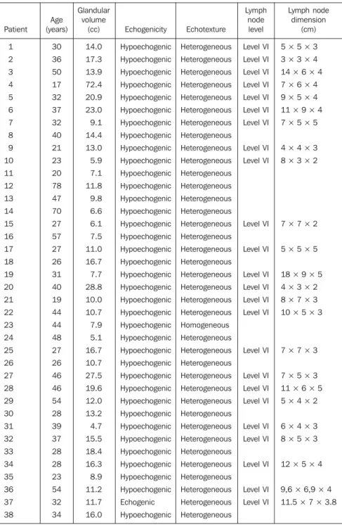
Figure 3. Heterogeneous thyroid gland with poorly defined hypoechogenic areas, and slightly lobular margins.
RIGHT LOBE
Figure 4. De Quervain’s thyroiditis. Hypoechogenic areas with poorly defined and irregular contours are observed, especially in subcapsular areas.
Figure 6. Postpartum thyroiditis. Note the diffuse thyroid gland hypoecho-genicity permeated by even more hypoechogenic areas.
Figure 7. Lymph nodes with reactional appearance (arrows) with loss of echo-genic central hilum in the pretracheal chain, confirmed by fine needle aspira-tion biopsy.
A B
Figure 5. Hypoechogenic areas with poorly defined and irregular contours are observed, with pseudo-nodules in the central region of hypovascularized thyroid gland.
and mainly regarding cervical chain lymph node involvement (level VI, pre-tracheal, the preferential site of thyroid drainage)(10).
Although these lymph nodes are not men-tioned in current studies, they may be con-fused with thyroid isthmus nodules. Fine needle aspiration biopsy demonstrated that these lymph nodes were of reactional in-flammatory origin (Figure 7).
Our casuistic matches the literature, the majority of our patients being female, with 97.3% of the thyroids evaluated presenting diffusely hypoechogenic and heteroge-neous at examination (with poorly defined, hypoechogenic foci and permeated with echogenic layers), as well as the presence of cervical chain lymph nodes in 73.6% of
cases. However, only 34.2% of the patients presented with goiter in the moment of ex-amination.
It is important to note that patients must be carefully evaluated in order to avoid that the presence of any nodule is missed in the middle of a heterogeneous parenchyma, since 20% of patients with Hashimoto thy-roiditis may develop malignant nodules or thyroid lymphomas(11). Lymph nodes
evaluation, especially by means of B-mode morphological imaging is also relevant, since these lymph nodes may correspond to previously unknown malignant pro-cesses(12). About 10%–15% of Hashimoto
thyroiditis course with lymphadenomegaly of questionable nature(9).
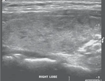
THYROID
RIGHT LOBE LEF T LOBE
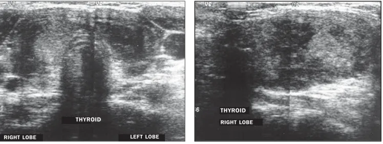
THYROID
RIGHT LOBE
LEF T LOBE
CONCLUSION
One may conclude that ultrasound find-ings of glandular heterogeneity and hypo-echogenicity, associated with cervical chain (level VI) lymph nodes, are extremely useful in the diagnosis of thyroiditis when correlated with laboratory and clinical tests.
REFERENCES
1. Goldman L, Bennet JC. Cecil Textbook of medi-cine. 21st ed. Philadelphia: WB Saunders, 2000. 2. Lazarus JH. Silent thyroiditis and subacute thy-roiditis. In: Braverman LE, Utiger RD, editors. The thyroid: a fundamental and clinical text. 7th ed. Philadelphia Lippincott-Raven, 1996;577–591. 3. Basgoz N, Swartz MN. Infections of the thyroid gland. In: Braverman LE, Utiger RD, editors. The thyroid: a fundamental and clinical text. 7th ed.
Philadelphia: Lippincott-Raven, 1996;1049– 1056.
4. Solbiati L, Charboneau JW, James EM, Hay ID. The thyroid gland. In: Rumack CM, Wilson SR, Charboneau JW, editors. Diagnostic ultrasound. 2nd ed. St. Louis: Mosby, 1998;703–729. 5. Bokhari R, Bhatara VS, Bandettini F, McMillin
JM. Postpartum psychosis and postpartum thy-roiditis. Psychoneuroendocrinology 1998;23: 643–650.
6. Chammas MC, Saito OC, Cerri GG. Tireóide. In: Saito OC, Cerri GG, organizadores. Ultra sono-grafia de pequenas partes. 2ª ed. Rio de Janeiro: Revinter, 2004;75–114.
7. Preziati D, La Rosa L, Covini G, et al. Autoim-munity and thyroid function in patients with chronic active hepatitis treated with recombinant interferon alpha-2a. Eur J Endocrinol 1995;132: 587–593.
8. Roti E, Minelli R, Giuberti T, et al. Multiple changes in thyroid function in patients with
chronic active HCV hepatitis treated with recom-binant interferon-alpha. Am J Med 1996;101: 482–487.
9. Roti E, Minelli R, Gardini E, Bianconi L, Braver-man LE. Thyrotoxicosis followed by hypothyroid-ism in patients treated with amiodarone. A pos-sible consequence of a destructive process in the thyroid. Arch Intern Med 1993;153:886–892. 10. Som PM, Brandwein. Lymph nodes. In: Som PM,
Curtin HD, editors. Head and neck imaging. 4th ed. St. Louis: Mosby, 2003;1880–1883. 11. Takashima S, Takayama F, Wang Q, Kobayashi
S, Sone S. Thyroid metastasis from rectal carci-noma coexisting with Hashimoto’s thyroiditis: gray-scale and power Doppler sonographic find-ings. J Clin Ultrasound 1998;26:361–365. 12. Senchenkov A, Staren ED. Ultrasound in head