PREDICTIVE VALUES OF BI-RADS CATEGORIES 3, 4 AND 5 IN
NON-PALPABLE BREAST MASSES EVALUATED BY MAMMOGRAPHY,
ULTRASOUND AND MAGNETIC RESONANCE IMAGING*
Decio Roveda Junior1
,Sebastião Piato2
, Vilmar Marques de Oliveira3
,José Francisco Rinaldi4 , Carlos Alberto Pecci Ferreira5
,Eduardo de Castro Faria Fleury6
OBJECTIVE: To evaluate the predictive value of BI-RADS™ categories 3, 4 and 5 in non-palpable breast masses assessed by mammography, ultrasound and magnetic resonance imaging. MATERIALS AND METHODS: Twenty-nine patients with BI-RADS categories 3, 4 and 5 non-palpable breast masses identified by mammograms were submitted to complementary ultrasound and magnetic resonance imaging studies, be-sides excisional biopsy. In total, 30 biopsies were performed. The lesions as well as their respective BI-RADS classification into 3, 4 and 5 were correlated with the histopathological results. The predictive values calcu-lation was made by means of specific mathematical equations. RESULTS: Negative predictive values for category 3 were: mammography, 69.23%; ultrasound, 70.58%; and magnetic resonance imaging, 100%. Positive predictive values for category 4 were: mammography, 63.63%; ultrasound, 50%; and magnetic resonance imaging, 30.76%. For category 5, positive predictive values were: mammography and ultrasound, 100%; and magnetic resonance imaging, 92.85%. CONCLUSION: For category 3, the negative predictive value of magnetic resonance imaging was high, and for categories 4 and 5, the positive predictive values of the three modalities were moderate.
Keywords: BI-RADS; Breast cancer; Mammography; Predictive value.
Valores preditivos das categorias 3, 4 e 5 do sistema BI-RADS em lesões mamárias nodulares não-palpáveis avaliadas por mamografia, ultra-sonografia e ressonância magnética.
OBJETIVO: Avaliar os valores preditivos positivo e negativo das categorias 3, 4 e 5 do sistema BI-RADS™ em lesões mamárias nodulares não-palpáveis avaliadas por mamografia, ultra-sonografia e ressonância mag-nética. MATERIAIS E MÉTODOS: Vinte e nove pacientes com achados mamográficos de lesões mamárias nodulares não-palpáveis, das classes 3, 4 e 5 do BI-RADS, que realizaram exames complementares de ultra-sonografia e ressonância magnética, além de biópsia excisional. Realizaram-se 30 biópsias e correlaciona-ram-se as lesões e suas respectivas classificações de 3 a 5 do BI-RADS com os resultados histopatológicos. O cálculo dos valores preditivos foi feito utilizando-se equações matemáticas específicas. RESULTADOS: O valor preditivo negativo da categoria 3 pela análise mamográfica foi de 69,23%, pela análise ultra-sonográ-fica foi de 70,58% e pela análise por ressonância magnética foi de 100%. O valor preditivo positivo da categoria 4 pela análise mamográfica foi de 63,63%, pela análise ultra-sonográfica foi de 50% e pela análise por resso-nância magnética foi de 30,76%. O valor preditivo positivo da categoria 5 foi de 100% pelas análises ma-mográfica e ultra-sonográfica e de 92,85% pela análise por ressonância magnética. CONCLUSÃO: O valor preditivo negativo da categoria 3 foi elevado na análise pela ressonância magnética e os valores preditivos positivos foram moderados na categoria 4 e elevados na categoria 5 pelos três métodos.
Unitermos: BI-RADS; Câncer mamário; Mamografia; Valor preditivo. Abstract
Resumo
* Study developed in the Department of Clinical Medicine and Department of Obstetrics & Gynecology of Faculdade de Ciências Médicas da Santa Casa de São Paulo, São Paulo, SP, Brazil.
1. Professor Instructor, Director for the Service of Diagnostic Imaging of the Department of Clinical Medicine of Faculdade de Ciências Médicas da Santa Casa de São Paulo, São Paulo, SP, Brazil.
2. Titular Professor, Head for the Gynecology Clinic – Depart-ment of Gynecology & Obstetrics of Irmandade da Santa Casa de Misericórdia de São Paulo, São Paulo, SP, Brazil.
3. Professor Instructor, Head for General Gynecology – Depart-ment of Gynecology & Obstetrics of Irmandade da Santa Casa de Misericórdia de São Paulo, São Paulo, SP, Brazil.
4. Assistant Professor, Head for the Mastology Division – De-partment of Gynecology & Obstetrics of Irmandade da Santa Casa de Misericórdia de São Paulo, São Paulo, SP, Brazil.
5. MD, Attending Physician, Coordinator for the Service of Breast Imaginology at Department of Clinical Medicine of Facul-dade de Ciências Médicas da Santa Casa de São Paulo, São Paulo, SP, Brazil.
INTRODUCTION
It is unquestionable that the programs of mammographic breast cancer screening have caused a significant decrease in the mortality by this disease thanks to the early diagnosis in a considerable number of
cases, as evidenced by several clinical in-vestigations(1–6).
However, the mammographic screening started being complemented by a great number of unnecessary biopsies, since a considerable part of lesions considered as suspect of malignancy have been found to be benign. Of 1,000,000 women submitted to breast biopsy in the USA as a result of abnormal mammographic findings in breast cancer screening programs, 700,000 to 850,000 presented negative results(7).
Aiming at improving the effectiveness of breast cancer screening programs, with
6. Attending Physician at the Service of Diagnostic Imaging at Department of Clinical Medicine of Faculdade de Ciências Médicas da Santa Casa de São Paulo, São Paulo, SP, Brazil.
Mailing address: Prof. Dr. Decio Roveda Junior. Rua Alves Guimarães, 1185, ap. 43. São Paulo, SP, Brazil, 05410 002. E-mail: [email protected]
an increase in quality of reports issued by radiologists; and recognizing the necessity of providing meaningful and unambiguous reports to allow a reliable data acquisition, the American College of Radiology, in a collaborative effort with the American Cancer Institute and American College of Surgeons, in 1992 developed a system for not only classifying mammographic im-ages, but also for structuring reports by means of lesions description and standard-ization of conclusions, and suggesting a course of action to be adopted depending on the final findings classification.
The result of such collaborative effort is the Breast Imaging Reporting and Data System (BI-RADS™), contemplating not only a classification of outcomes, but also the recommendation of a specific course of actions which, if adopted, will allow a higher efficacy of programs for early breast cancer detection. The system includes an introduction, a breast imaging lexicon and a reporting standardization and diseases coding system, besides a reliable method-ology for outcomes monitoring and follow-up(8).
Based on a descriptive lexicon of radio-logical lesions, the system classifies the findings into seven categories, aiming at facilitating the decision making on a spe-cific course of action by physicians in face of abnormal image findings. On its fourth and latest issue released in December 2003,
the BI-RADS Atlas, formerly restricted to the area of mammography, extends the standardization to the areas of ultrasonog-raphy and magnetic resonance imaging(8). The BI-RADS introduction raised the radiologists and breast specialists concern about the predictive values of categories 3, 4 and 5, aiming at improving the manage-ment of abnormal, non-palpable findings.
BI-RADS classification
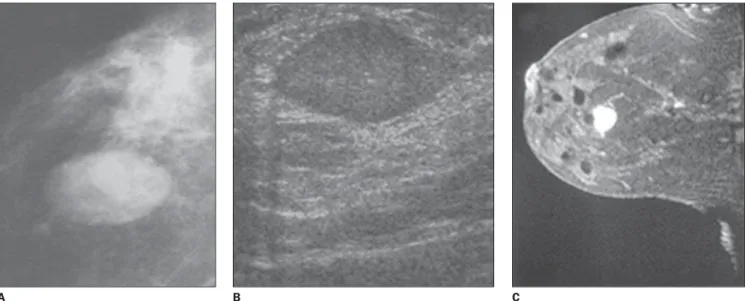
Category 3 –A finding in this category presents a high probability of benignity. However, considering a very low possibil-ity of malignancy, a short interval follow-up is recommended for evaluation of the lesion stability (Figure 1).
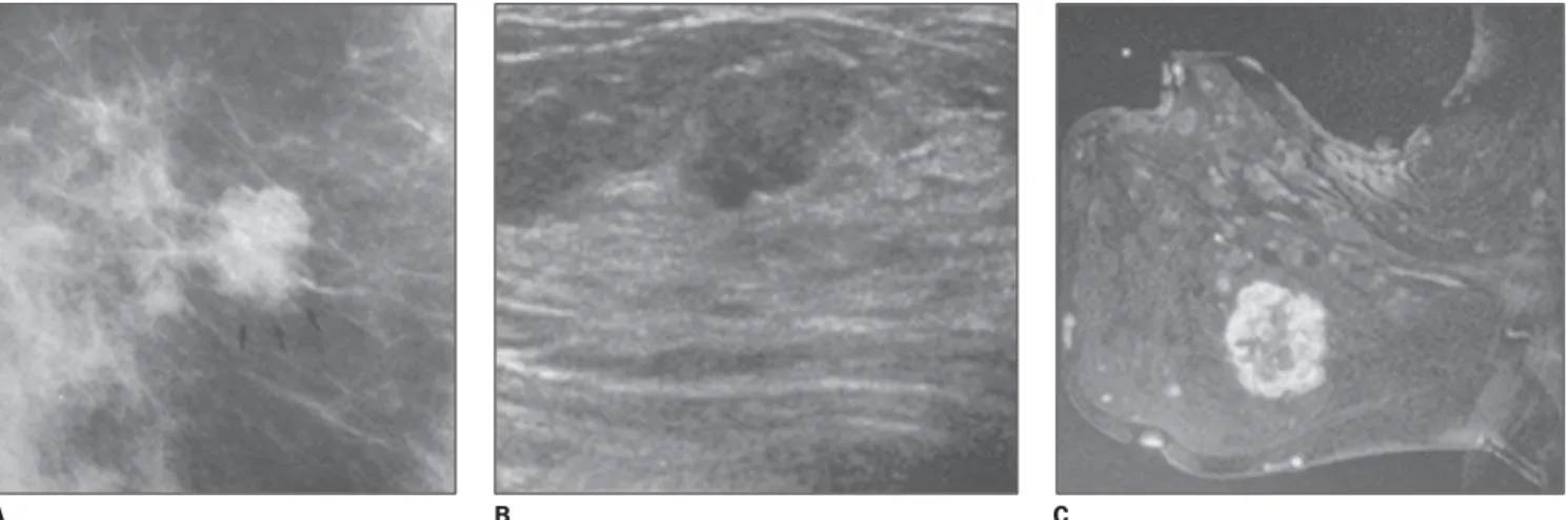
Category 4 – The lesions do not present any morphological characteristics typical of cancer, although with high probability of malignancy. The images raise sufficient concern to suggest a biopsy (Figure 2).
Category 5 – Lesions with morphologi-cal characteristics highly suggestive of ma-lignancy (Figure 3).
A review of the literature regarding the predictive values of BI-RADS categories 3, 4 and 5 has demonstrated the inexistence of studies on mammography exclusively related to non-palpable breast masses.
As regards breast ultrasound, Hong et al.(9) have studied 403 solid breast lesions, aiming at determining the positive predic-tive value (PPV) and negapredic-tive predicpredic-tive
value (NPV) of these findings, according to echographic characteristics and respec-tive histological diagnoses described in the new BI-RADS lexicon. They have found 141 (35%) positive cases with characteris-tics described by BI-RADS as malignant demonstrating high PPV. Solid lesions with spiculated margins presented 86% PPV (19 of 22); irregular lesions, 62% PPV (102 of 164); lesions with a non-parallel orienta-tion in relaorienta-tion to the costal grid, 69% PPV (75 of 109). As regards NPV, high values also have been observed for findings de-scribed by BI-RADS, such as circum-scribed margins in 90% (160 of 178), par-allel orientation in relation to the costal grid in 78% (228 of 294), and oval shape in 84% (200 of 237). These results show that the characteristics described in the new BI-RADS sonographic lexicon may be useful for differentiating between malignant and benign solid lesions.
Gokalp and Topal(10) have developed a study aiming at analyzing magnetic reso-nance imaging as method for evaluating supposedly benign lesions classified as BI-RADS category 3. They have studied 56 lesions present in 43 female patients, com-paring the studies with the respective his-tological results, and calculating sensitiv-ity, specificity and predictive values. The values found for lesions classified as prob-ably benign were: 100% for sensitivity, 94.6% for specificity, 33.3% for PPV, and
Figure 1. Examples of nodules classified as BI-RADS category 3. A: Mammographic image of an isodense, well-circumscribed ovoid nodule. B: Sonographic image of hypoechoic, well-circumscribed ovoid nodule, presenting parallel orientation and without posterior acoustic shadowing. C: Magnetic resonance image showing homogeneously contrast-enhanced round, well-circumscribed nodule.
B
100% for NPV, allowing us to conclude that the system may be useful in a conser-vative management of alterations classified as BI-RADS category 3.
A similar study developed by Sadowski and Kelcz(11)aimed at determining the chance of malignancy for breast lesions classified as probably benign, has retro-spectively evaluated 473 patients submit-ted to magnetic resonance imaging in the period between March 1994 and March 2002, and observed that 17% (79 of 473) were classified as probably benign. Of this group, 68 patients were followed-up dur-ing a minimum two-year period, and 6% (4 of 68) presented breast cancer in 14 to 18 months subsequent to the initial assess-ment. This study has led us to the conclu-sion that patients evaluated by means of magnetic resonance imaging and classified as BI-RADS category 3 are at higher risk
for breast cancer than those evaluated by mammography in the same category.
The present study was aimed at evalu-ating the PPV and NPV of BI-RADS cat-egories 3, 4 and 5 in non-palpable breast masses evaluated by mammography, ultra-sound and magnetic resonance imaging.
MATERIALS AND METHODS
Twenty-nine dossiers of patients with mammographic findings of nodular lesions in BI-RADS categories 3, 4 and 5 were evaluated. One of the patients presented with findings in both breasts, so the num-ber of lesions increased to 30. The patients also had their lesions evaluated by ultra-sound and magnetic resonance imaging.
The following exclusion criteria were taken into consideration for the casuistic selection: 1) abnormal mammographic
findings visualized on a single view; 2) abnormal findings with superficial or retroareolar localization; 3) patients with findings classified as BI-RADS categories 0, 1, 2 and 6 on complementary ultrasound and magnetic resonance imaging; 4) pa-tients previously submitted to radiotherapy, chemotherapy or hormone therapy.
The mammographic examinations were performed in a Philips M 3000 model equip-ment, with 0.1 and 0.3 mm microfocus, molybdenum anode, rhodium filter and automatic exposure meter.
All the patients were submitted to bilat-eral examination on craniocaudal and mediolateral oblique, 25° angle views; ad-ditional views with spot-compression and image magnification were made as neces-sary.
The images were analyzed in a dark room by means of a 4-compartment
negato-Figure 2. Examples of nodules classified as BI-RADS category 4. A: Mammographic image showing an isodense, irregular nodule with microlobulated margins.
B: Sonographic image showing hypoechoic, ovoid nodule with microlobulated margins and presenting non-parallel orientation. C: Contrast enhanced magnetic resonance image showing circumscribed, lobulated nodule, with a ring-like enhancement.
C B
A
Figure 3. Examples of nodules classified as BI-RADS category 5. A: Mammographic image showing a hyperdense, irregular nodule, with spiculated margins
B: Sonographic image showing hypoechoic, irregular nodule with spiculated margins, non-parallel orientation and without posterior acoustic shadowing. C:
Contrast enhanced magnetic resonance image showing an ovoid nodule with irregular margins and ring-like enhancement.
scope with the aid of a magnifying glass, and reports were elaborated by one of the authors of the present study, who is radi-ologist and specialist in breast imaging, and by another radiologist also experienced in breast imaging.
All the images were rated according to characteristics of the findings of non-pal-pable masses, based on the definitions of BI-RADS categories 3, 4 and 5.
Ultrasound studies were performed in a digital model 1500 HDI equipment, with a 7.5–10.0 MHz linear transducer.
All the patients underwent bilateral ex-amination, with radial, anti-radial and transverse scanning technique, and the documentation was elaborated in digital file. The same radiologists responsible for the mammographic reports elaboration per-formed the examinations.
All the sonographic images were clas-sified according to characteristics of the findings of non-palpable masses, based on the definitions of BI-RADS categories 3, 4 and 5.
The magnetic resonance imaging stud-ies were performed in a 1.0 tesla Philips, T10 NT model equipment, with a breast coil. The same radiologists responsible for the mammographic reports elaboration per-formed the examinations.
Initially, sagittal, T2-weighted se-quences were performed; and after, axial and sagittal, T1-wieghted sequences at a 5 mm interval, before and after intravenous paramagnetic contrast injection.
The contrast agent utilized was gado-linium-diethylenetriamine pentaacetic acid (Gd-DTPA), administered in 10 ml bolus. The images were analyzed by the same radiologists and classified according to characteristics of the findings of non-pal-pable masses, based on the definitions of BI-RADS categories 3, 4 and 5.
All of the non-palpable lesions were submitted to wire-guided surgical bi-opsy(12). After the radiological control, the specimens were sent for histopathological study, in plastic recipients containing formol at 10%, positioning the guide wire the nearest possible of the lesion for an easier identification.
The biopsy slides reading was per-formed in an ordinary optical microscope. The reports with results were issued in
compliance with the World Health Organi-zation standards.
PPV and NPV were calculated accord-ing to the methodology included in the BI-RADS chapter 5 – “Results Monitoring”. In this methodology, the classification of images and respective anatomopathologi-cal results are taken into consideration (Table 1).
PPV calculation – PPV is defined as the percentage of all the biopsies performed because of abnormal mammographic find-ings which have resulted in a diagnosis of cancer. The PPV calculation in the differ-ent BI-RADS categories was made by means of the following equation:
True-positive (TP) / true-positive (TP) + + false-positive (FP)
NPV calculation – The NPV is defined as the percentage of all the biopsies per-formed because of abnormal mammo-graphic findings which have not resulted in a diagnosis of cancer. The NPV calculation in the three BI-RADS categories was made by means of the following equation:
True-negative (TN) / true-negative (TN) + + false-negative (FN)
Statistical analysis – The statistical analysis was performed after the descrip-tive analysis tabulation of data included in explanatory tables and graphs. For testing the groups’ homogeneity as regards (posi-tive and nega(posi-tive) predic(posi-tive values, the Fisher’s exact test was employed for ex-pected frequencies of < 5. The null hypoth-esis rejection level was set at 5% (p < 0.05). The kappa concordance index was utilized for evaluating the concordance of the clas-sification of mammographic, sonographic and magnetic resonance imaging findings with the histopathological results(13).
RESULTS
The data indicate a predominance of be-nign results in patients with BI-RADS
cat-egory 3 findings in mammographic (69.23%), sonographic (70.58%) and mag-netic resonance imaging (100%) evalua-tions.
Among patients with BI-RADS cat-egory 4 findings, the cases of histopatho-logical malignancy increased progressively on mammographic, sonographic and mag-netic resonance imaging evaluations, rep-resenting respectively 63.63%, 50% and 30.76%. On the other hand, cases of histo-pathological benignity in BI-RADS cat-egory 4, constituted respectively 30.76%, 50% and 69.23%.
A similar phenomenon was observed in the evaluation of patients with results in the BI-RADS category 5. A progressive in-crease is observed on mammographic, sonographic and magnetic resonance imag-ing evaluations, representimag-ing 100% in the first two modalities, and 92.85% in the lat-est. On the other hand, the cases of histo-pathological benignity decreased to 0% in category 5 for mammographic and sono-graphic evaluations, and 7.15% for mag-netic resonance imaging.
For the mammographic analysis of 13 cases of supposedly benign findings in-cluded in category 3, 10 cases presented histopathological negative results for ma-lignancy, showing 69.23% NPV. For the sonographic analysis of 17 cases of suppos-edly benign findings included in category 3, 12 presented histopathological negative results for malignancy, showing 70.58% NPV. For the analysis by magnetic reso-nance imaging of three cases of supposedly benign findings included in category 3, all the cases presented histopathological nega-tive results for malignancy, showing 100% NPV.
For the mammographic analysis of 11 cases of supposedly malignant findings in-cluded in category 4, seven cases presented histopathological positive results for malig-nancy, showing 63.63% PPV. For the sonographic analysis of two cases of sup-posedly malignant findings included in
Table 1 Parameters for determination of true- and false-positive, and true- and false-negative results.
Positive mammogram BI-RADS 4 and 5
Negative mammogram BI-RADS 3
Biopsy positive for malignancy
Biopsy negative for malignancy
True-positive (TP)
False-negative (FN)
False-positivo (FP)
category 4, one case presented histopatho-logical positive results for malignancy, showing 50% PPV. For the analysis by magnetic resonance imaging of 13 cases of supposedly malignant findings included in category 4, four cases presented histo-pathological positive results for malig-nancy, showing 30.76% PPV.
For the mammographic analysis of six cases of supposedly malignant findings included in category 5, all the cases pre-sented histopathological positive results for malignancy, showing 100% PPV. For the sonographic analysis of 11 cases of suppos-edly malignant findings included in cat-egory 5, all the cases presented histopatho-logical positive results for malignancy, showing 100% PPV. For the analysis by magnetic resonance imaging of 14 cases of supposedly malignant findings included in category 5, four cases presented histo-pathological positive results for malig-nancy, showing 92.85% PPV (Tables 2 to 4). In a comparison between the different imaging modalities and the BI-RADS cat-egories, one may observe high PPV in the three modalities for the category 5 (100% doe mammography, and 92.85% for mag-netic resonance imaging). Magmag-netic reso-nance imaging presents a high NPV for category 3 (100%), while mammography and ultrasound present similar, intermedi-ate PPV (respectively 69.23% and 70.58%). Additionally, the results demonstrate that the three imaging modalities presented in-termediate PPV for category 4: mammog-raphy, 63,63%; ultrasound, 50%; and mag-netic resonance imaging, 65.96% (Tables 2 to 4).
DISCUSSION
As previously mentioned, BI-RADS category 3 includes lesions with high prob-ability of benignity. Notwithstanding the BI-RADS itself recommends not perform-ing biopsy in patients with lesions in cat-egory 3, this procedure is performed in a great number of cases. The main factors influencing the biopsy practice are: pa-tient’s anxiety, physician’s insecurity, and presence of risk factor for breast cancer.
The definition of BI-RADS categories 4 and 5 PPV, and category 3 NPV would be a contribution to aid breast specialists in
the decision making about submitting pa-tients to biopsies.
Studies in the literature evaluating the predictive values of these BI-RADS cat-egories for mammography cover all types of non-palpable breast lesions(14–16). In these studies, the NPV of category 3 ranged between 97% and 100%, while the PPV ranged between 23% and 34% for category 4, and between 81% and 97% for category 5.
Comparing the above mentioned results with those found by the present study ex-clusively about non-palpable breast masses, clear differences in predictive val-ues are observed. Such differences are
par-ticularly remarkable when BI-RADS cat-egory 3 is considered; in this catcat-egory we have found a lower NPV. This difference is explained by the fact that, because of their etiological and morphological diver-sity, the greatest part of calcifications are classified as probably benign.
Also, in the present study, with respect to category 4, we have observed a higher PPV compared with those found by other authors. Considerable differences have not been found in PPV for category 5, consid-ering that lesions in this category present typical features of malignancy in both groups, reducing the variability in the im-ages interpretation.
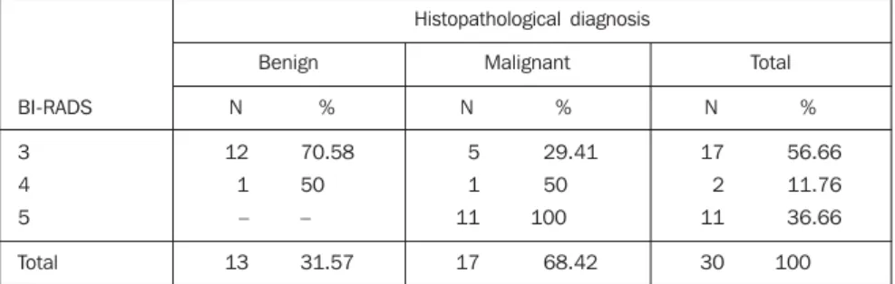
Table 2 Distribution of mammographic cases according to BI-RADS classification and histological diag-nosis of benignity or malignancy.
Histopathological diagnosis N 9 4 – 13
Benign Malignant Total
% 69.23 36.36 – 43.33 N 4 7 6 17 % 30.76 63.63 100 56.66 N 13 11 6 30 % 31.58 47.36 21.06 100 BI-RADS 3 4 5 Total
BI-RADS 3: NPV = 69.23%; BI-RADS 4: PPV = 63.63%; BI-RADS 5: PPV = 100%.
Table 3 Distribution of sonographic cases according to BI-RADS classification and histopathological diagnosis of benignity or malignancy.
Histopathological diagnosis N 12 1 – 13
Benign Malignant Total
% 70.58 50 – 31.57 N 5 1 11 17 % 29.41 50 100 68.42 N 17 2 11 30 % 56.66 11.76 36.66 100 BI-RADS 3 4 5 Total
BI-RADS 3: NPV = 70.58%; BI-RADS 4: PPV = 50%; BI-RADS 5: PPV = 100%.
Table 4 Distribution of cases of magnetic resonance imaging, according to BI-RADS classification and histopathological diagnosis of benignity or malignancy.
Histopathological diagnosis N 3 9 1 13
Benign Malignant Total
% 100 69.23 7.14 31.57 N – 4 13 17 % – 30.76 92.85 68.42 N 3 13 14 30 % 10 43.33 46.66 100 BI-RADS 3 4 5 Total
Analyzing the sonographic findings in BI-RADS categories 3, 4 and 5 as to their predictive value in relation to the malignant or benign nature of detected non-palpable breast masses, we have observed that the NPV of category 3 presented moderate lev-els; category 4, moderate PPV; and category 5, high PPV. Our results are similar to those presented by Hong et al.(9), emphasizing the capacity of predicting malignancy in cases of non-palpable breast lesions evaluated by ultrasound, if the BI-RADS is utilized, es-pecially in the category 5.
In our sampling, magnetic resonance imaging findings classified as BI-RADS categories 3, 4 and 5 were analyzed for a global evaluation of positive and negative predictive values. Correlating these find-ings with histopathological results from biopsy specimens, we have concluded that PPV have shown to be moderate for cat-egories 4 and 5 as whole, while category 3 NPV and category 5 PPV have shown to be high. Our results were similar to those from the study developed by Gokalp and To-pal(10) and Sadowski and Kelcz(11).
The present study, as well as other in-vestigations developed employing mam-mography, ultrasound and magnetic reso-nance imaging, is aimed at improving the prediction of malignancy or benignity of non-palpable breast lesions for a better management of the disease and improve-ment of the biopsies practice.
As regards category 4, the present study corroborates the systematic necessity of biopsy for non-palpable breast masses,
since the PPV observed for mammography, ultrasound and magnetic resonance imag-ing was higher than those reported by the international literature covering all the types of abnormal mammographic find-ings(14–16).
It is our opinion that the greatest con-tribution of the present study is related to non-palpable breast masses detected by mammography, ultrasound and magnetic resonance imaging and classified as BI-RADS category 3. Magnetic resonance imaging, because of the high NPV in this group of patients, should be considered as an important imaging method in the con-servative management of lesions classified as category 3, to avoid unnecessary biop-sies, according to the results found both by the present study and Gokalp and Topal(10).
Acknowledgement
The authors thank the Núcleo de Apoio à Publicação da Faculdade de Ciências Médicas da Santa Casa de São Paulo (NAP-SC), for the technical-scientific sup-port for publication of the present study.
REFERENCES
1. Shapiro S. Determining the efficacy of breast can-cer screening. Cancan-cer 1989;63:1873–1880. 2. Dodd GD. American Cancer Society guidelines
on screening for breast cancer. An overview. Can-cer 1992;69:1885–1887.
3. Hurley SF, Kaldor JM. The benefits and risks of mammographic screening for breast cancer.
Epidemiol Rev1992;14:101–130.
4. Smart CR, Hartmann WH, Beahrs OH, Garfinkel L. Insights into breast cancer screening of younger women. Evidence from the 14-year
fol-low-up of the Breast Cancer Detection Demon-stration Project. Cancer 1993;72:1449–1456. 5. Nystrom L, Rutqvist LE, Wall S, et al. Breast
can-cer screening with mammography: overview of
Swedish randomised trials.Lancet 1993;341:
973–978.
6. Smart CR. Highlights of the evidence of benefit for women aged 40-49 years from the 14-year follow-up of the Breast Cancer Detection Dem-onstration Project. Cancer 1994;74:296–300. 7. Hall FM, Storella JM, Silverstone DZ, Wyshak G.
Nonpalpable breast lesions: recommendations for biopsy based on suspicion of carcinoma at mam-mography. Radiology 1988;167:353–358. 8. Breast Imaging Reporting and Data System
(BI-RADSTM). 4th ed. Reston: American College of
Radiology, 2003.
9. Hong AS, Rosen EL, Soo MS, Baker JA. BI-RADS for sonography: positive and negative pre-dictive values of sonographic features. AJR Am J Roentgenol 2005;184:1260–1265.
10. Gokalp G, Topal U. MR imaging in probably be-nign lesions (BI-RADS category 3) of the breast. Eur J Radiol 2006;57:436–444.
11. Sadowski EA, Kelcz F. Frequency of malignancy in lesions classified as probably benign after dy-namic contrast-enhanced breast MRI examina-tion. J Magn Reson Imaging 2005;21:556–564. 12. Kopans DB, Lindfors K, McCarthy KA, Meyer JE. Spring hookwire breast lesion localizer: use with rigid-compression mammographic systems. Radiology 1985;157:537–538.
13. Rosner B. Fundamentals of biostatistics. 2nd ed. Boston: PWS Publishers, 1986.
14. Liberman L, Abramson AF, Squires FB, Glass-man JR, Morris EA, Dershaw DD. The Breast Im-aging Reporting and Data System: positive pre-dictive value of mammographic features and fi-nal assessment categories. AJR Am J Roentgenol 1998;171:35–40.
15. Lacquement MA, Mitchell D, Hollingsworth AB. Positive predictive value of the Breast Imaging Reporting and Data System. J Am Coll Surg 1999;189:34–40.