OctOber / December 2011
OrIGINALS ArtIcLeS
4
31
JACOMO AL, MARTINEZ CAR, SERRA MMP, AKAMATSU FE, PEREIRA JA, ANDRADE MFCD, MARGARIDO NF. Prognostic impact of the lymph node metastatic ratio on 5-year survival of patients with rectal cancer not submitted to preoperative chemoradiation. J coloproctol, 2011;31(4):311-324.
AbStrAct: Lymph node metastases are a major prognostic factor in colorectal cancer. Inadequate lymph node resection is related to shorter survival. the lymph nodes ratio (LNr) has been used as a prognostic factor in patients with colon cancer. Few studies have evaluated the impact of LNr on the 5-year survival of patients with rectal cancer. Objective: to evaluate the impact of LNr on the survival of patients with rectal cancer not submitted to preoperative chemoradiotherapy. methods: Ninety patients with rectal cancer excluding colon tumors, synchronous tumors, hereditary colorectal cancer and those undergoing preoperative chemoradiation. the patients were divided into three groups according to the LNr: LNr-0, no lymph nodes; LNr-1, 1 to 20% of compromised lymph nodes; and LNR-2, more than 21% of compromised lymph nodes. The cutoff identiication for the selected sample was obtained from the curve of receiver operating characteristics (rOc). Survival was assessed by Kaplan-meier test, the difference among groups by Cox-Mantel test and the correlation among variables by Pearson’s test, adopting a signiicance level of 5% (p≤ 0.05). Results: The 5-year survival was related to the Dukes classiication, TNM, number of metastatic lymph nodes and
Prognostic impact of the lymph node metastatic ratio on 5-year
survival of patients with rectal cancer not submitted
to preoperative chemoradiation
ALFREDO LUIZ JACOMO1, CARLOS AUGUSTO REAL MARTINEZ2, MARCIA MILENA PIVATTO SERRA3,
FLÁVIA EMI AKAMATSU4, JOSÉ AIRES PEREIRA5, MAURO FIGUEIREDO CARVALHO DE ANDRADE6,
NELSON FONTANA MARGARIDO7
1Associate Professor of Human Structural Topography, Surgery Department of the Medical School of Universidade de São
Paulo (USP) – São Paulo (SP), Brazil. 2Full Professor, Surgery Department of FMUSP – São Paulo (SP), Brazil; Adjunct Professor, Postgraduate Program in Health Sciences, Universidade São Francisco (USF) – Bragança Paulista (SP), Brazil. 3Assistant Professor, Doctor of Statistics, Medical Sciences at USF – Bragança Paulista (SP), Brazil. 4Professor and Doctor
of Human Structural Topography, Surgery Department of the Medical School of USP – São Paulo (SP), Brazil. 5Assistant Professor, Master in Pathology, Medical Sciences at USF – Bragança Paulista (SP), Brazil. 6Professor and Doctor of Human Structural Topography, Surgery Department of the Medical School of USP – São Paulo (SP), Brazil. 7Full Professor of Human
Structural Topography, Medical School of USP – São Paulo (SP), Brazil.
Study carried out at the LIM-02, Human Structural Topography, Surgery Department of the Medical School of Universidade de São Paulo (USP) – São Paulo (SP), Brazil. Postgraduate Program in Health Sciences, Universidade São Francisco (USF) – Bragança Paulista (SP), Brazil.
Financing source: none.
Conlict of interest: nothing to declare.
INtrODuctION
Colorectal cancer (CRC) is the third most preva-lent neoplasm in the world and the second cause of death related to cancer in western countries1. Epide-miological studies have shown a 2.4-fold increase in the incidence of CRC in the oriental countries2. In the last two decades, despite the increase in the number of proximal colon tumors, rectal tumors are still more prevalent3. Many clinical, histopathological, molecu-lar and genetic variables have been related to overall survival (OS) and disease-free survival (DFS) in pa-tients with CRC4. Despite the importance of all vari-ables, the parietal invasion, the lymph node involve-ment and the presence of metastases remain as the variables of more power to predict the OS, DFS and guide the adjuvant therapy indication5-7.
In 1932, Dukes8 developed the irst classiication system for colorectal (CR) staging. In this system, the
different stages of the disease were classiied accord -ing to the extent of rectal wall involvement and the presence (or not) of lymph node metastases. Later, several alterations were proposed to improve the OS
prediction capability of the original classiication9,10. Today, the TNM system recommended by the AJCC (American Joint Committee on Cancer) and the UICC (International Union Against Cancer), which stages neoplasms based on the tumor-lymph node (LN)-me-tastasis triad, is the most frequently used worldwide9-11. In the TNM system, the patients are divided into groups and subgroups, according to the extent of tu-mor invasion in the colon wall, presence and number of metastases in the LNs and the involvement of dis-tant organs11. Lymph node involvement is determined by using the number of metastatic LNs and subdivided into: N0 for no LN involvement; N1 for metastases in up to three LNs; and N2 for when four or more LNs are taken by neoplasm11.
The importance of lymph node involvement in the OS and DFS in CRC can be better evaluated by results
of studies showing that 80% of the patients without
metastases in regional LNs survive ive years, while
only 50% of those with compromised LNs survive for the same period12. The lowest OS of these patients re-quired complementary therapies associated with the surgical treatment to improve these rates, regardless of the number of compromised LNs4. Then, patients with only one compromised LN are submitted to the same complementary treatment protocol as those with more extensive lymph node involvement4.
The precise evaluation of the presence of lymp node metastases is possible only when a proper num-ber of LNs is examined5. Studies have shown that the DFS and OS in patients with CRC are directly related to the number of examined LNs12. Modern interna-tional guidelines recommend that the minimum num-ber of 12 LNs should be analyzed to enable proper staging12-15. However, the number of examined LNs
in the surgical specimen is inluenced by different variables. The number of identiied LNs is directly
related to the surgeon’s experience and practice, the histological technique used in the lymph node recov-ery (fresh dissection immediately after resection, fat clearing techniques for fast recovery) and the patholo-gist’s experience and patience to identify them16. The
neoplasm location – colon or rectum – can also inlu -ence the number of recovered LNs17. In colon cancer (CC), the number of dissected LNs is usually higher when compared to CRC18. Despite such peculiarities, the international guidelines recommend that the same number of LNs should be studied for a proper CRC staging19.
The dificult recovery of the minimum number
of LNs in CRS is even more critical when considering that many patients with CRC are submitted to neoad-juvant chemoradiation (NCR) protocols, in which the number of LNs is reduced by around 30%, further in-creasing the pathologist’s uncertainties regarding the correct disease staging20,21. A recent study quantifying the number of LNs recovered after the CRC resec-LNR. A difference was observed in 5-year survival between the different classes of resec-LNR. Patients classiied as LNR-0 had a survival rate of 85%, while classes LNr-1 and LNr-2, 73 and 19%, respectively (p=0.0001). conclusions: the results showed that the LNr has an impact on 5-year survival of patients with rectal cancer not submitted to neoadjuvant therapy.
tion, comparing patients submitted or not to the NCR, showed that the patients submitted to NCR had the mean value of recovered LNs of 6.29 per examined specimen, while those not submitted to NCR presented 13.5, i.e., half the minimum number recommended for a precise staging18. The importance of a proper lymph node resection is evident with the results of studies showing that the recovery of less than 12 LNs in the surgical specimen is directly related to lower OS and DFS in patients with CRC22,23.
In order to ind a variable that could improve the
accuracy of staging systems, especially in patients submitted to improper lymph node resection, the in-corporation of lymph node ratio (LNR) into staging systems as an additional variable has been proposed24.
LNR is deined as the relation between the total num -ber of examined LNs and the num-ber of compromised LNs. Initially, the prognostic value of LNR was eval-uated in patients with stomach25,26, bladder27, breast28 and pancreas29 cancer, presenting correlation with DFS and OS. In patients with gastric cancer submitted to improper lymphadenectomy, LNR presented great-er prognostic powgreat-er when compared to the numbgreat-er of comproimsed LNs, when using the current staging systems25. The routine incorporation of LNR into the staging systems was able to reduce the effects of stage migration4,30.
Berger et al.31 were the irst to analyze whether the LNR also related to OS and DFS in patients with CC. They observed that, after the curative resection, the LNR was an important prognostic variable, recom-mending its use in future studies to analyze adjuvant treatments31. Later, a series of studies conirmed the importance of LNR as a prognostic factor in patients with CC4,5,32-41. However, few studies have evaluated the importance of LNR as a variable related to OS in patient with CRC42-44. The evaluation in patients with
CRC is more dificult to be performed, because the
patients submitted or not to LNR protocols are usually
evaluated as a single group, which inluences the num -ber of recovered LNs in the surgical specimen.
It would be interesting to irst study the impact
of LNR, subdividing the patients into two groups: one of patients submitted and one of patients not
sub-mitted to the NCR, to conirm whether the LNR has
predictive power in OS in both groups. After that, the impact of LNR on a group of patients with CRC
submitted to NCR could be evaluated. However, ac-cording to our knowledge, no study has evaluated the impact of LNR on OS in patients with CRC not sub-mitted to NCR. If the LNR had any impact on OS, it could become a useless strategy to minimize the surgeon and the pathologist’s concern about substag-ing. For this reason, the purpose of this study was to evaluate the impact of LNR on OS of patients with CRC not submitted to NCR.
cASe rePOrt AND metHOD
The study was conducted according to all phases established by the Research Ethics Committee of the Universidade São Francisco and requirements of the Research Ethics Council of the Comissão Nacional de Ética em Pesquisa (CONEP), Ministry of Health (Resolution CNS196/96).
This is a retrospective study, a review in the da-tabase of the Coloproctology and Pathology Group of the Hospital Universitário São Francisco, Bragança Paulista. From total 348 patients with CRC monitored from 2001 to 2010, 90 were eligible for the study, with
conirmed histological diagnosis of rectal adenocarci
-noma, in any stage according to the TNM classiica -tion, and who had been submitted to complete resec-tion of primary tumor. The study excluded synchronic tumors, patients with suspicion of belonging to fami-lies with hereditary CRC (familial adenomatous poly-posis and non-polypoid CRC) or CRC associated with
intestinal inlammatory disease and patients submit -ted to NCR. The study considered as rectal tumors neoplasms located below the sacral promontory, ac-cording to data collected from the surgical descrip-tion. All patients were operated through laparotomy and none of them received drainage of lateral chain pelvic LNs. The mean follow-up period was 40.87 months (2-68 months). Thirty-two patients in stages III e IV received adjuvant chemotherapy, performed
in six cycles, with 5-luorouracil and leucovorin (5FU
450 mg/m2 + 20 mg/m2 leucovorin) repeated in
inter-vals of four to ive weeks. Twenty-six patients con -cluded the proposed adjuvant scheme.
moderate-ly and poormoderate-ly differentiated), type of neoplasm (mucus producer and non-producer), angiolymphatic invasion
(present or absent), Dukes classiications (A, B and C)
and TNM (I to IV), number of resected LNs (mean and median values), number of metastatic LNs (mean and median values), LNR (LNR-0, LNR-1 and LNR-2), follow-up period after the surgery (in months), death
date and ive-year survival. The OS, in months, was de
-ined using the death date or the period between the sur -gery date and the last doctor’s appointment.
The histological blades of each patient were he-matoxylin-eosin (HE) stained, then analyzed by the Pathology Department and reviewed by an pathologist with experience in digestive tract neoplasms to
con-irm the histopathological diagnosis and review the
considered variables. In the previous
anatomopatho-logical study, LN dissection was performed with ixed
specimen. No fat clearing method was used to en-hance LN recovery. Wall invasion was evaluated ac-cording to the involvement extent of mucosa, submu-cosa, muscularis propria, serous membrane, adipose tissue or adjacent organs. The review of neoplastic in-volvement of resected LNs was analyzed exclusively through the HE technique, not using the immunohis-tochemical method to study micrometastases.
The LNR calculation was performed using the ratio between the total number of compromised and examined LNs, categorizing the patients into three groups according to the LNR: LNR-0 for no LNs compromised by neoplasm; LNR-1: (0.01–0.20) for neoplastic involvement between 1% and 20% in the studied LNs; LNR-2: (0.21–1.0) when more than 21% of the analyzed LNs were compromised by neoplasm.
The ideal cut-off for the classiication of groups, con
-sidering the best speciicity and sensitivity values to
the selected sample, was obtained from the receiver
operating characteristics (ROC) curve, in order to ind the ideal LNR for the case classiication.
Descriptive statistics was used to describe the clinical characteristics of the selected case and the histopathological data of neoplasm. The correlations between variables used the Pearson’s test. The OS curves in a 5-year follow-up were determined using the Kaplan-Meyer method, with the Cox-Mantel test used in comparisons. The results obtained were ana-lyzed using SPSS for Windows version 13.0, adopting
the signiicance level of 5% (p<0.05) in all tests.
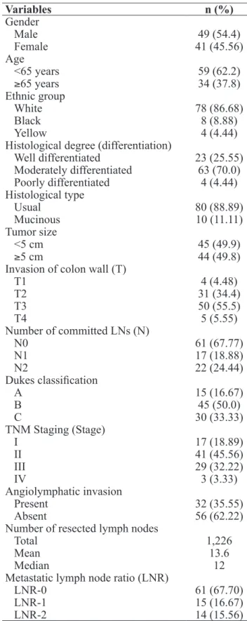
Variables n (%)
Gender
Male 49 (54.4)
Female 41 (45.56)
Age
<65 years 59 (62.2)
≥65 years 34 (37.8)
Ethnic group
White 78 (86.68)
Black 8 (8.88)
Yellow 4 (4.44)
Histological degree (differentiation)
Well differentiated 23 (25.55) Moderately differentiated 63 (70.0) Poorly differentiated 4 (4.44) Histological type
Usual 80 (88.89)
Mucinous 10 (11.11)
Tumor size
<5 cm 45 (49.9)
≥5 cm 44 (49.8)
Invasion of colon wall (T)
T1 4 (4.48)
T2 31 (34.4)
T3 50 (55.5)
T4 5 (5.55)
Number of committed LNs (N)
N0 61 (67.77)
N1 17 (18.88)
N2 22 (24.44)
Dukes classiication
A 15 (16.67)
B 45 (50.0)
C 30 (33.33)
TNM Staging (Stage)
I 17 (18.89)
II 41 (45.56)
III 29 (32.22)
IV 3 (3.33)
Angiolymphatic invasion
Present 32 (35.55)
Absent 56 (62.22)
Number of resected lymph nodes
Total 1,226
Mean 13.6
Median 12
Metastatic lymph node ratio (LNR)
LNR-0 61 (67.70)
LNR-1 15 (16.67)
LNR-2 14 (15.56)
reSuLtS
In total, 1,226 LNs were resected, mean of 13.6 (2–40) and median of 12. In the whole analysis, 140 compromised LNs, mean of 1.55 (minimum 1 and maximum 28), were found. The mean follow-up pe-riod was 40.87 months (2–68). Table 1 shows the se-lected patients’ clinical and histopathological charac-teristics.
A correlation was observed between the number of resected LNs and the number of compromised LNs (p=0.04; 95%CI 0.00–0.40). No correlation was ob-served between the number of resected LNs and the
LNR (p=0.46), but a signiicant correlation was ob -served between the number compromised LNs and the LNR (p=0.00001; 95%CI 0.50–0.75).
The evaluation of OS did not ind any signii -cant difference when analyzing age (p=0.08), gen-der (p=0.06), histological type (p=0.85), tumor size (p=0.053), extent of invasion in the rectal wall (p=0.06) and histological degree (p=0.07).
Fifteen patients (16.67%) were classiied as stage A in Dukes classiication, 45 (50%) as B and 30 (33.3%)
as C. Figure 1 shows the OS according to Dukes
clas-siication. Worsened OS is observed in more advanced stages in Dukes classiication (p=0,0001).
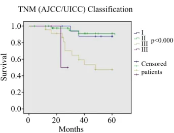
Seventeen patients (18.8%) were classiied as stage I in the TNM classiication, 41 (45.56%) as II,
29 (32.22%) as III and 3 (3.33%) as stage IV. Figure
2 shows the OS according to the TNM classiication.
The analysis showed that the patients in more advanced stages presented lower OS (p=0.0001). Sixty-one pa-tients (67.77%) did not present any compromised LN (N0), while 15 (16.67%) had less than three compro-mised LNs (N1) and 14 (15.56%) more than three LNs with metastasis (N2). Eighty per cent of the patients
classiied as N0 survived ive years, while 73% of the N1 patients no patient classiied as N2 survived for a
similar period. The OS was reduced when considering the number of compromised LNs (p=0.0003).
Sixty-one patients (67.70%) were classiied as
LNR-0 for not presenting any compromised lymph Dukes Classification
Survival
Months
Censored patients
p<0.0001
1.0
0 20 40 60
A B C
0.8
0.6
0.4
0.2
0.0
Figure 1. Five-year survival according to Dukes classiication.
A=Stage A; B=Stage B and C=Stage C. Kaplan-Meier Curve. Cox-Mantel Test. (p=0.0001).
TNM (AJCC/UICC) Classification
Survival
Months
Censored patients
p<0.0001
1.0
0 20 40 60
I II III III
0.8
0.6
0.4
0.2
0.0
Figure 2. Five-year survival according to TNM (AJCC/UICC) classiication. I=Stage I; II=Stage II; III=Stage III and IV=Stage IV. Kaplan-Meier Curve. Cox-Mantel Test (p=0.0001).
Metastatic Lymph Node Ratio (LNR)
Survival
Months
Censored patients
p<0.0001
1.0
0 20 40 60
0 1 2
0.8
0.6
0.4
0.2
0.0
Figure 3. Five-year survival according to LNR-0=no committed
LNs. LNR-1 = <20% committed LNs; LNR-2=≥21% committed
node, 15 (16.67%) as LNR-1 for having 20% or less and 14 (15.56%) as LNR-2 for having more than 21%
compromised LNs. Patients classiied as LNR-0 pre
-sented OS greater than 85%, while those classiied as LNR-1 presented 73% OS and, inally, the patients
with this index above 73% (LNR-2), it was less than 19%. Figure 3 shows the OS when considering the
LNR. The results conirm that the greater the LNR, the worse the prognosis (p<0.0001).
DIScuSSION
The number of resected and examined LNs is es-sential for the proper staging of patients with CRC. A considerable number of LNs in the surgical specimen ensures neoplasm staging certainty and suggests the surgical resection execution according to the recom-mended oncologic standards, demonstrating that the ex-tracted specimen had been submitted to a detailed his-topathological analysis5,45,46. Despite all precaution of surgeons and pathologists, other variables can interfere in the number of studied LNs. Proximal colon tumors
have shown to recover a signiicantly greater number
of LNs when compared to distal colon tumors47. These numbers are even more evident when comparing the number of resected LNs in CC and CRC12,15,19,22,23,48. These differences are attributed to the possibility of re-secting a greater number of lymph node chains in the proximal colon than in the distal colon and rectum47.
This possibility is conirmed by the results of a study
that analyzed 388 patients of CRC, showing that the mean value of recovered LNs in the right colon was higher than in the left colon (18.9 versus 12.6)47. An-other study, which evaluated 2,340 patients with CRC and compared the number of recovered LNs between patients with CC and CRC, showed that the mean value of recovered LNs among 1,314 patients with CRC was nine LNs, while among 1,026 patients with CC was 10
LNs, signiicant differences33. Our group also found similar results in a previous study, which analyzed only patients with CC and CRC located above the
perito-neal relection, with mean value of recovered LNs of
22.7(12–99). However, in this study, for which only pa-tients with CRC were selected, the mean value was 13.6 (2–40) and median was 12 LN4.
Recently, a study that dissected 12 cadavers of patients that died of diseases not related to the
diges-tive system conirmed these results and demonstrated
that the number of LNs in the rectum is changed de-pending on the site taken into account48. After removal of the entire rectum and mesorectum into a
‘mono-bloc’ before the specimens were ixed, the authors
counted the number of dissected LNs in each of the nine proportional axial cuts made in the upper, middle and lower thirds. They dissected total 412 LNs, with mean 34.3±2.1 LNs per cadaver, and conirmed that
the mean number they found signiicantly varied with
the cut height. They found on average 22.2 LNs in cra-nial cuts (upper rectum), 9.8 LNs in the intermediate cuts (middle rectum) and only 2.3 in caudal cuts (low-er rectum). Lat(low-er, oth(low-er authors, when dissecting 30 cadavers, counted the number of LNs in the mesorec-tum, comparing the lymph node recovery through manual dissection to the adipose tissue clearing tech-nique. The authors also divided the mesorectum into three segments (upper, middle and lower). The mean recovered LNs per cadaver was 6.2±1.3 (5–9); with 5.89±1.24 recovered in the group of manual dissec-tion and 6.60±1.29 in the group of fat clearing, a
dif-ference that was not statistically signiicant. However,
they point out the fact that, in the lower third of the mesorectum, the clearing technique enabled the re-covery of a greater number of LNs of small sizes49.
These indings are essential when the number of re -covered LNs is studied in patients with CRC
locat-ed below the peritoneal relection. As this region has
a lower number of LNs – exactly where the CRC is
more frequent – the pathologist inds it more dificult to recover a suficient number of LNs that enables to
establish the lymph node involvement with certainty and, consequently, the patient staging. In addition, it should be emphasized that the best recommendations for the CRC treatment propose the use of NCR exactly in the patients with tumors located in the middle and lower rectum, where the number of LNs is lower.
resection. However, they point out that these indings
are not completely applicable when using NCR, at NCR, besides reducing the number of recovered LNs, creates a group of patients without LN in the surgical specimen, changing the post-chemoradiation staging. From total 281 patients submitted to NCR, 32 (11%) did not present LNs in the surgical specimen51. They
suggest that the absence of LN may relect a better
response to NCR therapy, instead of worsened sur-gical radicality51. With these indings, they proposed that the surgical treatment could be avoided in patients with complete clinical, endoscopic and radiological response to NCR, with the indication of a rigorous postoperative follow-up only, and the surgical therapy could be indicated to cases of unsatisfactory response
or recurrence, identiied through clinical, endoscopic
and imaging exams during the follow-up52,53.
Howev-er, there are no suficient evidences, based on
well-conducted multi-center studies, that justify the indica-tion of non-surgical treatments to patients that present complete response after NCR54,55. All these evidences suggest that lymph node staging in patients with CRC
submitted to NCR, based on the number of identiied
LNs only, is controversial and should be interpreted as a precaution54.
The surgical and histological technique also
in-luences the number of recovered LNs in the surgical
specimen. A study analyzing 15 patients submitted to NCR, rectal resection with total excision of mesorec-tum and lateral chain LN dissection, showed the re-covery of 331 LNs. The study reported mean resected LNs per patient of 22.1, comprised of 258 perirectal, 73 pararectal and 27 lateral LNs. In this study, 20% of the patients showing no compromised LNs in the lat-eral chain at the conventional histological exam, when studied through immunohistochemistry to analyze cytokeratins (AE1/AE3), presented hidden microme-tastases, and one of them had presented complete re-sponse to the tumor56.
Surgeon training is also considered an impor-tant variable for a proper lymphadenectomy. A study evaluating total 371 patients showed that the number
of resected LNs signiicantly increases when the pa -tients are operated by surgeons specialized in CRC treatment57.This study showed that the number of LNs removed by the trained surgeons was, on average, of 19.9±10.6, while the number of LNs removed by non
trained surgeons was 14.8±10.6, signiicant differenc -es57. These differences increased even more in obese patients (BMI≥30), who presented a lower number of resected LNs (17.3±10.0 versus 19.9±11.5). All these arguments suggest that, when combining the proper surgeon training, histological technique, utilization of NCR and aspects related to the patient, such as obesity
and the fact of being a male, the number of identiied
LNs in the CRC specimens can be lower, further
ag-gravating the correct prognostic classiication18,20. Perhaps, the most important objective of a proper lymphadenectomy in patients with CRC is to select,
with superior accuracy, those who will beneit from a
complementary adjuvant treatment43. When comparing the OS of patients with CRC in relation to the number of extirpated LNs, it is observed that the improper
re-section signiicantly worsens the disease prognosis16,58. The ideal number of LN to be resected in the CRC is still a reason for controversies4,12,36. Most authors con-sider the range of 10 and 17 LNs as ideal12,36,59,60. In the USA, the National Quality Forum and other
organiza-tions have recently deined that, in patients with CRC,
the minimum number of 12 LNs should be resected, in this parameter, which is one of the most important in the quality analysis of a unit specialized in the disease treatment61. Then, resections with less than 12 LNs can be considered improper, not enabling the correct stag-ing. Many pathologists prefer to classify patients with
less than 12 LNs identiied in the surgical specimen
as NX or add a note to the anatomopathological study report emphasizing the risk of predicting the lymph node status based on the number of dissected LNs4,37.
Resections with insuficient number of LNs contribute
to the phenomenon of stage migration described by Feinstein et al. in 198530, known as Will Rogers phe-nomenon. In fact, it is a frequent phenomenon in on-cology, which occurs when the prediction of favorable survival is threatened by unfavorable progress4,30. The phenomenon is even more frequent in cases of CRC, in which, after NCR, the number of recovered LNs is usually still low.
with CC classiied as stages II and III and submitted
to adjuvant chemotherapy. They selected patients who presented the mean number of 13 LNs analyzed. From total 3,411 patients, 648 (19%) did not have lymph node metastases, 1,857 (54%) had up to three compro-mised LNs and 906 (27%) had more than three
meta-static LNs. When they classiied the same patients into groups according to the LNR (LNR<0.05; 0.05–0.19;
0.2–0.39 and 0.4–1.0), they observed that the LNR was related to the OS and DFS in patients with 10 to 15 or more than 15 resected LNs, but not in those with less than 10 LNs in the surgical specimen31. Un-like this study, De Ridder et al.40, when comparing the
TNM classiication to a system that included the LNR,
in a group of patients that presented mean 10 LNs, observed that the capability to establish with higher precision the prognostic stages using the LNR was 31
versus 25% only with the TNM classiication. They
concluded that the LNR is a variable that can improve staging in improper lymph node resection.
A multi-center study published by Wang et al.33 that evaluated 24,477 patients with CC in stage III
according to the TNM classiication, observed that
it was possible to recover more than 15 LNs in the surgical specimen in 7,469 (30.5%) of them. They categorized the patients into four groups according to the LNR (no involvement, 1/14, 1/4 and 1/2, re-spectively). They observed that the OS for patients in stages IIIA, IIIB and IIIC was 71.3, 51.7 and 34.0%,
respectively. No signiicant differences were found
in OS, according to the LNR, in the patients
classi-ied as stage IIIA. In the patients classiclassi-ied as stage IIIB, the OS according to the four classiication
classes (LNR-1 to LNR-4) was 63.5, 54.7, 44.4 and
34.2%, respectively, conirming that the higher the LNR, the worse the OS. In patients classiied as stage
IIIC, the OS according to LNR-2, LNR-3 and LNR-4
was 49.6, 41.7, and 25.2%, respectively, conirming
that the LNR was also a variable related to OS and DFS. Curiously, Derwinger et al.62 observed that the LNR could also be considered a prognosis factor in
136 patients with CRC classiied as stage IV accord -ing to the TNM system. They categorized the patients into three groups: LNR=0–0.15, LNR=0.16–0.65 and LNR=0.66–1. Through a univariate analysis, they re-ported that the LNR showed to be a more important prognostic factor to predict the OS and indication of
adjuvant chemotherapy than the number of compro-mised LNs. Rosenberg et al.35 studied the importance of LNR in 3,026 patients, 1,763 (58.2%) with CC and 1,263 (41.8%) with CRC. The rate of potentially cu-rative surgeries was 77.4% and the mean proportions of resected and metastatic LNs to each patient were 18.3 and 2.6, respectively. After the statistic study, they established that the best cohort level for the LNR
classiication was 0.17, 0.41 and 0.69. They observed
that the OS of patients without compromised LNs was 87%, while in patients with compromised LNs, it was 60.3, 34.4 and 17.6%, increasing around 5% when the
LNR classiication was adopted. When considering all
patients as a single group, they observed that the LNR presented a greater prognostic power than the num-ber of compromised LNs. When they categorized the patients into two groups – with CC and with CRC –, they observed that the LNR remained as an indepen-dent prognostic variable to both groups35. A series of studies performed later evaluated the importance of LNR in CRC4,38,39,43,63-65. In all of them, the LNR was considered an independent variable for OS and DFS of patients with CRC, especially in patients of stages
II and III in the TNM classiication.
In Brazil, to our knowledge, only two studies have evaluated the importance of LNR in patients with CRC36,37. In the irst, the authors analyzed 106 patients, most were males (53.8%). The median val-ue of dissected LNs per patient was 11.5 (3–45) and only 58.5% of the patients had more than 10 dissected LNs. The mean follow-up period was 25.05±15.21 months (2–64 months), and 32.1% of patients died of the disease. The univariate analysis showed that the OS of patients included in the study was related to LNR, disease staging and tumor recurrence. How-ever, in the multivariate analysis, they observed that the only independent factor related to OS was TNM.
Perhaps, these indings are related to the fact that more
than half the selected patients in the study (58.1%)
be-longed to stage IV according to the TNM classiication
period, small sample and non evaluation of presence of serious comorbidities and postoperative complica-tions36. In the second study published in Brazil, the au-thors studied 113 patients with CC and upper CRC37. They excluded patients with middle and lower CRC, as they had been submitted to NCR, and patients with less than 12 LNs in the surgical specimen. They cat-egorized the patients according to the LNR into three groups: LNR-0, with patients without lymph node in-volvement; LNR-1: involvement of max. 20% of ex-amined LN; and LNR-2: with neoplastic involvement in 21% or more of examined LNs. They found a
sig-niicant difference in OS when analyzing that in the
patients belonging to LNR-0, the OS was above 80%,
while in patients classiied as LNR-1 and LNR-2 the
OS was under 60% and 40%, respectively. With the multivariate analysis, they reported that the LNR can be considered an independent prognostic variable.
The results of all these studies were conirmed
by a recently-published systematic literature review that selected total 16 studies with good level of evi-dence. The authors included 33,984 patients with CC
or CRC classiied as stage III. The results showed that
the capability to predict OS as provided by the LNR was greater than that found only through the number of committed LNs66. They found the relative risk for OS of 2.36 (95%CI 2.14–2.61) and for DFS of 3.71 (95%CI 2.56–5.38).
However, most studies that have evaluated the LNR as a possible variable related to OS studied pa-tients with CC31-34,38-41 or with CC and CRC as a single group4,5,24,35-38,62,63. Few studies have evaluated whether the LNR could be related to OS in patients exclusively with CRC, and even so, these studies do not clearly state whether the patients were categorized according to the criterion of having been submitted to NCR or
not, which could inluence the LNR calculation42,44,64-66. Peng et al.42 studied, for the irst time in the litera -ture, the relation between LNR and OS in 318 patients with CRC previously submitted to curative-intention resection. With mean follow-up of 41 months and mean number of 12 resected LNs, they observed that OS and DFS were 58.82 and 59.8%, respectively. The multivariate analysis showed that, when considered as a continuous variable, LNR was the most important prognostic factor in OS. When they categorized the
pa-tients into three groups (LNR<0.4, between 0.14–0.40
and between 0.5–1), they observed that the OS was 72.19, 61.92 and 38.47%, respectively, statistically
signiicant differences. They concluded that the LNR
can also be considered an important prognostic factor in patients with CRC who presented positive LNs43. In the same year, Peschaud et al.44 also evaluated the prognostic power of LNR in patients with CRC. They analyzed OS, DFS and LNR in 307 patients with CRC and examined the mean number of 22 LNs. From the 307 patients, 178 (57.9%) did not show lymph node involvement, 67 (21.8%) had up to three committed LNs and 62 had more than three metastatic LNs. When they categorized the patients into four groups, accord-ing to: LNR=0 (no involvement), LNR=0.01 to 0.07, LNR>0.07 to 0.2 and LNR>0.2, they observed that the LNR was a variable related to OS, not the number of committed LNs. When they individually analyzed the patients with less than 12 examined 12 LNs, even so, the LNR was related to OS and DFS. They concluded that the LNR is the variable with the greatest prognos-tic power of both OS and DFS in patients with CRC, even in those whose surgical specimen presents less than 12 studied LNs.
the LNR is a better prognostic factor for OS than the number of recovered or metastatic LNs.
A study conducted by the Japanese Society of
Colorectal Cancer (JSCRC) to deine the impact of
LNR on patients with lower CRC in stage III, analyzed 501 patients submitted to curative resection with total excision of the mesorectum, coming from 12 institu-tions, and categorized them into four groups accord-ing to the LNR69. From total 501 patients, 381 were submitted to dissection of LNs from the lateral pelvic wall. The mean number of resected LNs in patients submitted or not to lateral dissection was 45 and 17, respectively. The study excluded 45 patients who had less than 12 LNs in the surgical specimen. Among the several clinical and pathological parameters analyzed, they observed that the number of committed LNs and LNR were variables related to the disease prognosis. When they added LNR to the seventh edition of TNM
classiication as a covariable, they observed that the
new staging system and LNR were independent prog-nostic variables in patients with CRC in stage III. The Japanese Society proposes to add the LNR concept to the staging system of AJCC to improve the accuracy of LN status in patients with lower CRC. This idea is defended by others who believe that the LNR can become a better method to select patients with CRC eligible to adjuvant therapy43,70,71. Recently, investiga-tors observed that the LNR is a variable that predicts the development of pulmonary metastases in patients with CRC72.
To our knowledge, in Brazil, the relation be-tween LNR and OS in patients with CRC submitted or not to NCR protocols has not been evaluated. In this study, the authors decided to study the impact of LNR and other variables on OS exclusively in patients with CRC. The study selected only patients that had been submitted to surgical resection, regardless of the number of recovered LNs in the surgical specimen, and that had not received NCR. The study excluded patients submitted to NCR to prevent the LNR-based categorization from having interferences related to the reduction of total and committed LNs due to NCR. The main purpose was to observe whether the system-atic surgery alone for CRC, with total excision of the mesorectum, and performed by a trained medical team
for the disease treatment, could conirm the relation
between LNR and OS. The review of
histopathologi-cal reports showing no involvement of proximal, dis-tal and circumferential margins in all selected spec-imens, and the mean number of 13.6 resected LNs, with median of 12 LNs combined with similar OS to other studied centers, allows to conclude that the pa-tients were submitted to surgical resection following the best oncologic principles.
In this study, when analyzing the clinical vari-ables, age, gender and ethnic group in relation to
OS, no signiicant differences were observed. When
the histological type of the tumor is considered, previous studies demonstrated that mucus-secret-ing tumors or with signet-rmucus-secret-ing cells present low-er OS, although accounting for 15% of the CRC cases73,74. They also demonstrated that the mean survival of these patients is 45.4 months, compared to 78.5 months in patients with tumors of tubular pattern73. In this study, no relation was found be-tween the histological type of the tumor and OS.
This inding is probably related to the small number
(8.98%) of mucus-secreting tumors in the studied cases. Perhaps this small number is due to the fact that only patients with CRC were selected, and in these patients, the proportion of this histological type is smaller when compared to CC patients, and due to the fact that the study excluded patients with suspicion of belonging to families with HNPCC, which present higher incidence of mucus-secreting tumors75. Although tumors with worse histological degree are also associated to worse OS, this study did not observe any relation between worse histo-logical degree and smaller OS. Perhaps the small number of little differentiated tumors found in the study (4.4%), just as it happened when considering
the histological type, inluenced the result.
Regarding the tumor size, the study observed that
tumor of more than ive centimeters presented mar
-ginal signiicance in relation to OS (p=0.053), while
the extent of colon wall invasion had no relation with worsened prognosis. Regarding the tumor size, the results obtained agree with those found in the litera-ture. Larger tumors usually present small chances of curative resection (R0), greater possibility of invasion of adjacent structures (prostate, bladder, vagina, pre-sacral fascia), locoregional recurrence and distant
related to OS, no relation with OS was found. Perhaps the high concentration of tumors TII and TIII, 89.94%
of the studied cases, may have inluenced the result. The Dukes classiication, the TNM system, the
number of committed LNs and the LNR were vari-ables related to OS. The study observed that the
pa-tients classiied as LNR-0 presented SG above 85%, while the patients classiied as LNR-1 presented 73%, and those classiied as LNR-3 presented OS below
19%. The results of this study showed that the greater the LNR, the worse the OS of patients with CRC, even
in those not submitted to NRC. The study conirmed
that the LNR has greater predictive power of OS than
the number of committed LNs identiied in the surgi -cal specimen and that it is similar to Dukes staging and LNR systems. It should be noted that the LNR could predict the OS even in patients with less than 12 recovered LNs (results not shown) and who would be properly categorized according to the best current directions for CRC staging.
Although the study evaluated only patients with CRC not submitted to NRC, the results obtained
conirmed the impact of LNR on OS of patients with
CRC. However, some considering should be point-ed out. Most authors that have evaluatpoint-ed the prog-nostic impact of LNR on OS have analyzed a small number of cases, patients with differences regarding
their biotype, gender, tumors in different locations of the rectum, of distinct histological types and de-grees, operated by surgeons with different levels of experience and, mainly, patients submitted to distinct protocols of adjuvant or neoadjuvant treatment. An-other factor of crucial importance is the absence of standardization at the cohort levels used to catego-rize the patients into groups according to the LNR in different studies. Only with the results of systematic reviews of multi-center, prospective studies, with an expressive number of cases, better standardization of inclusion criteria and that use standardized cohort
levels it will be possible to deinitively conirm the
prognostic impact of LNR and propose its inclusion as a useful variable to improve the staging systems currently available. Until then, due to the easy-to-use calculation of no additional cost, the routine use of LNR is recommended as a valid instrument to help the correct categorization of patients with CRC, re-ducing staging errors.
cONcLuSION
In the circumstances of this study, the results
ob-tained allow to conirm the impact of LNR on the
ive-year survival of patients with CRC not submitted to preoperative chemoradiation treatment.
reSumO: metástases linfonodais representam um dos principais fatores prognósticos no câncer colorretal. A ressecção linfonodal inadequada relaciona-se à menor sobrevida. A proporção entre linfonodos metastáticos (PLm) vem sendo utilizada como fator prognóstico em doentes com câncer de cólon. Poucos estudos avaliaram o impacto da PLm na sobrevida de doentes com câncer retal. Objetivo: Avaliar o impacto da PLm na sobrevida de doentes com câncer de reto não submetidos à quimioradioterapia pré-operatória. métodos: Foram incluídos 90 doentes com adenocarcinoma retal excluindo-se tumores de cólon, tumores sincrônicos, câncer colorretal hereditário e aqueles submetidos a tratamento radioquimioterápico pré-operatório. Os doentes foram dividi-dos em três grupos segundo a PLm: PLm-0, sem linfonodividi-dos comprometidividi-dos; PLm-1, 1 a 20% dividi-dos linfonodividi-dos comprometidividi-dos; e PLM-2, mais de 21% dos linfonodos comprometidos. A identiicação do ponto de corte da amostra selecionada foi obtida a partir da curva de características de operação do receptor (curva rOc). A sobrevida foi avaliada pelo teste de Kaplan-meier, a diferença entre os grupos pelo teste de Cox-Mantel e a correlação entre as variáveis pelo teste de Pearson, adotando-se um nível de signii -cância de 5% (p≤0,05). Resultados: A sobrevida em cinco anos relacionou-se à classiicação de Dukes, TNM, número de linfonodos metastáticos e PLM. Houve diferença na sobrevida ao compararem-se as diferentes classes de PLM. Doentes classiicados como PLm-0 apresentaram sobrevida de 85%, enquanto os pertencentes às classes PLm-1 e PLm-2, de 73 e 19%, respectivamente (p=0,0001). conclusão: Os resultados encontrados mostraram que a PLm tem impacto na sobrevida de doentes com câncer de reto não submetidos à neoadjuvância.
reFereNceS
1. Centers for Disease Control and Prevention (CDC). Vital signs: colorectal cancer screening, incidence, and mortality - United States, 2002-2010. MMWR Morb Mortal Wkly Rep 2011;60(26):884-9.
2. Coleman MP, Quaresma M, Berrino F, Lutz JM, De Angelis R, Capocaccia R, et al. Cancer survival in ive continents: a worldwide population-based study (CONCORD). Lancet Oncol 2008;9(8):730-56.
3. Boyle P, Ferlay J. Cancer incidence and mortality in Europe, 2004. Ann Oncol 2005;16(3):481-8.
4. Priolli DG, Cardinalli IA, Pereira JA, Alfredo CH, Margarido NF, Martinez CA. Metastatic lymph node ratio as an independent prognostic variable in colorectal cancer: study of 113 patients. Tech Coloproctol 2009;13(2):113-21. 5. Noura S, Ohue M, Kano S, Shingai T, Yamada T, Miyashiro I,
et al. Impact of metastatic lymph node ratio in node-positive colorectal cancer. World J Gastrointest Surg 2010;2(3):70-7. 6. Adjuvant therapy of colon cancer--results of a prospectively
randomized trial. Gastrointestinal Tumor Study Group. N Engl J Med 1984;310(12):737-43.
7. Sobin LH, Wittekind C. TNM classiication of malignant tumors. 6th ed. New York: Wiley-Liss; 2002.
8. Dukes CE. The classiication of cancer of the rectum. J Pathol 1932;35:323-32.
9. Astler VB, Coller FA. The prognostic signiicance of direct extension of carcinoma of the colon and rectum. Ann Surg 1954;139(6):846-52.
10. Nelson H, Petrelli N, Carlin A, Couture J, Fleshman J, Guillem J, et al. Guidelines 2000 for colon and rectal cancer surgery. J Natl Cancer Inst 2001;93(8):583-96.
11. Benson AB 3rd. New approaches to the adjuvant therapy of colon cancer. Oncologist 2006;11(9):973-80.
12. Wong JH, Severino R, Honnebier MB, Tom P, Namiki TS. Number of nodes examined and staging accuracy in colorectal carcinoma. J Clin Oncol 1999;17(9):2896-900.
13. Swanson RS, Copton CC, Stewart AK, Bland KI. The prognosis of T3N0 colon cancer is dependent of the number of nodes retrieved on resection. Ann Surg Oncol 2003; 10:65-71.
14. Compton CC, Fielding LP, Burgart LJ, Conley B, Cooper HS, Hamilton SR, et al. Prognostic factors in colorectal cancer. College of American Pathologists Consensus Statement 1999. Arch Pathol Lab Med 2000;124(7):979-94.
15. Cianchi F, Palomba A, Boddi V, Messerini L, Pucciani F, Perigli G, et al. Lymph node recovery from colorectal tumor specimens: recommendation for a minimum number of lymph nodes to be examined. World J.Surg 2002;26(3):384-9. 16. LeVoyer TE, Sigurdson ER, Hanlon AR, Mayer RJ,
MacDonald JS, Catalano PJ, et al. Colon cancer survival is associated with increasing number of lymph nodes analyzed: a secondary survey of intergroup trial INT-0089. J Clin Oncol 2003;21(15):2912-9.
17. Bilimoria KY, Palis B, Stewart AK, Bentrem DJ, Freel AC, Sigurdson ER, et al. Impact of tumor location on nodal evaluation for colon cancer. Dis Colon Rectum 2008;51(2):154-61.
18. Latkauskas T, Lizdenis P, Janciauskiene R, Pranys D, Tamelis A, Pavalkis D. Lymph node retrieval after resection of rectal cancer following preoperative chemoradiotherapy. Medicina (Kaunas) 2010;46(5):299-304.
19. Elferink MA, Siesling S, Lemmens VE, Visser O, Rutten HJ, van Krieken JH, et al. Variation in lymph node evaluation in rectal cancer: a Dutch nationwide population-based study. Ann Surg Oncol 2011;18(2):386-95.
20. Wichmann MW, Müller C, Meyer G, Strauss T, Hornung HM, Lau-Werner U, et al. Effect of preoperative radiochemotherapy on lymph node retrieval after resection of rectal cancer. Arch Surg 2002;137(2):206-10.
21. Ha YH, Jeong SY, Lim SB, Choi HS, Hong YS, Chang HJ, et al. Inluence of preoperative chemoradiotherapy on the number of lymph nodes retrieved in rectal cancer. Ann Surg 2010;252(2):336-40.
22. Norwood MG, Sutton AJ, West K, Sharpe DP, Hemingway D, Kelly MJ. Lymph node retrieval in colorectal cancer resection specimens: national standards are achievable, and low numbers are associated with reduced survival. Colorectal Dis 2010;12(4):304-9.
23. Murphy J, Pocard M, Jass JR, O’Sullivan GC, Lee G, Talbot IC. Number and size of lymph nodes recovered from dukes B rectal cancers: correlation with prognosis and histologic antitumor immune response. Dis Colon Rectum 2007;50(1):1526-34.
24. Tong LL, Gao P, Wang ZN, Song YX, Xu YY, Sun Z, et al. Can lymph node ratio take the place of pN categories in the UICC/AJCC TNM classiication system for colorectal cancer? Ann Surg Oncol 2011;18(9):2453-60.
25. Celen O, Yildirim E, Berberoglu U. Prognostic impact of positive lymph node ratio in gastric carcinoma. J Surg Oncol 2007;96(2):95-101.
26. Persiani R, Rausei S, Biondi A, Boccia S, Cananzi F, D’Ugo D. Ratio of metastatic lymph nodes: impact on staging and survival of gastric cancer. Eur J Surg Oncol 2008;34(5):519-24. 27. Herr HW. Superiority of ratio based lymph node staging for
bladder cancer. J Urol 2003;169(3):943-5.
28. Schiffman SC, McMasters KM, Scoggins CR, Martin RC, Chagpar AB. Lymph node ratio: a proposed reinement of current axillary staging in breast cancer patients. J Am Coll Surg 2011;213(1):45-52.
29. Bhatti I, Peacock O, Awan AK, Semeraro D, Larvin M, Hall RI. Lymph node ratio versus number of affected lymph nodes as predictors of survival for resected pancreatic adenocarcinoma. World J Surg 2010;34(4):768-75.
31. Berger AC, Sigurdson ER, Le Voyer T, Hanlon A, Mayer RJ, Macdonald JS, et al. Colon cancer survival is associated with decreasing ratio of metastatic to examined lymph nodes. J Clin Oncol 2005;23(34):8706-12.
32. Lee HY, Choi HJ, Park KJ, Shin JS, Kwon HC, Roh MS, et al. Prognostic signiicance of metastatic lymph node ratio in node-positive colon carcinoma. Ann Surg Oncol 2007;14(5):1712-7.
33. Wang J, Hasset JM, Dayton MT, Kulaylat MN. Lymph node ratio: role in the staging of node-positive colon cancer. Ann Surg Oncol 2008;15(6):1600-8.
34. Derwinger K, Carlsson G, Gustavsson B. A study of lymph node ratio as a prognostic marker in colon cancer. Eur J Surg Oncol 2007;34(7):771-5.
35. Rosenberg R, Friederichs J, Schuster T, Gertler R, Maak M, Becker K, et al. Prognosis of patients with colorectal cancer is associated with lymph node ratio: a single-center analysis of 3,026 patients over a 25-year time period. Ann Surg 2008;248(6):968-78.
36. Trufelli DC, Miranda VC, Palos CC, Ramos E, Abrão MN, Silva VA, et al. Positive/total dissected lymph nodes ratio as a prognostic factor in colorectal cancer. Rev Ass Med Bras 2007;53(6):539-42.
37. Priolli DG, Cardinalli IA, Alfredo CH, Spadari APP, Máximo FR, Margarido NF, et al. Proporção de linfonodos metastáticos como variável independente de prognóstico no câncer colorretal. J Coloproctol 2008;28(4):431-42. 38. Park IJ, Choi GS, Jun SH. Nodal stage of stage III colon
cancer: the impact of metastatic lymph node ratio. J Surg Oncol 2009;100(3):24-3.
39. Chin CC, Wang JY, Yeh CY, Kuo YH, Huang WS, Yeh CH. Metastatic lymph node ratio is a more precise predictor of prognosis than number of lymph nodes metastases in stage III colon cancer. Int J Colorectal Dis 2009;24(11):1297-302. 40. De Ridder M, Vinh-Hung V, Van Nieuwenhove Y, Hoorens
A, Sermeus A, Storme G. Prognostic value of lymph node ratio in node positive colon cancer. Gut 2006;55(11):1681. 41. Schumacher P, Dineen S, Barnett C Jr, Fleming J, Anthony
T. The metastatic lymph node ratio predicts survival in colon cancer. Am J Sur 2007;194(6):827-31.
42. Peng J, Xu Y, Guan Z, Zhu J, Wang M, Cai G, et al. Prognostic signiicance of the metastatic lymph node ratio in node-positive rectal cancer. Ann Surg Oncol 2008;15(11):3118-23.
43. Kim YS, Kim JH, Yoon SM, Choi EK, Ahn SD, Lee SW, et al. Lymph node ratio as a prognostic factor in patients with stage III rectal cancer treated with total mesorectal excision followed by chemoradiotherapy. Int J Radiat Oncol Biol Phys 2009;74(3):796-802.
44. Peschaud F, Benoist S, Julié C, Beauchet A, Penna C, Rougier P, et al. The ratio of metastatic to examined lymph nodes is a powerful independent prognostic factor in rectal cancer. Ann Surg 2008;248(6):1067-73.
45. Mönig SP, Baldus SE, Zirbes TK, Schröder W, Lindemann DG, Dienes HP, et al. Lymph node size and metastatic
iniltration in colon cancer. Ann Surg Oncol 1999;6(6): 579-81.
46. Wood TF, Saha S, Morton DL, Tsioulias GJ, Rangel D, Hutchinson W Jr, et al. Validation of lymphatic mapping in colorectal cancer: In vivo, ex vivo, and laparoscopic techniques. Ann Surg Oncol 2001;8(2):150-7.
47. Pappas AV, Lagoudianakis EE, Dallianoudis IG, Kotzadimitriou KT, Koronakis NE, Chrysikos ID, et al. Differences in colorectal cancer patterns between right and left sided colorectal cancer lesions. J BUON 2010;15(3): 509-13.
48. Miscusi G, di Gioia CR, Patrizi G, Gravetz A, Redler A, Petrozza V. Anatomical lymph node mapping in normal mesorectal adipose tissue. Dis Colon Rectum 2010;53(12):1640-4.
49. Thakur S, Somashekar U, Shiv Kumar Chandrakar, Sharma D. Anatomic study of distribution, numbers, and size of lymph nodes in mesorectum in indians: a autopsy study. Int J Surg Pathol 2011;19(3):315-20.
50. Marks JH, Valsdottir EB, Rather AA, Nweze IC, Newman DA, Chernick MR. Fewer than 12 lymph nodes can be expected in a surgical specimen after high-dose chemoradiation therapy for rectal cancer. Dis Colon Rectum 2010;53(7):1023-9. 51. Habr-Gama A, Perez RO, Proscurshim I, Rawet V, Pereira
DD, Sousa AH, et al. Absence of lymph nodes in the resected specimen after radical surgery for distal rectal cancer and neoadjuvant chemoradiation therapy: what does it mean? Dis Colon Rectum 2008;51(3):277-83.
52. Habr-Gama A, Perez RO, Wynn G, Marks J, Kessler H, Gama-Rodrigues J. Complete clinical response after neoadjuvant chemoradiation therapy for distal rectal cancer: characterization of clinical and endoscopic indings for standardization. Dis Colon Rectum 2010;53(12):1692-8. 53. Habr-Gama A, Perez RO, São Julião GP, Proscurshim I,
Gama-Rodrigues J. Nonoperative approaches to rectal cancer: a critical evaluation. Semin Radiat Oncol 2011;21(3):234-9. 54. Singh-Ranger G, Kumar D. Current concepts in the
non-operative management of rectal cancer after neoadjuvant chemoradiation. Anticancer Res 2011;31(5):1795-800. 55. Baxter NN, Morris AM, Rothenberger DA, Tepper AJ. Impact
of preoperative radiation for rectal cancer on subsequent lymph node evaluation: a population-based analysis. Int J Radiat Oncol Biol Phys 2005;61(2):426-31.
56. Coy CS, Meirelles LR, Leal RF, Ayrizono ML, Góes JR, Fagundes JJ. Evaluation of lateral lymph node metastases in advanced distal rectal cancer. Hepatogastroenterology 2010;57(104):1363-6.
57. Barbas A, Turley R, Mantyh C, Migaly J. Advanced fellowship training is associated with improved lymph node retrieval in colon cancer resections. J Surg Res 2011;170(1):e41-6. 58. Sigurdson ER. Lymph node dissection: Is it diagnostic or
therapeutic? J Clin Oncol 2003;21(6):965-7.
for adenocarcinoma: Trends over time and a recommendation for a minimum number of lymph nodes to be recovered. Am J Clin Pathol 1996;106(2):209-16.
60. Chen SL, Bilchik AJ. More extensive nodal dissection improves survival for stages I to III of colon cancer: a population-based study. Ann Surg 2006;244(4):602-10. 61. Wong SL, Ji H, Hollenbeck BR, Morris AM, Baser O,
Birkmeyer JD. Hospital lymph node examination rates and survival after resection for colon cancer. JAMA 2007;298(18):2149-54.
62. Derwinger K, Gustavsson B. A study of lymph node ratio in stage IV colorectal cancer. World J Surg Oncol 2008;6:127. 63. Vather R, Sammour T, Kahokehr A, Connolly AB, Hill AG.
Lymph node evaluation and long-term survival in stage II and Stage III colon cancer: a national study. Ann Surg Oncol 2009;16(3):585-93.
64. Moug SJ, Saldanha JD, McGregor JR, Balsitis M, Diament RH. Positive lymph node retrievel ratio optimises patient staging in colorectal cancer. Br J Cancer 2009;100(10): 1530-3.
65. Vaccaro CA, Im V, Rossi GL, Quintana GO, Benatti ML, Perez de Arenaza D, et al. Lymph node ratio as prognostic factor for colon cancer treated by colorectal surgeons. Dis Colon Rectum 2009;52(7):1244-50.
66. Ceelen W, Van Nieuwenhove Y, Pattyn P. Prognostic value of the lymph node ratio in stage III colorectal cancer: a systematic review. Ann Surg Oncol 2010;17(11):2847-55. 67. Kang J, Hur H, Min BS, Lee KY, Kim NK. Prognostic
impact of the Lymph node ratio in rectal cancer patients who underwent preoperative chemoradiation. J Surg Oncol 2011;104(1):53-8
68. Klos CL, Bordeianou LG, Sylla P, Chang Y, Berger DL. The prognostic value of lymph node ratio after neoadjuvant chemoradiation and rectal cancer surgery. Dis Colon Rectum 2011;54(2):171-5.
69. Kobayashi H, Mochizuki H, Kato T, Mori T, Kameoka S, Shirouzu K, et al. Study Group for Rectal Cancer Surgery of the Japanese Society for Cancer of the Colon and Rectum. Lymph node ratio is a powerful prognostic index in patients with stage III distal rectal cancer: a Japanese multicenter study. Int J Colorectal Dis 2011;26(7):891-6.
70. Yalman D, Demirci S, Bolukbasi Y, Caliskan C, Ozkok S. Postoperative radiotherapy in rectal cancer: long-term results of 290 patients. Hepatogastroenterology 2010;57(102-103): 1099-105.
71. Dekker JW, Peeters KC, Putter H, Vahrmeijer AL, van de Velde CJ. Metastatic lymph node ratio in stage III rectal cancer; prognostic signiicance in addition to the 7th edition of the TNM classiication. Eur J Surg Oncol 2010;36(12):1180-6. 72. Watanabe K, Saito N, Sugito M, Ito M, Kobayashi A,
Nishizawa Y. Predictive factors for pulmonary metastases after curative resection of rectal cancer without preoperative chemoradiotherapy. Dis Colon Rectum 2011;54(8):989-98. 73. Nissan A, Guillem JG, Paty PB, Wong WD, Cohen AM.
Signet-ring cell carcinoma of the colon and rectum: a matched control study. Dis Colon Rectum 1999;42(9):1176-80. 74. Börger ME, Gosens MJ, Jeuken JW, van Kempen LC, van de
Velde CJ, van Krieken JH, et al. Signet ring cell differentiation in mucinous colorectal carcinoma. J Pathol 2007;212(3):278-86. 75. Martinez CAR, Priolli DG, Cardinalli IA, Pereira JA, Portes
AV, Margarido NF. Inluência da localização do tumor na expressão tecidual da proteína p53 em doentes com câncer colorretal: estudo de 100 casos. Rev Col Bras Cir 2008;35(4):235-43.
correspondence to:
Carlos Augusto Real Martinez Rua Rui Barbosa, 255, ap. 32