Original article
carcinoma of the breast
Uma análise multivariada de fatores prognósticos para o carcinoma lobular de mama
Hugo Fontan Köhler
I, Maria do Socorro Maciel
II, Juan Donoso Collins
III, Renato de Lima Rozenowicz
I, Mário Mourão Netto
IVDepartment of Breast Surgery, Hospital A. C. Camargo, São Paulo, Brazil
IMD. Former staff member, Department of Breast Surgery, Hospital A. C. Camargo, São Paulo, Brazil. IIMD, PhD. Attending physician, Department of Breast Surgery, Hospital A. C. Camargo, São Paulo, Brazil. IIIMD. Attending physician,Department of Breast Surgery, Hospital A. C. Camargo, São Paulo, Brazil.
ABSTRACT
CONTEXT AND OBJECTIVE: Lobular carcinoma is the second most common type of breast neoplasia and has unique clinical and pathological features. Our aim was to evaluate prognostic factors for this type of breast cancer.
DESIGN AND SETTING: Retrospective study at a tertiary oncological institution.
METHODS: 162 patients diagnosed and treated between January 1985 and January 2002 were included. The inclusion criteria were: absence of previous treatment, histological diagnosis of lobular carcinoma, no previous history of breast cancer and minimum follow-up of 36 months.
RESULTS: In univariate analysis, the following factors were statistically signiicant: clinical stage T (P = 0.0005), clinical stage N (P = 0.0014), neoadjuvant chemotherapy (P = 0.0008), primary tumor size (P < 0.0001), vascular invasion (P < 0.0001), lymphatic invasion (P = 0.0004), neural invasion (P = 0.0004), skin invasion (P < 0.0001), capsular transposition (P = 0.0008), lymph node ratio (P < 0.0001), estrogen receptor expression (P = 0.0186), progesterone receptor expression (P = 0.0286), pathological stage T (P < 0.0001), pathological stage N (P < 0.0001), adjuvant chemotherapy (P < 0.0001) and postoperative hormone therapy (P = 0.0367). After grouping the variables, multivariate analysis was performed. Presence of lymph node metastases, capsular transposition, lymph node ratio and postoperative hormone therapy remained signiicant.
CONCLUSION: In this series, the most important prognostic factors for lobular carcinoma of the breast seemed to relate to lymph node status and presence of capsular transposition. Factors relating to axillary involvement, capsular transposition and hormone therapy were signiicant for survival.
RESUMO
CONTEXTO E OBJETIVO: O carcinoma lobular é o segundo tipo de neoplasia mais frequente na mama e tem características clínicas e patológicas próprias. Nosso objetivo foi avaliar fatores prognósticos para esse tipo de câncer de mama.
TIPO DE ESTUDO E LOCAL: Estudo retrospectivo em instituição terciária oncológica.
MÉTODOS: 162 pacientes diagnosticadas e tratadas entre janeiro de 1985 e janeiro de 2002 foram incluídas. Os critérios de inclusão foram: ausência de tratamento prévio, diagnóstico histológico de carcinoma lobular, ausência de história prévia de câncer de mama e acompanhamento mínimo de 36 meses.
RESULTADOS: Em análise univariada, os seguintes fatores foram estatisticamente signiicativos: estágio T clínico (P = 0,0005), estágio N clínico (P = 0,0014), quimioterapia neoadjuvante (P = 0,0008), tamanho do tumor primário (P < 0,0001), invasão vascular (P < 0,0001), invasão linfática (P = 0.0004), invasão neural (P = 0,0004), invasão de pele (P < 0,0001), transposição capsular (P = 0,0008), relação linfonodal (P < 0,0001), expressão de receptor estrogênico (P = 0,0168), expressão de receptor de progesterona (P = 0,0286), estágio T patológico (P < 0,0001), estágio N patológico (P < 0,0001), quimioterapia adjuvante (P < 0,0001) e hormonioterapia pós-operatória (P = 0.0367). Agrupando-se as variáveis, realizou-se análise multivariada. Presença de metástases linfonodais, transposição capsular, razão linfonodal e hormonioterapia pós-operatória permaneceram signiicantes.
CONCLUSÃO: Nesta série, os fatores prognósticos mais importantes para carcinoma lobular de mama parecem ser aqueles relacionados com status linfonodal e presença de transposição capsular. Fatores relacionados ao comprometimento axilar, transposição capsular e terapia hormonal foram signiicativos para sobrevida.
KEY WORDS: Carcinoma, lobular. Prognosis. Lymph node excision. Drug therapy. Radiotherapy.
PALAVRAS-CHAVE:
INTRODUCTION
Invasive lobular carcinoma (ILC) is the second most common type of breast cancer after invasive ductal carcinoma (IDC), accounting for 5-15% of all breast cancer cases.1 Its incidence rates increased from 1987
to 1999, and predominantly in postmenopausal women, in contrast to IDC rates, which remained largely constant throughout this period. he proportion of breast cancers with a lobular component increased from 9.5% in 1987 to 15.6% in 1999.2
he morphological features of lobular carcinoma difer from those of ductal carcinoma. ILC is characterized by small, round cells that are bland in appearance and have sparse cytoplasm. hese cells iniltrate the stroma in single ile and surround benign breast tissues in a tar-geted manner. Iniltration typically does not destroy anatomical struc-tures or induce a substantial connective tissue response. By virtue of their distinctive growth pattern and biology, lobular carcinomas often fail to form distinct masses that can easily be diagnosed by palpation or mammography.3,4
here are clinical and pathological diferences between ILC and IDC. A large retrospective study compared the clinical and biological features of 4,140 patients with ILC with those of 45,169 patients with IDC.5 Compared with IDC, ILC occurred signiicantly more
frequent-ly in older patients, was larger in size and was frequentfrequent-ly more estrogen receptor and progesterone receptor-positive. Moreover, ILC had a lower S-phase fraction and tended to be diploid and HER-2, p53 and epider-mal growth factor receptor-negative.5-7
Multivariate analysis has not identiied any prognostic diferences associated with ILC and IDC. he same standard prognostic factors (tu-mor size, axillary nodal status, hormone receptors, S-phase and age) are applicable both to lobular and to ductal carcinoma.5
OBJECTIVE
he purpose of this paper was to report on a multivariate analy-sis of ILC using a single institution’s experience, and to attempt to de-ine prognostic factors for ILC that are not based on comparisons with IDC.
PATIENTS AND METHODS
Patients with a histological diagnosis of ILC of the breast who were treated between January 1985 and January 2002 were selected for this study. All these patients were enrolled at a single private tertiary cancer center. he inclusion criteria were absence of previous treatment, con-irmed histology with review at our institution, no previous history of breast cancer and minimum follow-up of 36 months after conclusion of treatment, except for patients who died due to treatment complica-tions or other causes. he histological diagnosis included optical micros-copy examination of samples from all the patients, while immunohis-tochemical studies were indicated at the pathologist’s discretion. Patients with distant metastasis at diagnosis were excluded from this series. From these criteria, 162 patients were identiied and included in this study.
he patients were staged in accordance with the 2002 TNM classi-ication.8 he following data were collected: age, race, symptoms,
previ-ous gynecological history, clinical and pathological staging, treatment, pathological characteristics of the tumor, hormone receptor status of the primary tumor, presence of c-erb-B2, recurrence (if present) and status at last follow-up.
he statistical analysis was performed using the Statistical Pack-age for the Social Sciences (SPSS) packPack-age, version 11 for MacOS X. P values greater than 0.05 were considered non-signiicant through-out the study. Initially, a univariate analysis was performed and the fac-tors identiied as signiicant were included in a multivariate analysis. his was performed using chunkwise testing methods with a backward elimination procedure. A set of logically related predictors of equal im-portance constituted the irst chunk and backward elimination was ap-plied. hen, respectively, new factors were added to the chunks and the backward elimination was reapplied, while signiicant factors were kept throughout the steps.9 Survival curves were constructed and compared
using the Kaplan-Meier and Cox methods.
RESULTS
All the patients were female. he age at diagnosis ranged from 28 to 87 years (mean of 55.89 years and median of 55.50 years). For sta-tistical purposes, the patients were divided according to a cutof point of 45 years: 36 patients (22.2%) were younger than 45 years and 126 (77.8%) were above the cutof point. According to race, 134 were white and 28 were nonwhite.
In 142 patients (87.7%), clinical symptoms were present at the time of diagnosis, while in 20 (12.3%), only radiological indings were indic-ative of breast disease. Forty-ive patients (27.8%) reported previous use of exogenous hormones as contraceptives (32 patients, 19.8%) or hor-mone replacement (13 patients, 8.0%). hirty patients (18.5%) had a family history of breast cancer.
he treatment was based on the protocols used at the time of diag-nosis. Preoperative chemotherapy was indicated for 13 patients (8.0%). Mastectomy was performed on 124 patients (76.5%), while breast-con-serving surgery was performed on 38 patients (23.5%). In the postoper-ative setting, chemotherapy was administered for 87 patients (53.7%), radiotherapy for 100 patients (61.7%) and hormone therapy for 74 pa-tients (45.7%). At the time of this study, trastuzumab was not used as routine chemotherapy, and no patients received this drug.
he duration of follow-up ranged from 1.45 to 147.78 months (mean, 75.87 and median, 56.53 months). here were six cases of local recurrence (3.7%), four of regional recurrence (2.5%) and 50 of distant recurrence (30.9%). At the last contact, 105 patients (64.8%) were alive and without evidence of active disease, one patient (0.6%) had active disease, 50 patients (30.9%) had died due to disease progression and six patients (6.7%) had died from other, unrelated causes.
Initially, univariate analysis was performed to enable identiication of the factors that were signiicant for survival. he variables that showed sig-niicance in the analysis were: clinical stage T (P = 0.0005), clinical stage N (P = 0.0014), neoadjuvant chemotherapy (P = 0.0008), size of the
pri-Table 1. Multivariate analysis on prognostic factors
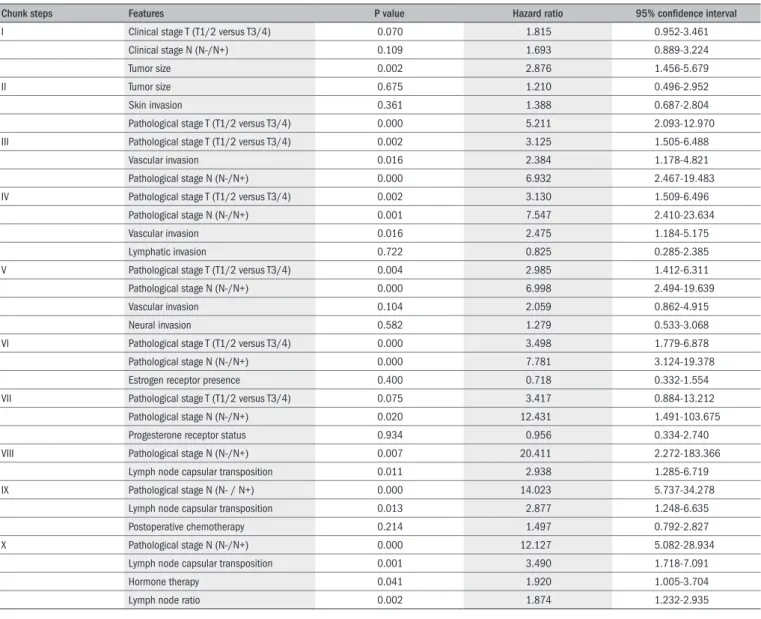
Chunk steps Features P value Hazard ratio 95% conidence interval
I Clinical stage T (T1/2 versus T3/4) 0.070 1.815 0.952-3.461
Clinical stage N (N-/N+) 0.109 1.693 0.889-3.224
Tumor size 0.002 2.876 1.456-5.679
II Tumor size 0.675 1.210 0.496-2.952
Skin invasion 0.361 1.388 0.687-2.804
Pathological stage T (T1/2 versus T3/4) 0.000 5.211 2.093-12.970
III Pathological stage T (T1/2 versus T3/4) 0.002 3.125 1.505-6.488
Vascular invasion 0.016 2.384 1.178-4.821
Pathological stage N (N-/N+) 0.000 6.932 2.467-19.483
IV Pathological stage T (T1/2 versus T3/4) 0.002 3.130 1.509-6.496
Pathological stage N (N-/N+) 0.001 7.547 2.410-23.634
Vascular invasion 0.016 2.475 1.184-5.175
Lymphatic invasion 0.722 0.825 0.285-2.385
V Pathological stage T (T1/2 versus T3/4) 0.004 2.985 1.412-6.311
Pathological stage N (N-/N+) 0.000 6.998 2.494-19.639
Vascular invasion 0.104 2.059 0.862-4.915
Neural invasion 0.582 1.279 0.533-3.068
VI Pathological stage T (T1/2 versus T3/4) 0.000 3.498 1.779-6.878
Pathological stage N (N-/N+) 0.000 7.781 3.124-19.378
Estrogen receptor presence 0.400 0.718 0.332-1.554
VII Pathological stage T (T1/2 versus T3/4) 0.075 3.417 0.884-13.212
Pathological stage N (N-/N+) 0.020 12.431 1.491-103.675
Progesterone receptor status 0.934 0.956 0.334-2.740
VIII Pathological stage N (N-/N+) 0.007 20.411 2.272-183.366
Lymph node capsular transposition 0.011 2.938 1.285-6.719
IX Pathological stage N (N- / N+) 0.000 14.023 5.737-34.278
Lymph node capsular transposition 0.013 2.877 1.248-6.635
Postoperative chemotherapy 0.214 1.497 0.792-2.827
X Pathological stage N (N-/N+) 0.000 12.127 5.082-28.934
Lymph node capsular transposition 0.001 3.490 1.718-7.091
Hormone therapy 0.041 1.920 1.005-3.704
Lymph node ratio 0.002 1.874 1.232-2.935
mary tumor, using 3.50 centimeters as the cutof point (P < 0.0001), vas-cular invasion (P < 0.0001), lymphatic invasion (P = 0.0004), neural inva-sion (P = 0.0004), skin invainva-sion (P < 0.0001), capsular transposition (P = 0.0008), estrogen receptor (ER) expression (P = 0.0186), progesterone re-ceptor (PgR) status (P = 0.0286), pathological stage T (P < 0.0001), path-ological stage N (P < 0.0001), lymph node ratio (P < 0.0001), postopera-tive chemotherapy (P < 0.0001) and postoperapostopera-tive hormone therapy (P = 0.0367). he following factors were found to be nonsigniicant: age group (P = 0.497), race (P = 0.8770), previous history of hormone therapy (P = 0.7825), previous family history (P = 0.0901), type of surgery (P = 0.0587), presence of desmoplasia (P = 0.2720), inlammatory iniltrate (P = 0.0567), comedocarcinoma (P = 0.3681), histological grading (P = 0.1800), bilateral disease (P = 0.285) and postoperative radiotherapy (P = 0.3037).
Figure 3. Overall survival rates for patients with and without postoperative hormone therapy.
1.00
0.75
0.50
0.25
0.00
0 50 100 150 200
Duration of follow-up (months) Suvival according to adjuvant hormone therapy
Hormone treatment No hormone treatment
1.00
0.75
0.50
0.25
0.00
0 50 100 150 200
Duration of follow-up (months)
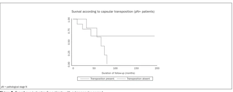
Suvival according to capsular transposition (pN+ patients)
Transposition present Transposition absent
pN = pathological stage N
Figure 2. Overall survival rates for patients with extracapsular spread.
1.00
0.75
0.50
0.25
0.00
0 50 100 150 200 250 Duration of follow-up (months)
Suvival according to pN status
pN0 pN+
Figure 1. Overall survival rates for patients with and without metastatic axillary nodes. pN = pathological stage N.
DISCUSSION
In this report, we analyzed the prognostic factors for ILC in a man-ner not performed before in the literature, with multivariate analysis per-formed through chunkwise testing methods with a backward elimination procedure. It allowed us to diminish the number of variables analyzed at
each step. One major limitation of this study was its retrospective nature, but lobular carcinomas of the breast are not as common as ductal carcino-mas and prospective studies are diicult to conduct because of the length of time involved in amassing a signiicant number of patients.
age (P = 0.0039), stage T (T1/2 versus T3, P = 0.0099), lymph node status (N0/1 versus N2, P = 0.0009), and grade (I/II versus III, P = 0.0128) afected the prognosis, in a multivariate model.10 he role of
lymph node status, capsular transposition and the size of the metastat-ic deposits have also been demonstrated in other reports.11 Histological
grading has also been shown to be a signiicant prognostic factor. In a re-port from the Danish Breast Cancer Cooperative Group, patients with grade III tumors showed a higher proportion of non-classical subtypes and signiicantly worse prognosis for overall and disease-free survival, in comparison with patients with grade I/II carcinomas.12 In another
report, histological grading was correlated with other factors for poor prognosis, such as increasing tumor size, positive lymph nodes, vascu-lar invasion and estrogen receptor negativity. Multivariate analysis has shown that histological grading is a signiicant factor indicating shorter disease-speciic and disease-free survival.13 he role of bilateral disease as
a prognostic factor has also been assessed. In a previous report, it was shown that patients with synchronous, bilateral ILC had a signiicant worse prognosis than did patients with unilateral or metachronous bi-lateral disease (P < 0.02).14
Information on prognostic factors for ILC may be also found in reports that had the original intent of comparing this histological type with IDC. In a report originally intended to compare the prognostic niicance of lobular histology, the following factors were found to be sig-niicantly linked to overall survival in cases of ILC: node status, age, pri-mary tumor size, ER status and S-phase.5 Another study also compared
the prognosis using histological analysis and concluded that for ILC, the following factors were signiicant in multivariate analysis: age at diag-nosis, tumor size, lymph node status and pathological stage. In another report analyzing the prognostic factors for 217 patients with ILC, the following factors were found to be signiicant: tumor size (P < 0.0001), axillary nodal metastasis (P < 0.0001), absence of tumor necrosis (P < 0.0001), low mitotic count (P < 0.0001), low histological grading (P = 0.001) and S-phase fraction lower than the median (P = 0.005).15
Age at diagnosis, tumor size, pathological stage and lymph node status were shown to be independent prognostic indicators for 10-year sur-vival, with no signiicant prognostic diference between ILC and IDC.16
he lymph node ratio has been shown to be an important predictor for survival among patients with stage I and II breast cancer.17
In our analysis, the number of factors found to be signiicant was smaller than in these other series. Age at diagnosis was found to be non-signiicant at diferent cutof points and as a continuous variable, and tumor size was only signiicant in the univariate analysis, and thus was excluded from the multivariate analysis. No signiicance was found in the multivariate analysis for ER status or histological grading, which were signiicant in previous reports.5 On the other hand, postoperative
hormone therapy was signiicant for survival.
CONCLUSIONS
From the data in this report, we conclude that the most important prognostic factors for lobular carcinoma of the breast seem to relate to
lymph node status and presence of capsular transposition. he pres-ence of estrogen receptors and the inclusion of postoperative hormone therapy was also associated with a signiicant improvement in survival. However, further prospective studies would be needed to conirm this evidence.
REFERENCES
1. Berg JW, Hutter RV. Breast cancer. Cancer. 1995;75(1 Suppl):257-69.
2. Li CI, Anderson BO, Daling JR, Moe RE. Trends in incidence rates of invasive lobular and ductal breast carcinoma. JAMA. 2003;289(11):1421-4.
3. Fisher ER, Gregorio RM, Fisher B, et al. The pathology of invasive breast cancer. A syllabus derived from indings of the National Surgical Adjuvant Breast Project (protocol no. 4). Cancer. 1975;36(1):1-85.
4. Martinez V, Azzopardi JG. Invasive lobular carcinoma of the breast: incidence and variants. Histopathology. 1979;3(6):467-88.
5. Arpino G, Bardou VJ, Clark GM, Elledge RM. Iniltrating lobular carcinoma of the breast: tumor characteristics and clinical outcome. Breast Cancer Res. 2004;6(3):R149-56. 6. Ferlicot S, Vincent-Salomon A, Médioni J, et al. Wide metastatic spreading in iniltrating
lobular carcinoma of the breast. Eur J Cancer. 2004;40(3):336-41.
7. Cocquyt VF, Schelfhout VR, Blondeel PN, et al. The role of biological markers as predic-tors of response to preoperative chemotherapy in large primary breast cancer. Med Oncol. 2003;20(3):221-31.
8. Greene FL, Compton CC, Fritz AG, Shah J, Winchester DP. AJCC Cancer staging atlas. New York: Springer; 2002.
9. Kleinbaum DG, Kupper LL, Muller KE, Nizam A. Applied regression analysis and multivariate methods. Paciic Grove: Books/Cole; 1998.
10. Moreno-Elola A, Aguilar A, Roman JM, et al. Prognostic factors in invasive lobular carcinoma of the breast: a multivariate analysis. A multicentre study after seventeen years of follow-up. Ann Chir Gynaecol. 1999;88(4):252-8.
11. Ladekarl M, Sørensen FB. Prognostic, quantitative histopathologic variables in lobular carci-noma of the breast. Cancer. 1993;72(9):2602-11.
12. Talman ML, Jensen MB, Rank F. Invasive lobular breast cancer. Prognostic signiicance of histological malignancy grading. Acta Oncol. 2007;46(6):803-9.
13. Rakha EA, El-Sayed ME, Menon S, Green AR, Lee AH, Ellis IO. Histologic grading is an inde-pendent prognostic factor in invasive lobular carcinoma of the breast. Breast Cancer Res Treat. 2008;111(1):121-7.
14. Dixon JM, Anderson TJ, Page DL, Lee D, Duffy SW, Stewart HJ. Iniltrating lobular carcinoma of the breast: an evaluation of the incidence and consequence of bilateral disease. Br J Surg. 1983;70(9):513-6.
15. Toikkanen S, Pylkkänen L, Joensuu H. Invasive lobular carcinoma of the breast has better short- and long-term survival than invasive ductal carcinoma. Br J Cancer. 1997;76(9): 1234-40.
16. Jayasinghe UW, Bilous AM, Boyages J. Is survival from iniltrating lobular carcinoma of the beast different from that of iniltrating ductal carcinoma? Breast J. 2007;13(5):479-85. 17. van der Wal BC, Butzelaar RM, van der Meij S, Boermeester MA. Axillary lymph node ratio
and total number of removed lymph nodes: predictors of survival in stage I and II breast cancer. Eur J Surg Oncol. 2002;28(5):481-9.
Sources of funding: None
Conlict of interest: None
Date of irst submission: October 27, 2008
Last received: May 11, 2010
Accepted: May 11, 2010