Balsamo F, Lima RFC, Batitsta RR, Formiga GJS. Intestinal obstruction due to malign breast neoplasm and peritoneal carcinomatosis: a case report. J Coloproctol, 2012;32(2): 188-192.
ABSTRACT: Peritoneal carcinomatosis due to breast cancer is rare and gastrointestinal tract involvement is also unusual. Symptoms are unspeciic and can begin many years after the primary tumor. Investigation of carcinomatosis origin is mandatory as breast cancer carcinoma -tosis can relieve partially or totally with chemo and hormonal therapy. A case of colonic obstruction due to carcinoma-tosis secondary to breast cancer is reported, emphasizing its diagnostic aspects and treatment.
Keywords: intestinal obstruction; /secondary; breast neoplasms; carcinoma; abdomen, acute.
RESUMO: A carcinomatose peritoneal secundária ao câncer de mama é entidade rara e o comprometimento do trato gastrointestinal é pouco frequente. A sintomatologia bastante inespecíica diiculta o diagnóstico e os sintomas podem surgir vários anos após o aparecimento do tumor primário. O diagnóstico da origem da carcinomatose é fundamental, pois quando a doença é secundária à neoplasia de mama, pode ocorrer remissão parcial e até total da doença com quimio e hormonioterapia. Relata-se caso de obstrução colônica devido a carcinomatose peritoneal secundária a neoplasia maligna de mama, com ênfase em seu diagnóstico e tratamento.
Palavras-chave: obstrução intestinal; /secundário; neoplasias da mama; carcinoma; abdome agudo.
Intestinal obstruction due to malign breast neoplasm and
peritoneal carcinomatosis: a case report
Flávia Balsamo1, Rafael Ferreira Correia Lima2, Rodrigo Rocha Batitsta2, Galdino José Sitonio Formiga3
1Assistant, Service of Coloproctology, Hospital Heliópolis – São Paulo (SP), Brazil. Permanent Member, Sociedade Brasileira
de Coloproctologia. 2Resident, Service of Coloproctology, Hospital Heliópolis – São Paulo (SP), Brazil. 3Head of the Service of
Coloproctology, Hospital Heliópolis – São Paulo (SP), Brazil; Permanent Member, Sociedade Brasileira de Coloproctologia.
Study carried out at the Service of Coloproctology, Hospital Heliópolis – São Paulo, SP, Brazil.
Conlict of interest: nothing to declare. Financial source: none.
Submitted on: 02/25/2011 Approved on: 08/25/2011
INTRODUCTION
Breast cancer is the most frequent malignant tu -mor in women1-4, but peritoneal carcinomatosis due
to breast cancer is rare1, accounting from 6 to 8% of
breast adenocarcinoma cases4,5.
Metastatic lesion of breast cancer usually affects lymphatic ganglions, bones, lungs, brain and liver1-3.
More rarely, it may also affect the gastrointestinal
tract, peritoneum and genital organs1,4.
Its very unspeciic symptoms make diagnosis more dificult and they may appear several years after the primary tumor1,3. The investigation of carcinoma
-tosis origin is mandatory, as breast cancer
carcinoma-tosis can relieve partially or totally with proper chemo and hormonal therapy1.
The lobular type of the disease is more associat
-ed with metastases in the gastrointestinal tract, genital
organs and peritoneum1-4.
The purposes of this report were to describe a case of colonic obstruction due to peritoneal carcino -matosis secondary to malignant breast neoplasm and
make a literature review.
CASE REPORT
-pothyroidism, treated with Puran T4 100 mcg/day and right mastectomy and hormonal therapy due to malignant breast neoplasm ten years before. The physical examination detected globus and tympanic abdomen, a little distended, no pain at supericial
and deep palpation, no palpable mass and bowel
sounds were present and normal. The proctologic exam was performed until 20 cm from the anal ca
-nal, with normal mucosa. The colonoscopic study was performed until the transverse colon with lu
-minal stenosis and preserved mucosa, blocking the device progression. Biopsies were made with sam
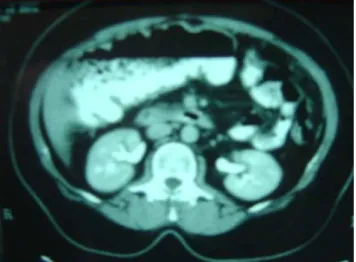
-ples from the site, whose histopathological result showed unspeciic chronic inlammatory process. Abdominal tomography showed liver without alter
-ations and colonic dilatation, with abrupt interrup
-tion at the transverse colon level, as well as great
-er omentum thickening (Figure 1). Tomography of thorax was normal (Figure 2).
As illustrated in Figure 3, the opaque enema showed free progression of rectal contrast until distal transverse colon, with inadequate illing at this level.
High digestive endoscopy until the second du
-odenal portion showed mild enanthematous gastri
-tis. The serum level of carcinoembryonic antigen (CEA) was 2.7 ng/dL.
The patient was submitted to exploratory lapa
-rotomy, which showed peritoneal carcinomatosis (Figure 4) and involvement of distal transverse colon, with the omentum full of carcinomatosis nodules.
Figure 3. Opaque enema with inadequate illing in transverse colon.
A
B
Figure 1. Abdominal tomography showing colonic dilatation.
A small portion of the omentum was resected for derivative transversostomy proximally to stenosis. The histopathological analysis of the omentum showed ad
-enocarcinoma iniltrating adipose tissues (Figure 5) and the immunohistochemical analysis showed 90% of the
Figure 4. Peritoneal carcinomatosis with distal transverse colon obstruction.
A
B
C
Figure 5. Histopathological analysis: adenocarcinoma iniltrating
adipose tissues.
A
B
Figure 6. Immunohistochemical analysis with positive markers of breast cancer.
A
B
cells positive to markers SP1 (estrogen receptor) and PgR636 (progesterone receptor), as well as presence of positiveness to cytokeratin 7 (CK7), showing primary breast adenocarcinoma (Figure 6).
The patient was submitted to adjuvant chemo
-therapy with Adriamycin 60 mg/m2 and Paclitaxel
175 mg/m2 and hormonal therapy with Anastrozole
(Arimidex®) 1 mg/day, showing improved general
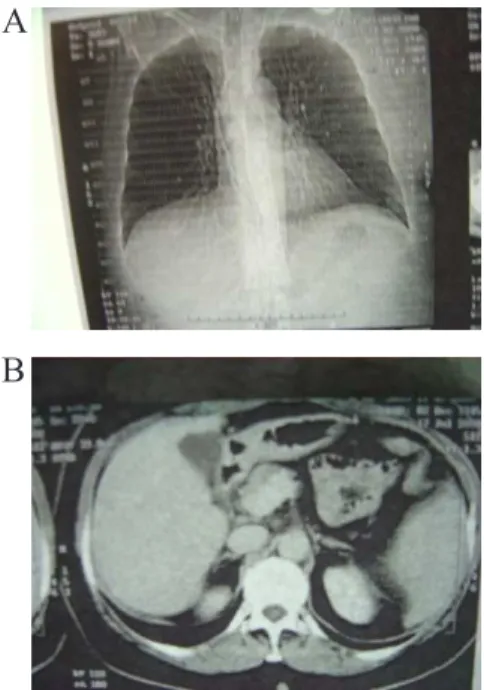
conditions and weight gain. Control exams showed absence of omentum thickening, normal liver and un
-speciic distribution of intestinal loops (Figure 7) In Figure 8, the opaque enema shows persistent stenosis of transverse colon, with dificult progression of rectal contrast until the colostomy.
The patient was submitted to a new explorato
which found the cicatricial stenosis of transverse co
-lon, and a segmental colectomy was performed in
-volving the area of stenosis, as well as colostomy with reconstruction of intestinal low and termino-terminal
colonic anastomosis (Figures 9 and 10).
The histopathological study of the specimen showed no evidence of neoplasm, only cicatricial tissue.
The patient presented good progress and was discharged from hospital six days after the surgery. Today, the patient is in 27-month follow-up, without symptoms and free from the disease.
Figure 7. (A) thorax X-ray and (B) abdominal tomography with normal aspect after chemotherapy and hormonal therapy.
A
B
Figure 8. Free progression of contrast, with dificult progression
until the stoma.
DISCUSSION
The infrequent occurrence of peritoneal carcino -matosis due to breast adenocarcinoma and its
unspe-ciic symptoms make its diagnosis more dificult4.
In the case reported, despite the endoscopic and contrast radiological and tomographic investigations in the preoperative period, it was not possible to diag
-nose it on this occasion, but only conirm the site with the probable obstruction, which has also been found by other authors1. The deinitive diagnosis was only
achieved after the histopathological and immunohis
-tochemical analysis of a fragment of the omentum ob
-tained through exploratory laparotomy.
In cases of history of breast neoplasm with any gastrointestinal tract manifestation, the secondary involvement due to breast neoplasm should be con
REFERENCES
1. Priego PJ, Rodriguez GV, Reguero MEC, Cabañas JM, Lisa EC, Peromingo RF, et al. Carcinomatosis peritonial secundaria a carcinoma lobulillar de mama. Rev Chil Cir 2007;59(3):223-8. 2. Araújo LHL, Melo AC, Moreira MML,Gomes CAS,
Noronha Jr H, Cunha WML, et al. Metástase gástrica de câncer de mama: relato de caso e revisão de literatura. Rev Bras Cancerol 2007;53(3):365-8.
3. Fillmann LS, Pinho CM, Fillmann HS, Fillmann EEP. Relato de caso: metástase de carcinoma de mama para o intestino grosso. Rev Bras Coloproct 2007;27(4):50-2.
4. McLemore EC, Pockaj BA, Reynolds C, Gray RJ, Hernandez JL, Grant CS, et al. Breast cancer: presentation and intervention in women with gastrointestinal metastasis and carcinomatosis. Ann Surg Oncol 2005;12(11):886-94. sidered as one of the hypotheses and, whenever pos
-sible, conirm the inding with immunohistochemical exams6. The immunohistochemical markers that can
help in this diagnosis are the expression of cytokera
-tin 7, estrogen and progesterone receptors and nega
-tivity to cytokeratin 201,6, also used in this case.
The interval between the primary disease appear
-ance and the peritoneal carcinomatosis may vary from months to 30 years, average interval of 6 years1; in this
case, this interval was 10 years.
It was not possible to identify the histological type of breast adenocarcinoma that affected the pa
-tient in question and, therefore, we cannot correlate the inding of carcinomatosis to lobular or ductal type.
Surgical interventions are used only to resolve
com-plications, considering that this is a disease with systemic
dissemination4, as performed here, with exploratory lap
-arotomy required for intestinal derivation due to partial bowel occlusion and to help achieve the diagnosis.
The patient remains fully asymptomatic, 27 months after the secondary disease diagnosis, as she presented a good response when submitted to chemotherapy and hormonal treatment. This inding agrees with the average survival presented in these cases, which is 24 to 36 months4.
CONCLUSION
In cases of peritoneal carcinomatosis and history of breast adenocarcinoma, the investigation of the car -cinomatosis origin is essential, as good alleviation can
be obtained with a speciic treatment.
Chemotherapy associated with conservative sur
-geries can also provide good quality of life.
5. Hewitt MJ, Hall GD, Wilkinson N, Perren TJ, Lane G, Spencer JA. Image-guided biopsy in women with breast cancer presenting with peritoneal carcinomatosis. Int J Gynecol Cancer 2006;16 Suppl 1:108-10.
6. Schwartz RE, Klimstra DS, Turnbull ADM. Metastatic breast cancer masquerading as gastrointestinal primary. Am J Gastroenterol 1998;93(1):111-4.
Correspondence to: Flávia Balsamo