279
Impact of Malnutrition and Moderate Aerobic
Training on the Structure of Arterial
Wall in Aging Rats
Vitor Caiaffo Brito1
Danielle Augusta de Sá Xerita Maux2
Belisa Duarte Ribeiro de Oliveira3
Rita de Cássia Silva Costa4
Camilla Rodrigues Souza e Silva4
Silvana Tavares Paes5
Sílvia Regina Arruda de Moraes6
1. Physiotherapist, Professor at the Animal Morphology and Physiology Department , Recife/ PE – UFRPE
2. Physiotherapist, M.A. in Pathology, Recife/PE – UFPE 3. Physiotherapist, M.A. in Physilogy and Biochemistry, Recife/PE – UFPE 4. Physiotherapist, Recife/PE – UFPE 5. M.A. in Pathology, Recife/PE – UFPE 6. Physiotherapist, Professor of the Anatomy Department, Recife/ PE – UFPE
Mailing address:
Rua Marquês do Paraná, 160, ap. 1.202 – Espinheiro – 52021-050 – Recife, PE
E-mail: [email protected]
ABSTRACT
We evaluated the late effects of multi-needy malnutrition imposed during lactation and the pos-sible effects and / or changes to both the aging process as of moderate aerobic training on the luminal diameter and the tunica media area of the left common carotid artery, horizontal aorta and thoracic aorta of rats in the aging process. Twenty-four Wistar male rats were separated according to diet and physical training: SNG – Sedentary Nourished Group, TNG - Trained Nourished Group, SMG- Sedentary Malnourished Group and TMG – Trained Malnourished Group. At 10 months, the TNG and TMG were submitted to aerobic training in water for 8 weeks. Then the mice were anesthetized and sacrificed and the arterial fragments were collected for histological processing. The area of the tunica media was mea-sured by the difference between external and internal areas of the arterial wall and luminal diameter was measured from the average of two diameters measured from four points marked on the internallayer of the vessel. The area of tunica media decreased in the horizontal aorta when compared groups SNG x SMG (p = 0.015) and increased in left common carotid artery between groups SNG and TNG (p = <0.001). The thoracic aorta showed an increase in luminal diameter when comparing the SMG with the TMG (p = 0.041). Multi-needy malnutrition induced both to partial changes in the horizontal aorta wall, as well as physical training was able to promote changes in the tunica media area of the left common carotid artery and luminal diameter of the thoracic aorta.
Keywords: aerobic moderate training, arterial remodeling, aorta, carotid artery.
INTRODUCTION
The tissues and organs have “critical” development periods(1),
which are characterized by fast growth, replication, cellular diffe-rentiation and maturation of organs and systems, and are decisi-ve for the determination of the morphofunctional characteristics in adults(2). Disorders occurred during this period may determine
permanent or long-term alterations in different organs(3). Among
these disorders, we can stress the nutritional deficiencies which, associated with unfavorable socieconomical conditions(4), may be
related to high risk of development of defects in metabolism, as well as trigger diseases such as type diabetes, obesity(5), systemic
arterial hypertension (SAH), as well as cardiovascular diseases in more advanced ages(6,7).
The type, duration as well as phase of development at which malnutrition occurs may determine the type of interference on the organic structure(3,5). Thus, malnutrition has been mentioned as one
of the main non-genetic factors which can interfere in the normal development of the organs(2). During the neonatal development,
malnutrition may reflect on alterations such as growth delay(8), low
weight(9), alterations in the arteries straight muscle tissue(10),
hemo-dynamic alterations(11) as well as cardiovascular problems(8,12).
Seve-ral experimental studies showed that a hypoprotein and hypocaloric diet or food restriction in initial phases of life, may cause adverse remodeling of the tunica media of the throacic aorta, hyperplasia of the straight muscle cells and consequently, increase of wall thi-ckness of this artery(7), endothelial dysfunctions such as increase
of basal tonus of the left internalthoracic artery(13), increase of the
contraction strength of the femoral arteries(14), insufficiency in the
relaxation of the vascular endothelium(15) leading to increase of
systolic blood pressure(7).
Some studies have shown that the cardiovascular function may be significantly improved by the practice of physical training (16,17),
which contributes to reduction on the cardiovascular morbimor-tality indices(16).
Physical training generally speaking, increases the energetic need of the body, causing expressive alterations in the cellular metabolism, implying in a series of physiological responses in the body systems, and especially in the cardiovascular system(18).
Frequent and regular exposure to physical training result in adap-tations to the organism typically represented by alterations in the heart rate. The increase in venous return and improvement in myocardic contrability(16,19), induced by physical training, can
result in relative rest bradycardia(19). This alteration of heart rate
is also related to improvement in cardiac chronotropism due to alterations occurred in the atrioventricular conduction system(16).
The peripheral arteries adapt with alterations in their structure induced by physical training in a trial to correspond to the meta-bolic needs of the skeletal musculature supplied by these vessels. These adaptations are represented by the increase of the vessels diameter as well as in the arterial walls diameter(20).
Thus, the study aims to analyse the late effects of the altera-tions early caused by a multi-needy malnutrition status on the vascular structure of the left common carotid, horizontal aorta and thoracic aorta arteries. Additionally, to demonstrate how ae-robic physical training (AT), with moderate intensity can interfere in this process.
EXERCISE AND SPORTS SCIENCES
280
METHODS
All experiments were performed according to the guidelines suggested by the Brazilian Committee of Animal Experimentation (COBEA). The study was submitted to and approved by the Ethics in Animal Experimentation Committee (CEEA) of the Center of Biologi-cal Sciences of UFPE (protocol # 21931/2008 – 29).
24 male albino Wistar rats from the animal facility of the Nutrition Department of the Federal University of Pernambuco-UFPE were used. The animals were randomly sorted in two groups: Nourished group N (n = 12), composed of pups which received LABINA diet®
and Malnourished group – M (n = 12), composed of pups which received basic regional diet (RBD) form the first to the 21st day
of life, and after this period, also started being fed with standard diet (LABINA®
).
At 10 months, the animals were randomly distributed accor- ding to performance or not of physical training, in four subgroups: Sedentary Nourished Group (SNG), Trained Nourished Group (TNG), Sedentary Malnourished Group (SMG) and Trained Malnour-ished Group (TMG). Each experimental subgroup was composed of six animals.
The animals from TNG and TMG were submitted to the aerobic physical training protocol with moderate intensity(21). The training
modality sued was swimming in a plastic round tank with water temperature kept between 30 and 32°C through a digital thermostat. The trained animals were submitted to an adaptation period which lasted 10 minutes on the first day. On the following days, 10 minutes were added to the training until total time would reach 40 minutes on the fourth day and remaining with this duration until the end of the first week. Subsequently, 40min/day were kept until the eighth training week. The animals of the SNG and SMG limited to activities inside their cage.
At the end of the training period, the animals received intra-muscular anesthesia of Xylazine Chloridate (Rompum®
– Bayer) and Ketamine. The thoracic and abdominal cavities were then opened and the hearts were perfused with cannulation of the left ventricle for introduction of 250ml of saline solution at 0.9% with 1ml of heparin per liter of saline solution. The right atrium was opened for draining of the perfusion solutions. After washing of the system, the tissues were treated with glutaraldehyde 4% diluted in phosphate buffer at 0.5 molar with pH = 7.2. Fragments of the left common carotid artery (5mm proximal to the horizontal aorta) and horizon-tal aorta (portion between the arterial brachiocephalic trunk and the left common carotid artery) were dissected for removal of the adhering tissues, treated with alcohol 70% and transversal cuts of 3µm were stained by Verhoeff-Van Gienson method.
The histological preparations of the left common carotid ar-tery were submitted to histomorphometric analysis through pho-tomicrograpy performed with help of a system composed of the Tv Turner Application software, a Samsung video camera (model SHC-410NAD) attached to the Olympus optical microscope (model Bx-50) with final increase of 40x, all integrated to a micro computer. The photomicrografies of the horizontal aorta and thoracic arteries were performed with the aid of a Sony digital camera (model DSC-H7) directly attached to the lenses of an Olympus BX-50 optical microscope with final increase of 40x.
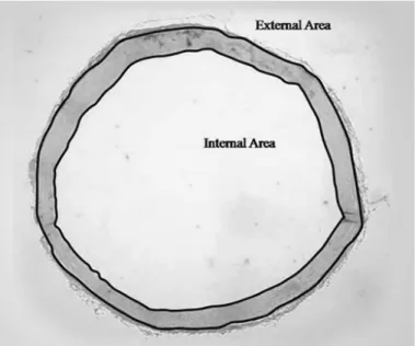
Having these photomicrografies as starting point, the difference between the value of the external and internal areas of the vessel wall was performed for determination of the area of the tunica media of the arterial wall (figure 1).
The 0o, 90º, 270º and 360o points were determined for
measure-ment of the luminal diameter of the vessels. Two straight lines were drawn in a way they crossed each other and formed a 90 degree angle from these points. Subsequently, the diameter values were summed and divided by two, so that a mean value could be found for the luminal diameter of the vessels(22) (figure 2).
Measurement of both luminal diameter and area of tunica me-dia of the arteries was performed with the Scion Image software for Windows (Beta 4.0.3 version for Windows). A scale of 0.564 pixels/ micrometer was used for the left common carotid artery values and for the horizontal and thoracic aorta arteries, the scale of 0.279 pixels/micrometers was used.
Figure 1. Illustration of the measurement of the tunica media area of the left com-mon carotid artery wall, horizontal aorta and thoracic with aid of the Scion Image software for Windows. Increase of 40x.
281
Histomorphometric analysis of the arteries was done with the SPSS 11.5 program for Windows and, for comparison between groups, the Student’s t test for independent and parametric sam-ples as well as Mann-Whitney test for non-parametric samsam-ples, with significance level of p < 0.05 were used; data were presented in mean ± standard deviation.
RESULTS
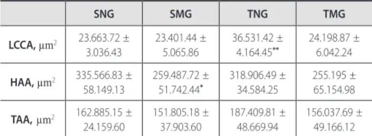
Concerning the area of the tunica media, the horizontal aorta artery presented decrease when SNG and SMG are compared (335.566.83 ± 58.149.13µm2 and 259.487.72 ± 51.742.44µm2,
respectively, p = 0.015), demonstrating partial influence of nu-tritional deficiency, since neither the left common carotid nor the thoracic aorta arteries presented significant differences. Still concerning the area of the tunica media, the left common carotid artery presented significant increase between TNG (36.531.42 ± 4.164.45µm2) and SNG (23.663.72 ± 3.036.43) (p ≤ 0.001),
indica-ting a partial effect of physical training over the structure of the arterial wall. The remaining arteries under study did not present alterations in the normonourished animals (table 1).
Regarding the luminal diameter, only the thoracic aorta artery presented significant increase when the SMG (798.98 ± 69.83µm2)
and TMG (980.35 ± 154.62µm2) were compared (p = 0.041),
de-monstrating that aerobic physical training with moderate intensity partially influenced the structure of the wall of this artery. The left common carotid and horizontal aorta did not duffer influence of the moderate aerobic training on the luminal diamater (table 2).
SNG SMG TNG TMG
LCCA, µm2 23.663.72 ±
3.036.43
23.401.44 ± 5.065.86
36.531.42 ± 4.164.45**
24.198.87 ± 6.042.24
HAA, µm2 335.566.83 ±
58.149.13
259.487.72 ± 51.742.44*
318.906.49 ± 34.584.25
255.195 ± 65.154.98
TAA,µm2 162.885.15 ±
24.159.60
151.805.18 ± 37.903.60
187.409.81 ± 48.669.94
156.037.69 ± 49.166.12
Table 1. Mean values concerning area of the arterial tunica media. Data as mean ± SD.
*Difference between groups SNG and SMG, p = 0.015 – Student’s t test. **Difference between groups SNG and TNG, p ≤0.001 – Student’s t test. LCCA: left common carotid artery, HAA: horizontal aorta artery, TAA: thoracic aorta artery and µm2: micrometer to the square.
SNG SMG TNG TMG
LCCA, µm2 358.16 ± 29.73 331.41 ± 43.98 367.02 ± 26.44 355.64 ±18.32
HAA, µm2 1.041.63 ± 67.56 934.15 ± 53.44 1.107.95 ± 56.73 1.080.92 ±
166.28
TAA,µm2 843.23 ± 42.02 798.98 ± 69.83 892.41 ± 89.70 980.35 ±
154.62 ***
Table 2. Mean values concerning the luminal diameter of the arteries. Data as mean± SD.
***Difference between groups SMG and TMG, p = 0.041 – Mann-Whitney test. LCCA: left common carotid artery, HAA: horizontal aorta artery, TAA: thoracic aorta artery and µm2: micrometer to the square.
which may interfere in the development phases, such as malnu-trition, may favor the onset of cardiovascular pathologies in late phases of life. The mechanisms which promote such alterations have not been totally elucidated(3), although such vascular
patho-logies have already been related to alterations in the structure of vessels, notably to alterations on the arterial wall(24).
The multi-needy malnutrition used in this study during the lactation period promoted reduction in the area of the tunica media of the horizontal aorta artery of 12-month rats, demonstra-ting that, although malnutrition has occurred in a very early phase of the animal’s life, the alterations caused by it remained, reducing hence the thickness of the arterial wall. A study with rats submit-ted to protein malnutrition during gestation and lactation also demonstrated reduction in the thickness of the tunica media of the aorta artery(23). However, this same author in another study(24),
observed increase of thickness of arterial wall of the abdominal aorta in neonatal pups, attributing this divergence in results to the different models of fetal malnutrition. In addition to this, a previous study carried out in our laboratory in rats with 80 days of life whose mothers were fed with hypoprotein diet during the gestational and neonatal periods, demonstrated reduction of thickness of the left common carotid artery(25). It has also been demonstrated that
in humans and adults (mean of 58 years) whose mothers were not fed with hypoprotein diet presented reduction of thickness of the left common carotid artery(26). According to Barker and Hanson(27),
these data divergences may occur due to the structural differences of the artery walls. For example, the coronary arteries are much more sensitive to alterations of the structure components (pro-teins, straight muscle tissue) derived form nutritional aggression, which may imply in increase of thickness of their walls as well as reduction in luminal diameter, increasing hence the risk of cardio-vascular diseases, especially of ischemic order(27). These structural
alterations may have not occurred with the arteries of our study, demonstrating that the multi-needy diet may have caused loss of structural components, ended with reduction of thickness of the arterial wall found in our study.
Khorram et al.(28) using rats with one day of life and whose
mothers had been submitted to food restriction (50% of food intake compared to control group) on the 10th Day of gestation, and Pires and Mandarim-de-Lacerda(7), using animals with 36
we-eks submitted to protein restriction during lactation, on the other hand observed increase of thickness of tunica media of aorta ar-tery of these animals. This increase of arterial thickness could be related to the increase of systolic blood pressure found by these authors in their studies.
At basal conditions, the vascular endothelium secretes relaxing factors (nitric oxide, NO; prostacyclins) and contractile factors (en-doperoxides, thromboxane A2 and endothelins, ET)(29). The increase
of contraction strength of the arteries as well as the insufficiency in the endothelial relaxation are related to decrease of NO availa-bility in the vascular endothelium(14,15). NO induces to relaxation
of the straight muscle layer of the arterial wall through increase of conduction of K+ ions to the cellular interior, hyper polarizing
the cells and inhibiting the intracellular flow of Ca++ ions, altering
hence the contraction mechanism of the straight musculature of the arteries. Rats submitted to protein restriction in initial pha-DISCUSSION
282
ses of life, presented disorders in the endothelial relaxation of the thoracic aorta(14,15), implying in increase of vascular tonus, which
could explain the fact these animals present increase in thickness of arterial wall.
Cardiovascular diseases, such as arteriosclerosis and SAH, may be related to increase of vascular tonus derived from disorders in endothelial relaxation(29). The natural aging process can also
indu-ce to alterations in the vascular endothelium(30). Large arteries as
the aorta are mainly constituted by straight muscle tissue, which, among other functions, produces the components of the extracellu-lar matrix(31). Alterations on the arterial wall consequent from aging
may occur due to alterations in the components of the extracellular matrix such as decrease of elastin biosynthesis, increase of elastane enzyme activity, collagen deposition, hypertrophy and hyperplasia of the straight muscle cells(32). Thus, the arterial wall becomes more
rigid, due to higher concentration of collagen fibers, increasing even more the probability of cardiovascular diseases in older subjects(33).
The maternal nutrition status during gestation and lactation are more important and decisive to the body development than the genetic constitution itself(34). Gestational and neonatal malnutrition
causes negative impact on the development of the organs and systems which cannot be reverted in the long term, even with suitable diet after the critical period of development(35), which, in
rats, comprehends the 21 first days of life(2). Our study
demonstra-ted that the multi-needy diet administered in the neonatal period induced alterations in the vascular system which persisted during the entire life of the animal, despite they have been fed immediately after weaning with a balanced diet.
Physical training has been recommended as an excellent pre-vention factor of cardiovascular diseases(36) for promoting
he-modynamic and autonomous adaptations, structural alterations on the arterial vessel walls as well as decrease in sympathetic to-nus(17,18,21). However, in the present study, aerobic physical training
with moderate intensity using swimming did not influence on the tunica media area of the wall of the horizontal and thoracic aorta arteries. Maux et al.(25) did not observe alterations on the arterial
wall of horizontal aorta of rats submitted to protein malnutrition in gestation and lactation which performed physical training from the age of 60 days, during eight weeks. Likewise, Huonker et al.(20),
using athletes of different sports modalities, demonstrated there are not dimension alterations in the aorta artery, possibly due to the predominance of elastic fibers in this artery. According to the same authors, the aorta artery is a central vessel responsible for collecting the blood flow ejected by the left ventricle, through a passive dilatation caused by the expansion of the elastic structures of the artery. Such expansion allows an elastic detachment of the vessel wall during the ventricular diastole(20). Since the aorta arterial
wall is highly elastic, it may not give away to the increased blood flow due to cardiac adaptations derived from physical exercise(18),
which could explain the fact that we had not found difference in thickness of the horizontal and thoracic aorta arteries.
However, swimming physical training promoted increase of the tunica media of the left common carotid artery. Similar data have been demonstrated by Maux et al.(25) using the left common carotid
artery of malnourished rats in the gestational and neonatal phases with hypoprotein diet which performed swimming training during eight weeks. On the other hand, the study by Tanaka et al.(37) did not
demonstrate differences in the thickness of the arterial wall of the
common carotid in young (18-37 years), adults (38-57 years) and older (58-77 years) sedentary individuals and who have practiced regular physical training for three months. These authors attribute the lack of differences to the intensity of the used training protocol. Aerobic physical training works as a stimulus to the proliferation of endothelial progenitor cells and of straight muscle fibers, leading to increase in the endothelial and muscular layers of the artery, and can result in increase of its thickness(26). Moreover, the present
study used animals in aging process, which are more sensitive to present increase of thickness of the arterial wall.
It is believed that the natural aging process associated with nu-tritional aggressions may promote increase in stiffness and thickness of the arterial wall. Such alterations may lead to reduction of luminal diameter of some arterial vessels(38). Conversely, the present study
observed increase in luminal diameter of the thoracic aorta artery in the malnourished animals which practiced training compared with the sedentary malnourished animals. Maux et al.(25), also using
swimming as physical training, observed increase in the diameter of left common carotid artery in malnourished animals. Huonker et al.(20) obtained different results concerning the luminal diameter of
different arterial vessels: while the aortic arch and the abdominal aorta in trained and non-trained athletes did not present differences, the subclavian artery presented wider diameter in the dominant limb of athletes from tennis modality.
The tunica media of the muscular arteries is predominantly constituted by straight muscular cells, with scattered elastic mem-branes and few collagen fibers(38). Due to this display, the arterial
wall would be more prone to alterations in its lumen caused by the alterations in the tonus of the straight muscle cells of the tunica media according to the variation in the demand flow of a given or-gan, such as the skeletal muscle during training(20). Moreover, since
the arterial wall in malnourished animals is normally thinner than in normonourished animals, it would be more vulnerable to distension and consequently, increase of demand promoted by the exertion resulting from physical training could cause increase in the vessel diameter. This fact could explain the increase in luminal diameter of the thoracic aorta artery in the malnourished animals submitted to physical training with moderate intensity observed in the present study. We speculate that during swimming training the animals use more the lower limbs, and need greater blood flow for this region and thus can cause more pressure on the thoracic aorta walls and increase in the luminal diameter of this artery.
The nutritional aggression occurred during the lactation period was efficient in partly inducing alterations in the structure of the arterial wall, which remained during the entire life of the animals. However, aerobic physical training with swimming in later phases of the malnourished animals lives during lactation was partially a factor which can contribute to revert the alterations caused by malnutri-tion since exercising promotes increase of luminal diameter of the thoracic aorta. Nevertheless, it was not able to revert the structural alterations derived from malnutrition and the aging process on the horizontal aorta artery wall.
283
REFERENCES
1. Barker DJP. Fetal programming of coronary heart disease. TRENDES in Endocrinology & Metabolism. 2002;13:364-8.
2. Morgane PJ, Austin-LaFrance R, Bronzino J, Tonkiss J, Díaz-Cintra S, Cintra L, et al. Prenatal malnutrition and development of the brain. Neurosci Biobehav Rev. 1993;17;91-128.
3. Ashton N. Perinatal development and adult blood pressure. Braz J Med Biol Res. 2000;33;731-40. 4. Fernandes MTB, Sesso R, Martins PA, Sawawa AL. Increased blood pressure in adolescents of low
socioeconomic status with short stature. Pediatr Nephrol. 2003;18:435-9.
5. Bonfim AS, Mandarim-de-Lacerda, CA. Programação pré-natal de hipertensão arterial na vida adulta. Revista da SOCERJ. 2005;18(6).
6. Paixão ADO, Maciel CR, Teles MBB, Figueiredo-Silva JL. Regional Brazilian diet-induced low birth weight is correlated with changes in renal hemodynamics and glomerular morphometry in adult age. Biol Neonate. 2001;80:239-46.
7. Pires KMP, Mandarim-de-Lacerda CA. Restrição proteica na lactação como causa de hiperplasia da tunica media da aorta em ratos adultos. Revista da SOCERJ. 2005;18(3).
8. Barker DJP. In utero programming cardiovascular disease. Theriogenology. 2000;53:555-74. 9. Sawawa AL, Solimos GMB, Menezes TM, Martins PA. Os dois Brasis: quem são, onde estão e como
vivem os pobres brasileiros. Estudos Avançados. 2003;17(48).
10. Paixao ADO, Alessio MLM, Martins JPC, Leger CL, Monnier L, Pare’s-Herbute N. Regional Brazilian diet induced pre-natal malnutrition in rats is correlated with the proliferation of culture vascular smooth muscle cells. Nutr Metab Cardiovasc Dis. 2005;15:302-9.
11. Hoy W, Ress M, Kile E, Mathews JD, Wang Z. A new dimension to the Barker hypothesis:Low birthweight and susceptibility renal disease Kidney Int. 1999;56:1072-7.
12. Ericksson JG, Forsén T, Tuomilehto J, Osmond C, Barker DJP. Early grow and coronary heart disease in later life: longitudinal study. Br Med J. 1999;318:427-31.
13. Khan OA, Torrens C, Noakes DE, Poston L, Hanson MA, Green LR, et al. Effects of pre-natal and ear-ly post-natal undernutrition on adult internal thoracic artery function. Eur J Cardiothorac Surg. 2005;28:811-15.
14. Ozaki T, Nishina H, Hanson MA, Poston L. Dietary restriction in pregnant rats causes gender-related hypertension and vascular dysfunction in offspring. J Physiol. 2001;530:141-52.
15. Brawley L, Itoh S, Torrens C, Barker A, Bertram C, Poston L, et al. Dietary protein restriction in preg-nancy induces hypertension and vascular defects in rat male offspring. Pediatr Res. 2003;54:83-90. 16. Almeida MB, Araujo CGS. Efeitos do treinamento aeróbico sobre a freqüência cardíaca. Rev Bras Med
Esporte. 2003;9(2): 113-120.
17. De Angelis K, Wichi RB, Jesus WRA, Moreira ED, Morris M, Krieger EM. Exercise training changes autonomic cardiovascular balance in mice. J Appl Physiol. 2004;96:2174-8.
18. Monteiro MF, Sobral Filho DC. Exercício físico e o controle da pressão arterial. Rev Bras Med Esporte. 2004;10(6): 513-516.
19. Souza HC, Penteado DM, Martin-Pinge MC, Barbosa Neto O, Teixeira Vde P, Blanco JH, et al. O bloqueio da síntese do óxido nítrico promove aumento da hipertrofia e da fibrose cardíaca em ratos submetidos a treinamento físico. Arq Bras Cardiol. 2007;82:99-104.
20. Huonker M, Schmid A, Schimidt-Trucksäß D, Grathwohl D, Keul J. Size and blood flow of central
and peripheral arteries in highly able-bodied and disabled athletes. J Appl Physiol. 2003;95:685-91. 21. Medeiros A, Oliveira EM, Gianolla R, Casarini DE, Negrão CE, Brum PC. Swimming training increase cardiac vagal activity and induces cardiac hypertrophy in rats. Braz J Med Biol Res. 2004;37:1909-17. 22. Aguila MB, Mandarim-de-Lacerda CA. Aorta wall quantitative alterations due different long-term high
fat diet in rats. Food Chem Toxicol. 2003;41:1391-97.
23. Skilton MR, Gosby AK, Wu BJ, Ho LM, Stocker R, Caterson ID, et al. Maternal undernutrition reduces aortic wall thickness and elastin content in offspring rats without altering endothelial function. Clin Sci (Lond). 2006;111:281-7.
24. Skilton MR, Evans N, Griffiths KA. Aortic wall thickness in newborns with intrauterine growth restriction. Lancet. 2005;365:1484-86.
25. Maux DASX, Araujo TN, Viana MT, Andrade MA, Paes ST, Moraes SRA. Influência do treino físico mo-derado sobre a estrutura da parede arterial de ratos submetidos à desnutrição protéica gestacional e neonatal. Rev Bras Med Esporte. 2009;15:334-7.
26. Painter RC, de Rooij SR, Hutten BA, Bossuyt PM, de Groot E, Osmond C, et al. Reduced intima media thickness in adults after prenatal exposure to the Dutch famine. Atherosclerosis. 2007;193:421-7. 27. Barker DJ, Hanson MA. Altered regional blood flow in the fetus: the origins of cardiovascular disease?
Acta Paediatric. 2004;93:1559-60.
28. Khorram O, Khorram N, Momeni M, Han G, Halem J, Desai M, et al. Maternal undernutrition inhibits angiogenesis in the offspring: a potential mechanism of programmed hypertension. Am J Physiol Regul Integr Comp Physiol. 2007;293:745-53.
29. Waldron GJ, Ding H, Lovren F, Kubes P, Triggle CR. Acetylcholine-induced relaxation of peripheral arteries isolated from mice lacking endothelial nitric oxide synthase. Br J Pharmacol. 1999;128:653-8. 30. Robert L. Aging of the vascular wall and atherogenesis: role of the elastin-laminin receptor. Athe-
Athe-rosclerosis. 1996;123:169-79.
31. Santhiago V, Silva ASR, Gobatto CA, Mello MAR. Treinamento físico durante a recuperação nutricional não afeta o metabolismo muscular da glicose de ratos. Rev Bras Med Esporte. 2006;12(2): 76-80. 32. Lakata EG, Levy D. Arterial and Cardiac Aging: Major Shareholders in Cardiovascular Disease Enterprises:
Part I: Aging Arteries: A “Set Up” for Vascular Disease. Circulation 2003;107:139-46. 33. O’Rourke MF. Arterial aging:pathophysiological principles. Vasc Med. 2007;12:329-42.
34. Dubos R, Savage D, Schaedler R. Biological Freudianism: lasting effects of early environmental influ-ences. Pediatrics. 1966;38:789-800.
35. Blackwell NM, Blackwell RQ, Yu TTS, Weng YS, Chow BF. Further studies on growth and feed utilization in progeny of underfed mother rats. J Nutrition. 1968;97:79-4.
36. Myers J, Prakash M, Froelicher V, Do D, Partington S, Atwood E. Exercise capacity and mortality among men referred for exercise testing. N Engl J Med. 2002;346:793-01.
37. Tanaka H, Seals DR, Monahan KD, Clevenger CM, De Souza CA, Dinenno FA. Regular aerobic exercise and the age-related increase in carotid artery intima-media thickness in healthy men. J Appl Physiol. 2002;92:1458-64.