Action of ascorbic acid on the healing
of malnourished rats
’
skin wounds
1Ação do ácido ascórbico na cicatrização de
feridas cutâneas de ratos desnutridos
Gisele Alborghetti NAI2 Louise Maria Freitas MANZOLI2 Tayane Carvalho Isidoro da SILVA3 Larissa de Queiroz MAMEDE4
Mary Ellen de Oliveira Martins DISCONZI4 Rogério GIUFFRIDA2
A B S T R A C T
Objective
To evaluate the action of ascorbic acid on the healing of malnourished rats’ cutaneous wounds compared with normal weight rats.
Methods
We used 92 adult, male Wistar rats divided into four groups: 24 normal weight rats given only water and chow; 24 normal weight rats given vitamin C by gavage (340 mg/kg 12/12 hours); 22 malnourished rats given only water and chow; and 22 malnourished rats given vitamin C by gavage (340 mg/kg 12/12 hours). Malnutrition was induced by feeding the animals half of their daily energy requirement for 30 days. Two incisions were made, one sutured (healing by primary intention) and one left unsutured (healing by secondary intention). The rats were euthanized on the third, seventh, and fourteenth days of the experiment.
Results
The following parameters differed significantly between the groups (p>0.05): granulation of the wound edge in the primary and secondary intention; extent of injuries on day 7 for primary intention and on day 3 for secondary intention; reepithelialization on day 7 for primary intention; fibrin-leukocyte scab on day 14 for
1
Article based on the master’s thesis of LMF MANZOLI intitled “Ação do ácido ascórbico na cicatrização de feridas cutâneas de ratos desnutridos”. Universidade do Oeste Paulista; 2013.
2
Universidade do Oeste Paulista, Programa de Pós-Graduação em Ciência Animal. R. José Bongiovani, 700, 19050-680, Presidente Prudente, SP, Brasil. Correspondência para/Correspondence to: GA NAI. E-mail: <[email protected]>.
3
Universidade do Oeste Paulista, Faculdade de Odontologia de Presidente Prudente, Curso de Odontologia. Presidente Prudente, SP, Brasil.
4
primary intention; amount of neovascularization and concentration of macrophages, fibroblasts, and collagen fibers for primary and secondary intention.
Conclusion
The use of vitamin C in malnourished and normal weight rats increases fibroblast proliferation and collagen deposition in the tissue, which helps to improve healing both by primary and secondary intention.
Indexing terms: Ascorbic acid. Malnutrition. Skin. Vitamins. Wound healing.
R E S U M O
Objetivo
Avaliar a ação do ácido ascórbico na cicatrização de feridas cutâneas de ratos desnutridos e de eutróficos.
Métodos
Utilizaram-se 92 ratos Wistar, adultos, machos, divididos em quatro grupos: 24 ratos eutróficos que receberam somente água e ração; 24 ratos eutróficos que receberam vitamina C por gavagem (340 mg/kg de 12/12 horas); 22 ratos desnutridos que receberam somente água e ração; 22 ratos desnutridos que receberam vitamina C por gavagem (340 mg/kg de 12/12 horas). A desnutrição foi realizada fornecendo-se aos animais metade da ração diária durante 30 dias. Realizaram-se duas incisões, uma suturada (cicatrização por 1ª intenção) e outra não suturada (cicatrização por 2ª intenção). Os ratos foram sacrificados no 3º (D3), 7º (D7) e 14º (D14) dia do experimento.
Resultados
Observou-se diferença estatística (p<0.05) entre os grupos para os parâmetros: granulação na borda da ferida para 1ª e 2ª intenção; medida das lesões por 1ª intenção em D7 e para cicatrização por 2ª intenção em D3; reepitelização das lesões em D7 para 1ª intenção; crosta fibrinoleucocitária em D14 para 1ª intenção; quantidade de neovascularização, concentração de macrófagos e concentração de fibroblastos e de fibras colágenas para 1ª e 2ª intenção.
Conclusão
O uso de vitamina C em desnutridos assim como em eutróficos aumenta a proliferação fibroblástica e depósito de colágeno no tecido, o que contribui para melhorar a cicatrização de 1ª e 2ª intenção.
Termos de indexação: Ácido ascórbico. Desnutrição. Pele. Vitaminas. Cicatrização.
I N T R O D U C T I O N
A chronic or complex wound may be an important source of morbidity in many individuals in critical condition or in patients with infection or pressure ulcers1.
Wound healing involves a perfect and coordinated cascade of cellular and molecular events that interact to repave and rebuild tissue2. It is a complex but ordered phenomenon coordinated by chemical mediators, such as Vascular Endothelial Growth Factor (VEGF) and Transforming Growth Factor Beta (TGF-β), among others, that involves the following process: induction of the inflammatory process in response to the initial injury with removal of the damaged
tissue; proliferation and migration of parenchymal and connective tissue cells; angiogenesis and formation of granulation tissue; synthesis of extracellular matrix proteins and collagen deposition; tissue remodeling; wound contraction and acquisition of wound strength2.
Secondary intention healing relies on granulation and wound contraction for joining the edges2.
The factors that affect the healing process can be local (infection, foreign body, mechanical factors) and/or systemic (metabolic condition, nutrition, circulatory condition), and they may increase or reduce the scar. Hemodynamic instability, metabolic stress, and nutritional status may interfere with the healing stages3.
Although the role of nutritional supplementation on wound treatment is still unclear4, and the nutrients that promote proper healing are unknown, it is certain that proteins, vitamins, arginine, glutamine, iron, zinc, and selenium have significant beneficial effects on healing and the immune system5.
Ascorbic acid hydroxylates the lysine and proline in protocollagen necessary for the cross-links between collagen fibers because they maintain the prosthetic iron (cofactor) of hydroxylase enzyme in the ferrous form (reduced) while maintaining enzymatic activity. For this reason ascorbic acid is important in maintaining normal connective tissue and in healing by participating in the synthesis of the collagen matrix6.
Protein malnutrition is associated with poor healing by reducing fibroblast production, decreasing angiogenesis and collagen synthesis, and lowering tissue remodeling capacity7.
In third world countries, Protein-Energy Malnutrition (PEM) remains a common problem, but it also occurs in the richest societies and impoverished communities7. Additionally, PEM is associated with surgical procedures like bariatric surgery5. Despite recent evidence of reduced rates of protein-calorie malnutrition in Brazil, especially in the Northeast, malnutrition remains the most important endemic deficiency in our country, placing a high demand on health services and causing high mortality rates8-10.
The framework of malnutrition can impair the process of tissue repair, and ascorbic acid deficiency decreases fibroblast synthesis of collagen. No studies in the literature have
evaluated the effect of ascorbic acid on the wound healing of malnourished rats.
This study aimed to determine the action of ascorbic acid on the healing of skin wounds in malnourished versus normal weight rats.
M E T H O D S
This study was approved by the Animal Research Ethics Committee of Universidade do
Oeste Paulista under Protocol nº 1.103, on June
1, 2012.
For this study, we used 96 adult, male Wistar rats (Ratus Norvegicus albinus) weighing 200 to 250 g. The rats were separated and placed in individual small and rectangular boxes measuring 30x20x13 cm, the accommodation recommended for adult rats. The rats were kept in an air-conditioned vivarium with controlled humidity and temperature and a 12:12-hour light-dark cycle. The animals were weighed weekly until euthanized.
All animals (n=96) received the same chow (Supralab Comércio e Serviço Ltda, São Leopoldo, Brazil). Malnutrition was induced in 48 animals by feeding them 50% of the recommended daily chow for 30 days11, equivalent to 15 to 20 g, based on their usual daily intake12. Malnutrition was assessed by weighing the animals weekly. Animals with a weight loss of 15 to 20% were considered malnourished.
acetaminophen was administered postoperatively at a dose of 25-75 mg/250 g of body weight orally (in the drinking water), 4/4 hours, for two days13.
Four malnourished animals died after the incisions were made as there was no recovery from anesthesia.
The animals were then divided into four groups: E (normal weight rats) - 24 normal weight rats with free access to food and water, EVC (normal weight rats given vitamin C) - 24 normal weight rats given vitamin C (Cewin drops, Sanofi Aventis Pharmaceuticals, São Paulo, Brazil) by gavage at a dose of 340 mg/kg at 12/12 hours (7:30 hours a.m. and 7:30 hours p.m.) diluted in 1 mL of 0.9% saline15, M (malnourished rats) -22 malnourished with free access to water and 50% of the recommended daily amount of chow, MVC (Malnourished Rats Given Vitamin C) - 22 malnourished rats given vitamin C (Cewin drops, Sanofi Aventis Pharmaceuticals, São Paulo, Brazil) by gavage at a dose of 340 mg/kg at 12/12 hours (7:30 hours a.m. and 7:30 hours p.m.) diluted in 1 mL of 0.9% saline and half the recommended daily amount of chow.
Eight rats from each group were euthanized on the third (D3), seventh(D7) and fourteenth (D14) days of the experiment16. The animals were killed in a CO2 chamber (Industry Seaside, São Paulo, Brazil). After euthanasia, the area containing the skin wound was removed.
We observed the following morphological parameters with their respective scores color of the wound (1 pale pink, 2 yellow, 3 pale, 4 -cyanotic); wound edges (1 - without granulation, 2 - little granulation, 3 - much granulation); scab (0 - absent, 1 - small, 2 - moderate, 3 - large); scab features (1 - serous, 2 - hematic, 3 - purulent); and sensitivity, assessed by reaction to touch (1 - no pain, 2 - with pain).
At euthanasia the lesion was measured with a vernier caliper (Digimess Precision Instruments Ltd., São Paulo, Brazil).
All parameters were evaluated only at euthanasia (D3, D7, and D14), except body weight.
The specimens were fixed in 10% formalin (Cinética Indústria Química, São Paulo, Brazil) for 24 hours, embedded in paraffin (Dinâmica Reagentes Analíticos, São Paulo, Brazil), sectioned in 5 ìm sections, and stained with Hematoxylin and Eosin (HE) (Dolles, São Paulo, Brazil) and the Masson’s trichrome stain (Merck, Darmstadt, Germany) for better identification of collagen fibers17.
Blind histological analysis was performed by a trained and experienced observer who evaluated and scored the following parameters using an optical microscope: skin re-epithelialization (0 - absent, 1 - partial, 2 - total), fibrin-leukocyte scab (0 - absent, 1 - present), neovascularization (0 - absent, 1 - mild, 2 - moderate, 3 severe), intensity of inflammatory process (0 -no inflammation, 1 - mild, 2 - moderate, 3 - severe), type of inflammatory cell (neutrophils, lymphocytes, or mixed), concentration of macrophages (0 - absent, 1 - mild, 2 - moderate, 3 - severe), concentration of fibroblasts (0 - absent, 1 - few, 2 - moderate, 3 - many) and concentration of collagen fibers (0 - absent, 1 - few , 2 - moderate, 3 - many) in the incised area.
The nonparametric Kruskal-Wallis test with contrasts using the Dunn’s test were used for determining whether the variable scores differed by experimental group and day. The paired t-test compared the weights of the rats before and after. Analysis of Variance (Anova) with contrasts using the Tukey test verified whether lesion sizes differed between groups and days. The data were analyzed by the software Biostat 5.0 as described by Ayres et al.18 with a significance level of p<0.05.
R E S U L T S
The malnourished group (M) had a mean initial weight of 242.80 g (SD=±19.69 g) and final weight of 187.20 g (SD=±15.64 g) (p<0.0001) and the malnourished group given vitamin C (MVC) had a mean initial weight of 245.28 g (SD=±0.07 g) and final weight of 188.84 g (SD=±16.99 g) (p<0.0001). While the animals in groups E and EVC gained weight, the animals in groups M and MVC lost about 20% of their body weight.
Wound dehiscence did not occur in any animal. The wounds of all animals were pale pink regardless of intention or experimental day.
On day 3 the animals in the normal weight groups (given or not vitamin C) had no granulation on the wound edges of the primary and secondary intention lesions, unlike the malnourished groups (given or not vitamin C), which presented slight granulation on the wound edges of the primary and secondary intention lesions (p<0.0001). The lesions of the four groups healed by primary or secondary intention had no granulation on the edge of the lesions by days D7 and D14, respectively. (Não foi possível entender o original)
Most cases (85%) had small amounts of hematic scabs on D3 and D7. On D14, only two M animals had scab on the primary intention lesion. The presence and type of scab in the primary and secondary intention lesions did not differ, regardless of experimental day and group (p>0.05).
Only two animals (groups EVC and M) experienced pain in both lesions on D3 (p>0.05).
Lesion size healed by primary intention did not differ by group on D3 and D14, but did on D7 between the groups E and EVC, E and M, and E and MVC (p<0.05). Lesion size healed by secondary intention did not differ by group on D7 and D14, but did on D3 between groups E and M (p<0.01), and B and C (p<0.05).
On day 3, only one animal in the group EVC showed any re-epithelialization of the primary intention lesion. However, on D7, most
animals (87.5%), regardless of group, presented complete re-epithelialization of the wound, and epithelialization of the primary intention lesions differed by group (p=0.0197). The re-epithelialization of secondary intention lesions did not differ by experimental day or group (p>0.05) (Figures 1, 2, and 3).
On day 3, all animals had fibrin-leukocyte scab on both lesions, and on D14, this was present only in the primary intention lesion of two animals of group C. Most animals in groups EVC and MCV (87.5%) no longer had fibrin-leukocyte scab on D7, while most animals in groups E and M (87.5%) did. On D7, the presence of fibrin-leukocyte scab only differed in the primary intention lesions (p=0.0093) (Figures 1, 2, and 3).
Only two animals (groups EVC and MVC) had no neovascularization in the primary intention wounds by D3, and four animals (three from group E and one from group M) still had slight neovascularization in their secondary intention wounds by D14. The amount of neovascularization in the primary intention lesions on D3 (p=0.0445) and D7 (p=0.0109) differed, but not on D14 (p=0.9422). The amount of neovascularization in the secondary intention lesions differed only on D3 (p=0.0009), particularly between groups E and EVC, and E and M (Figures 1, 2 and 3).
Only two animals (one from group E and one from M) had no inflammation in the primary intention lesions on D3. On D7, most animals (75.0%) in group EVC had no inflammation in either lesion, while most animals in the other groups (E - 100.0%; M - 75.0%; EVC - 62.5%) had mild inflammation. On D14, only animals of group M (62.5%) still had mild inflammation in both lesions. However, healing of the primary intention lesion did not differ by day (p>0.05), and healing of the secondary intention lesion only differed on D7 (p=0.0393) (Figures 1, 2, and 3).
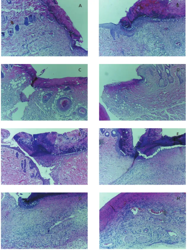
Figure 1. Histological analysis on D3. Right healing by primary intention and left healing by secondaryintention. Note the presence of fibrin-leukocyte scab and inflammatory infiltrate in all images. A and B: from Group E. C and D: from Group EVC. E and F: from Group M. G and H: from Group MVC (Hematoxylin-eosin, 200x magnification). Presidente Prudente (SP), Brazil, 2013.
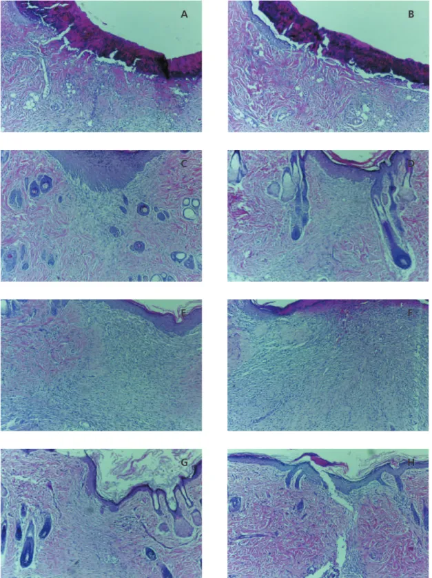
Figure 2. Histological analysis on D7. Right healing by primary intention and left healing by secondaryintention. Note the presence of fibrin-leukocyte scab only in A, B, and F, and more exuberant inflammatory infiltrate in E and F. A and B: from Group E. C and D: from Group EVC. E and F: from Group M. G and H: from Group MVC (Hematoxylin-eosin, 200x magnification). Presidente Prudente (SP), Brazil, 2013.
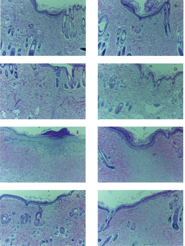
Figure 3. Histological analysis on D14. Right healing by primary intention and left healing by secondaryintention. Note the presence of fibrin-leukocyte scab and inflammatory infiltrate only in E. A and B: from Group E. C and D: from Group EVC. E and F: from Group M. G and H: from Group MVC (Hematoxylin-eosin, 200x magnification). Presidente Prudente (SP), Brazil, 2013.
regardless of the type of lesion, presented mixed inflammatory infiltrate with polymorphonuclear and mononuclear cells (lymphocytes). On D14, lymphocytes prevailed in the inflamed lesions of the animals in group M.
On day 3 and day 14, macrophages were not present in any lesion of any animal. On D7, the concentration of macrophages in primary intention lesions (p=0.0004) and secondary intention lesions (p<0.0001) differed: while all
animals in group EVC had no macrophages in either lesion, all animals in groups E and M, and most of group MVC (62.5%) had a small concentration of macrophages in both lesions. Healing by primary and secondary intention differed between groups E and EVC, EVC and M, and EVC and MVC (p<0.05).
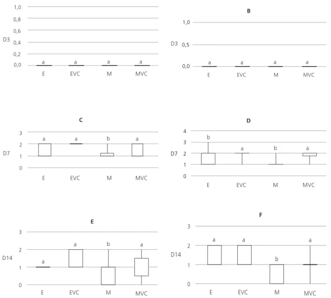
The concentration of fibroblasts in both lesions of all animals was higher (small to moderate) on D7; and in most animals on D14 (E
Figure 4. Concentration of fibroblasts in the four study groups on different study days (expressed as scores). Presidente Prudente (SP), Brazil, 2013.
Note: The central line indicates the median, the upper and lower edges delimit the interquartile range, and the outer vertical lines indicate maximum and minimum values. Results with different superscripts differ significantly (p<0.05).
-100.0%; EVC -100.0%; M e MVC - 87,5%). The groups differed for this parameter only on D7 for healing by primary intention (p=0.0243); and on D7 (p=0.0219) and D14 (p=0.0119) for healing by secondary intention (Figures 1, 2, 3, and 4).
By D3, the concentration of collagen fibers had not yet increased in any group. On D7, most animals (E, EVC, and MVC - 100.0%; M - 87.5%) had a small or moderate amount of collagen
fibers in the tissue. On D14, all animals in Group EVC and most in Group MVC (85.7%) had great concentration of collagen fibers in both lesions, while most animals from the other groups (E - 62.5%; M - 71.4%) had only moderate amounts. The groups differed for this parameter only on D14 for healing by primary (p=0.0001) and secondary(p<0.0001) intention (Figures 1, 2, 3 and 5).
Figure 5. Concentration of collagen fibers in the four study groups at different moments of analysis (expressed in scores). Presidente Prudente (SP), Brazil, 2013.
Note: The central line indicates the median, the upper and lower edges delimit the interquartile range, and the outer vertical lines indicate maximum and minimum values. Results with different superscripts differ significantly (p<0.05).
D I S C U S S I O N
The healing process in animals of group M was delayed, and vitamin C contributed to fibroblast proliferation and collagen deposition in the primary and secondary intention wounds of the animals in group MVC. However, the use of this vitamin did not affect the decrease in macrophage migration in the malnourished rats and only decreased lymphocyte migration in this group in the final stages of healing.
Healing involves several growth factors, such as Epidermal Growth Factor (EGF), TGF, VEGF, Platelet Derived Growth Factor (PDGF), Fibroblast Growth Factor (FGF), which act by stimulating the proliferation of keratinocytes, fibroblasts, and endothelial cells; and the chemotaxis of polymorphonuclear macrophages and fibroblasts2. Cytokines are also involved, such as Tumor Necrosis Factor (TNF), interleukins, and interferons, which activate macrophages, have chemotactic activity for polymorphonuclear cells, stimulate angiogenesis, and even inhibit fibroblast proliferation and extracellular matrix synthesis2.
Furthermore, the inflammatory process that occurs primarily in the initial phase of the healing process leads to the activation of various chemical mediators (histamine, bradykinin, prostaglandins, leukotrienes, nitric oxide, cytokines, etc.)2.
After exposure to microorganisms, chemokines, and immune complexes in the extracellular medium or after phagocytic activation, leukocytes can release oxygen free radicals2. These free radicals can injure the endothelium and parenchymal cells and inactivate antiproteases. However, the influence of oxygen free radicals on any inflammatory reaction depends on the balance between production and inactivation of these metabolites by cells and tissues2. Since ascorbic acid is an antioxidant, it could help to minimize the effects of free radicals on inflamed tissues. Oxidative damage can be further reduced by giving patients preoperatively
a diet rich in proteins (e.g, glutamine and arginine) and antioxidants, especially ascorbic acid19.
The diary recommendation for vitamin C is 90 mg/day for males and 75 mg/day for females20. In the present study, we administered a daily dosage of 136 mg of vitamin C. Será que não tem algum erro nos números? Uma dose de 340 g/kg num rato de 250 g seria uma dose de 85 mg. Essa mesma dose num humano de 70 kg equivaleria a 23,8 g de vitamina C.
Malnutrition is one of the major global health problems, affects between 20 and 60% of hospitalized patients, and relates closely to higher morbidity and mortality21 since it changes immune function and causes hypoalbuminemia, low hemoglobin, impaired healing, and more complications22. An experimental study feeding malnourished rats half of their chow requirement found that the animals had impaired healing, increasing mortality by 15% compared with a control group11.
Mild, moderate, and severe malnutrition are defined as body weight losses of 10%, 10-20%, and >10-20%, respectively23. In the present study, the animals in groups M and MVC can be classified as moderately malnourished because most of them lost about 20% of their body weight.
Both wounds healed by primary and secondary intention were beefy red/pale pink on all study days. Although malnourished patients have a higher risk of infection and wound dehiscence, this was not observed in this study. Also, very few animals experienced pain in the lesions and when they did, it was only on D3, confirming the absence of infection in the wounds.
cells in animals consuming inadequate protein24. The same was observed in a study on oral wound healing, where rats fed a diet containing specific nutrients presented better healing clinically and histologically25. On D7 primary intention lesion sizes differed significantly, as did secondary intention lesion sizes on D3, regardless of nutritional status or vitamin C supplementation (p>0.05). Likewise, scab and hematic type occurred only on D3 and D7 regardless of group, corroborating the abovementioned data on adequate wound retraction. On D3, no animal had wound re-epithelialization, but on D14, all of them did. On D7, most animals had complete re-epithelialization and some partial, with statistical difference only for healing by primary intention regardless of nutritional status or vitamin C supplementation, a finding that disagrees with the literature In this study, the absence of infection in addition to the absence of purulent scab may have contributed to proper wound contraction and re-epithelialization.
Studies indicate that vitamin C associated with pantothenic acid and protein is important for patients with pressure ulcers and poor surgical wound healing26. Topical use of ascorbic acid in rats decreases the number of macrophages, increasing the amount of fibroblasts and promoting collagen deposition and wound organization, which results in better tissue repair and shorter healing time16.
Healing is divided into an inflammatory, a proliferative, and a remodeling phase. In the inflammatory phase, there is an increase in capillary permeability that allows the migration of specific cells to the wound, such as neutrophils (6 to 48 hours), macrophages (72 hours), and lymphocytes (5 days)2. In this study, we observed the expected pattern of inflammatory cells during the healing process. Both wounds healed by primary and secondary intention had a predominance of neutrophils on D3 and mixed inflammatory infiltrate (neutrophils and lymphocytes) on D7; only group M still had
lymphocytes in both lesions on D14. This suggests that vitamin C in malnourished rats (Group MVC) may have contributed to the absence of lymphocytes on D14. Although vitamin C was given orally, our results agree with those of Lima
et al.16, who found an anti-inflammatory effect
of topical vitamin C. On D7, unlike most animals in Group EVC, most animals had inflammatory infiltrate. This datum shows that vitamin C decreases inflammation intensity in normal weight animals sooner than in malnourished ones, probably by decreasing inflammatory cell migration. Moreover, vitamin C promoted the absence of fibrin-leukocyte scab in healing by primary intention; on D7 most animals in groups EVC and MVC no longer had this scab, contrary to most animals in groups E and M (p<0.05).
Macrophages were present on D7, but absent on D3 and D14. However, vitamin C decreased macrophage migration only in the normal weight groups, corroborating the literature16. These groups also did not have macrophages on D7, unlike the malnourished groups. These data indicates that vitamin C does not reduce macrophage migration in malnourished animals.
shows that the influence of malnutrition on healing cannot be related to neovascularization.
Fibroblasts produce ground substance (composed of glycosaminoglycans, fibronectin, and hyaluronic acid) and collagen, which will reconstitute the destroyed tissue6. The number of fibroblasts increased on D7: most of group E, all of group EVC, half of group MVC, but only a few in group M (p<0.05) had a moderate amount of fibroblasts in the lesion healed by primary intention. On D14, all animals in group E had few fibroblasts, some of group EVC had some fibroblasts, most of group MVC had few to some fibroblasts, and most of group C did not have fibroblasts (p<0.05). With respect to healing by secondary intention, most animals in groups E, EVC, and MVC had moderate amounts of fibroblasts, while animals in group M had a slight increase on D7 (p<0.05). On D14, most animals in group EVC and some in group E had a moderate amount of fibroblasts (p<0.05). These data show that vitamin C increases the proliferation of fibroblasts in the tissue of even malnourished animals, which may improve wound healing but at different rates depending on intention.
Deposition, clustering, and remodeling of collagen tissue and endothelial regression occur on the third stage of healing, called remodeling. The collagen tissue is present in the form of long fibrils. After many substances are placed disorderly on the scar, including collagen, the wound is restructured, or ordered. More fibroblasts and collagen fibers appear in the lesion27. Ascorbic acid is important in this process because hydroxylases do not function in its absence and consequently, hydroxyproline, hydroxylysine, and collagen are not formed27. Collagen fibers increased from D7, as expected. All animals in group EVC and most in group MVC had high levels of collagen fibers in both lesions on D14, unlike the animals without vitamin C supplementation, which had only moderate levels (p<0.05) Thus, even in malnourished rats, vitamin C can increase collagen deposition and improve the healing process.
C O N C L U S I O N
In conclusion, vitamin C increases fibroblast proliferation and collagen deposition and reduces lymphocyte migration in the final stages of healing, speeding the healing of primary and secondary intention wounds regardless of nutritional status. However, early inflammation and macrophage migration subsidence only occur in normal weight animals.
A C K N O W L E D G M E N T
We thank José Luiz Santos Parizi, Ms., professor of the Department of Pathology of Universidade do Oeste Paulista, for guidance and helping us make the incisions, and Giovana de Carvalho Katayama, undergraduate student of the School of Dentistry of Presidente Prudente, Universidade do Oeste Paulista, for helping us conduct the study.
C O N T R I B U T O R S
All authors helped to collect, analyze, and discuss the data and to write the article; all read and approved the final version.
R E F E R E N C E S
1. Stechmiller JK. Understanding the role of nutrition and wound healing. Nutr Clin Pract. 2010; 25(1):61-8. doi: 10.1177/0884533609358997 2. Kumar V, Abbas AK, Fausto N, Mitchell RN, editores.
Robbins: patologiabásica. 8ª ed. Rio de Janeiro: Elsevier; 2008.
3. Wild T, Rahbarnia A, Kellner M, Sobotka L, Eberlein T. Basics in nutrition and wound healing, Salzburg. Nutrition. 2010; 9(26):862-6. doi: 10.1016/j.nut. 2010.05.008
4. Brown KL, Phillips TJ. Nutrition and wound healing. Clin Dermatol. 28(4):432-9. doi: 10.1016/j. clindermatol.2010.03.028
6. Yang BW, Lin YM, Wang SY, Yeh DC. The study of absorption efficiency and restoring effects of collagen and ascorbic acid on aged skin by fluorescence and reflection spectroscopy. Guang Pu Xue Yu Guang Pu Fen Xi. 2012; 32(12):3299-303. doi: 10.1016/j.bbagen.2013.03.014
7. Otranto M, Souza Netto I, Águila MB, Monte Alto Costa A. Male and female rats with severe protein restriction present delayed wound healing. Appl Physiol Nutr Metab. 2009; 34(6):1023-31. 8. Falbo AR, Alves GB. Desnutrição grave: alguns
aspectos clínicos e epidemiológicos de crianças hos-pitalizadas no Instituto Materno Infantil de Pernam-buco (IMIP), Brasil. Cad Saúde Pública. 2002; 18(5):1473-7.
9. Monteiro CA, Conde WL, Popkin BM. Is obesity replacing or adding to undernutrition? Evidence from different social classes in Brazil. Public Health Nutr. 2002; 5(1A):105-112. doi: 10.1079/PHN 2001281
10. Rissin A, Filho M. B. A transição nutricional no Brasil: tendências regionais e temporais. Cad Saúde Pública. 2003; 19(Supl 1):181-91.
11. Ferreira MM, Scialom JM, Campos AD, Ramalho LLZ, Marchini JS, Féres O, et al. Efeito da desnutrição na cicatrização de anastomoses colônicas: estudo experimental em ratos. Rev Bras Coloproct. 2006; 3(26):239-43.
12. Andersen ML, Tufik S. Animal models as tools in ethical biomedical research. São Paulo: Unifesp; 2010.
13. Paiva FP, Mafilli VV, Santos ACS. Curso de mani-pulação de animais de laboratório. Rio de Janeiro: Fundação Osvaldo Cruz; 2005 [acesso 2011 mar 22]. Disponível em: <http://www.bioteriocentral. ufc.br/arquivos/apostilha_manipulacao.pdf>.
14. Schirato GV, Monteiro FMF, Silva FO, Filho JLL, Leão AMA, Porto ALF. O polissacarídeo do Anacardium Occidentale L. na fase inflamatória do processo cicatricial de lesões cutâneas. Ciênc Rural. 2006; 36(1):149-54.
15. Pace D, Campos AC, Graf R. Efeito de substâncias antioxidantes (vitamina C, vitamina E e Gingko biloba) na viabilidade de retalho cutâneo dorsal em ratos. Rev Soc Bras Cir Plast. 2006; 2(21):77-81. doi: 10.1590/S0100-69912013000100009 16. Lima CC, Pereira APC, Silva JRF, Oliveira LS, Resck
MCC, Grechi CO, et al. Ascorbic acid for the healing of skin wounds in rats. Braz J Biol. 2008; 69(4):
1195-201. doi: 10.1590/S1519-6984200900050 0026
17. Michalany J. Técnica histológica em anatomia pato-lógica com instruções para o cirurgião, enfermeira e citotécnico. 3ª ed. São Paulo: Michalany; 1998. 18. Ayres M, Ayres Júnior M, Ayres DL, Santos AA.
BIOESTAT: aplicações estatísticas nas áreas das ciências biomédicas. Belém: ONG Mamiraua; 2007. 19. Campos ACL, Groth AK, Branco AB. Assessment and nutritional aspects of wound healing. Curr Opin Clin Nutr Metab Care. 2008; 11(3):281-8. doi: 10.1177/0022034509359125
20. Valdés F. Vitamina C. Actas dermosifiliogr. 2006; 97(9):557-68.
21. Azevedo LC, Medina F, Silva AA, Campanella ELS. Prevalência de desnutrição em um hospital geral de grande porte de Santa Catarina/Brasil. Arq Catarinenses Med. 2006; 35(4):89-96.
22. Kavalukas SL, Barbul A. Nutrition and wound healing: An update. Plast Reconstr Surg. 2011; 1(127):38S-43S. doi: 10.1111/j.1524-475X.2012.0 0833.x
23. Ramos Fernández CP. Malnutrición: conceptos, repercusión en los distintos órganos y sistemas. Indicaciones del suporte nutricional. 2000 [acceso 2013 mayo 27]. Disponible em: <http:1.www. multimania.com/trenchei.opin.1201.httn>. 24. Zacharias DPM, Waitzsberg DL, Schmidt B, Oliveira
ASB, Gonçalves EL. Efeito da desnutrição protéica sobre a resposta cicatricial ao trauma: aspectos histológicos, histoquímicos e contração cicatri-cional. Acta Cir Bras. 1991; 33(6):97-102. 25. Galvão CBC, Pinheiro ALB, Pessoa DCNP.
Compa-ração da cicatrização de feridas bucais em animais submetidos às dietas: DBR e dieta balanceada acres-cida de suplementação alimentar. Rev Bras Med. 2000; 57(8):844-50.
26. Ellinger S, Stehle P. Efficacy of vitamin supplementation in situations with wound healing disorders: Results from clinical intervention studies. Curr Opin Clin Nutr Metab Care. 2009; 12(6):588-95. doi: 10.10 97/MCO.0b013e328331a5b5
27. Choi HI, Park JI, Kim HJ, Kim DW, Kim SS. A novel L-ascorbic acid and peptide conjugate with increased stability and collagen biosynthesis. BMB Rep. 2009; 42(11):743-6.