Rev Odontol UNESP. 2014 Jan-Feb; 43(1): 72-76 © 2014 - ISSN 1807-2577
CLINICAL REPORT
Doi: http://dx.doi.org/10.1590/S1807-25772014000100012
Surgical management of palatine Torus - case series
Tratamento cirúrgico de Tórus palatino - série de casos
haís Sumie Nozu IMADA
a, Kellen Cristine TJIOE
a, Marcelo Bonifácio da Silva SAMPIERI
a,
José Endrigo TINOCO-ARAUJO
a, Izabel Regina Fischer RUBIRA-BULLEN
a,
Paulo Sérgio DA SILVA SANTOS
a, Eduardo Sanches GONÇALES
aaFaculdade de Odontologia, USP – Universidade de São Paulo, Bauru, SP, Brasil
Resumo
Introdução: Torus palatinus é um nome específico usado para identificar exostoses no palato duro ao longo da sutura palatina mediana. Apesar de não ser considerado uma condição patológica, sua presença requer atenção e conhecimento no que diz respeito ao seu tratamento. A remoção cirúrgica de exostoses é indicada quando o paciente traumatiza frequentemente a área do Torus palatinus durante a mastigação e a fala, ou quando for necessária a reabilitação da arcada dentária superior com próteses totais. Objetivo: O objetivo deste trabalho é apresentar três casos de Torus palatinus e discutir os seus respectivos tratamentos. Caso clínico:O primeiro caso era de um homem leucoderma e com maxila edêntula que procurou reabilitação dentária, porém apresentou um nódulo no palato duro. O segundo caso era de uma mulher, leucoderma e de 40 anos que foi encaminhada devido ao trauma frequente na mucosa do palato durante a mastigação, insatisfação com a estética e desconforto causado pelo trauma na língua. O terceiro caso era de uma mulher de 45 anos de idade, leucoderma com uma lesão no palato e dificuldade pra engolir. Uma vez que o Torus palatinus estava prejudicando as funções fisiológicas básicas dos pacientes, todos os casos foram cirurgicamente tratados, melhorando a qualidade de vida dos mesmos. Consideração final: O dentista deve estar preparado para selecionar a técnica cirúrgica mais indicada para cada caso buscando o melhor resultado e evitando possíveis complicações.
Descritores: Exostose; palato duro; diagnóstico bucal.
Abstract
Introduction:Torus palatinus is a specific name to identify exostoses developed in the hard palate along the median palatine suture. Despite of not being a pathological condition, its presence requires attention and knowledge regarding its management. Surgical removal of exostoses is indicated when the patient frequently traumatizes the area of palatine torus during mastication and speech or when it is necessary for the rehabilitation of the upper arcade with complete dentures. Objective: The aim of this article is to present three cases of Torus palatinus and to discuss the management of them. Case report: In the first case, a 57-year-old Caucasian man sought oral rehabilitation of his edentulous maxilla but presented a hard nodules in the hard palate; in the second case, a 40-year-old Caucasian woman was referred for frequent trauma of palatal mucosa during mastication, aesthetic complaint, and discomfort caused by the trauma of her tongue in this area; and in the third case, a 45-year-old Caucasian woman presented with a lesion on the palate that caused difficulty swallowing. When the Torus palatinus was impairing the basic physiological functions of the patients, all cases were surgically treated, improving the patients’ quality of life.
Final consideration: The dentist should be properly prepared to choose the best from among the existing surgical approaches for each individual lesion in order to improve the results and avoid possible complications.
Descriptors: Hyperostosis; palate, hard; diagnosis, oral.
INTRODUCTION
Torus palatinus (TP) is a specific name to identify
exostoses developed in the hard palate along the median palatine suture. It is constituted by normal compact and cancellous bone1.About 12-30% of the population has TP and
it is often accidentally detected in young adults and middle-aged patients2,3. Despite not being considered a pathological
condition, the detection of a palatine torus requires attention
and knowledge of its management. Surgical removal of exostoses is indicated when the patient traumatizes the area of TP during the mastication and speech or when it is necessary for the rehabilitation of the upper arcade with complete dentures.
he aims of this article are to report three distinct cases of
CASE REPORT
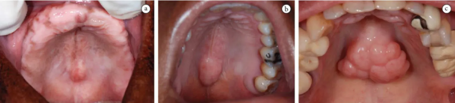
CASE 1: A 57-year-old Caucasian man sought oral rehabilitation of his edentulous maxilla. Oral examination revealed a hard nodule at the midline of hard palate of approximately 1.5 cm, covered by healthy mucosa (Figure 1A).
Medical history did not reveal any comorbidity. he presence of the TP was impairing the confection of the upper complete denture, so surgical removal of the exostosis under local anesthesia (articaine 4% with epinephrine 1:100.000) was perfromed. A single “Y” incision was performed to expose the bone, followed by segmental osteotomy under plentiful irrigation, removal of bone fragments with chisel, nylon sutures, and compression. Microscopical examination of the specimen conirmed the diagnosis of Torus palatinus. Post-operatory was uneventful. Four months later, the patient did not experience any sign of recurrence and he was rehabilitated with complete dentures.
CASE 2: A 40-year-old Caucasian woman was referred for frequent trauma of palatal mucosa during mastication, aesthetic complaint and discomfort caused by the trauma of her tongue in this area. Oral examination revealed a nodular and hard swelling covered by healthy mucosa at the midline of the hard palate extending from the height of the irst molars to the middle of the third ones, with an approximated dimension of 2 cm (Figure 1B). Medical records were not contributory. he clinical diagnosis was palatine torus. Due to the functional impairment, we decided on surgical excision under local anesthesia with the same technique employed in the irst case (Figures 2A-D). Microscopical analysis of the removed specimen conirmed the diagnosis of Torus
palatinus. he four-month follow-up was uneventful.
CASE 3: A 45-year-old Caucasian woman presented with a lesion on the palate that caused diiculty to swallow. Oral examination revealed a lobulated and hard nodule with a 5 cm diameter, located in the midline of the palate and covered by healthy mucosa (Figure 1C). he swelling was painless and presented slow growth, without signs of inlammation. Medical history did not reveal any comorbidity. Total maxilla occlusal radiography was performed to rule out the presence of neoplasia and to examine the shape and size of the bony prominence. It showed a radiopaque lobular lesion on the hard palate midline (Figure 3).
Due to the functional discomfort, we decided for surgical excision under local anesthesia with the same technique employed in the previous cases. Microscopical analysis of the specimen conirmed the diagnosis of Torus palatinus. he post-operative period of 4 weeks showed good healing of the surgical area.
DISCUSSION
Some lesions of the oral mucosa do not receive the necessary attention because of its high frequency and outward indolent behavior. Torus palatinus (TP) is an exostoses of the hard palate usually discovered during a routine clinical exam. It may present signiicant growth, impair swallowing and prosthesis itting2. herefore, it is important to highlight and discuss the
management of TP.
he irst report of exostotic changes of the hard palate was written by Fox in 18144. Although this anatomical variation had
been described earlier under various names, the term Torus
palatinus was determined by Kupfer and Bessel-Hagen in 18795.
TP etiology remains unclear6,7. Eforts to link its occurrence
to third molar agenesia8, bone density9 and elongation of styloid
process10 have been made, but these relationships remain a source
of debate. Nowadays, the most widely accepted theory is that TP represents genetic traits6,7,11-15, but it has not always been possible
to show the autosomal dominant nature of these structures15.
Others16,17 have considered that the development of TP results
from an interplay of genetic and environmental factors, especially those related to occlusal stress2.
he average age of our patients was 47.3 years old, similar to other studies, which show incidence of palatine torus at age ranging from 30 to 50 years old14,15. MacInnis et al.18 asserted that
TP appears during puberty and slowly grows until adulthood, with the possibility of continuing growth until the seventh decade. his slow development of palatine torus associated to the asymptomatic nature of this variation can explain the relatively old age of diagnosis.
In the presented study, we reported one male and two female cases of TP. he literature shows the majority frequency of TP in women19,20, probably because it seems to be a dominant type of
TP linked to the X chromosome2.
he TP may display a wide variety of shapes. It can be lat, nodular, spindle and lobular or spindle-shaped2,21. In all of our
cases the TPs were nodular and 1.5-2 cm in size (Figures 1A-C). his corroborated with Haugen22 who reported that the most
common shape was nodular. However, Sisman et al.10 (2009)
showed that the lat TP is the most common type. hese diferences might be due to diferent classiications of palatine torus employed by the authors though comparisons are diicult to conduct.
he prevalence of the tori varies from 12.3 to 14.6%14,15. In
the majority of the studies, palatine torus is more frequent than
mandibular torus6,11,14,20,22,23 and concurrence appears to happen
in about 2-3% of cases20.
he diagnosis of TP is usually incidental, during clinical examination, due to its asymptomatic nature2,14. In some
situations, however, the patient may present speech and masticatory disturbances, traumas and ulcerations of the mucosa, prosthetic instability, and even cancerophobia. In these cases, the patient should be reassured and depending on the severity, surgical removal should be studied. he removal of the tori is indicated when functional prejudice is detected2.he most
frequent cause of exeresis is the need for prosthetic treatment2,7
as presented in Case 1. A palatine torus may interfere with the design, retention and function of a complete denture. In addition, a TP under a prosthesis constitutes an additional source of trauma. In cases 2 and 3, the exeresis was indicated because the patient frequently traumatized the TP area during mastication and had aesthetic discomfort. Such situation was impairing her quality of life and the surgical approach was chosen. Despite being a widely accepted indication for surgery, some authors do not recommend the removal of tori except in very extreme cases2.
hey advocate the accommodation of the prosthesis in these areas or relining them with sot acrylic resin24. Another option to
avoid the removal of TP is the use of dental implants2. he bone
of a torus may be source of autogenous cortical bone for grats in periodontal, cyst and implant surgeries2,7, although long-term
stability of the grats remains uncertain2.
Figure 2. Surgical technique. A. Single Y incision, B. Trans-operatory view of the segmental osteotomy, C. View of the hard palate ater the surgical removal of the palatine torus, D. Seven-days post-operatory.
Unlike some reported excisions conducted in a hospital environment under general anesthesia25,26, the TP excisions in our
cases were conducted in the ambulatory, under local anesthesia. Diferent incisions can be made in order to remove the TP. he most common type of incision is the double Y incision, one linear incision at the middle line of the torus and two oblique anteroposterior at its both borders. Another technique used is the single Y incision which difers from double Y incision because the oblique incisions are made on just one side of the middle incision corner2. We chose the single Y incision for all cases because it
prevents injury of the nasopalatine and anterior palatine nerves2.
he excision should be done carefully because the mucosa that covers the torus is very thin and easy to tear2.
Periotome was used for the detachment until the nodule was exposed. In cases of pedunculate base, the palatine torus can be easily removed with a hand osteotome by chisel2. However, in all
presented cases, the base was sessile so a segmental osteotomy with high speed rotation drill cooled with normal saline solution was performed irst (Figure 2B) as advocated by García-García et al.2.
Although, there is the low risk of emphysema, we chose this technique because the use of a chisel and hammer involves a major risk of iatrogenic injury, and also to avoid bumping the patient with the chisel2.
Mattress or simple sutures should not be too tight and surgical cement can be used to protect the wound during the healing process2. In the presented cases, we chose simple sutures
and did not use surgical cement. he patients were advised about post-operative cares and common signs and symptoms during this period (edema, hematoma, mild pain) and were medicated with analgesics and anti-inlammatory.
Complications can occur as a result of iatrogenic maneuvers of the surgeon such as perforation of the nasal cavities, nerve damage, bone necrosis due to poor refrigeration during surgical drilling, hemorrhage due to section of palatine arteries, dilacerations of the palatine mucosa, fracture of the palatine bone2. Post-operative complications include hematoma, edema,
suture opening, infection, bone or mucosa necrosis, neuralgia and poor scaring. herefore, the dentist should be properly prepared as to the management of these surgical approaches and possible complications.
he present three cases demonstrate the surgical technique approach for TP exeresis in order to improve the quality of life of the patients. Despite of being a common lesion, the management of palatine torus is not widely known and requires attention in order to avoid complications.
REFERENCES
1. Vidic B. Incidence of torus palatinus in Yugoslav skulls. J Dent Res. 1966;45:1511–5. http://dx.doi.org/10.1177/00220345660450054101
2. García-García AS, Martínez-González JM, Gómez-Font R, Soto-Rivadeneira A, Oviedo-Roldán L. Current status of the torus palatinus and torus mandibularis. Med Oral Patol Oral Cir Bucal. 2010;15(2):e353-60. PMid:19767716. http://dx.doi.org/10.4317/medoral.15.e353
3. Raldi FV, Nascimento RD, Sá-Lima JR, Tsuda CA, Moraes MB. Excision of an atypical case of palatal bone exostosis: a case report. J Oral Sci. 2008;50:229-31. PMid:18587217. http://dx.doi.org/10.2334/josnusd.50.229
4. Fox J. Natural history and diseases of human teeth. E. Cox: London; 1814.
5. Kupfer C, Bessel-Hagen F. Verhandlungen der berliner gesellschat fur anthropologie, ethonologie und urgeschichte. Zeitschrit fur Ethnologie. 1879;11:70–1.
6. Sirirungrojying S, Kerdpon D. Relationship between oral tori and temporomandibular disorders. Int Dent J. 1999;49:101-4. http://dx.doi. org/10.1111/j.1875-595X.1999.tb00516.x
7. Sonnier KE, Horning GM, Cohen ME. Palatal tubercles, palatal tori, and mandibular tori: prevalence and anatomical features in a U.S. population. J Periodontol. 1999;70:329-36. PMid:10225550. http://dx.doi.org/10.1902/jop.1999.70.3.329
8. Cagirankaya LB, Kansu O, Hatipoglu MG. Is torus palatinus a feature of a well-developed maxilla? Clin Anat. 2004;17:623-5. PMid:15494968. http://dx.doi.org/10.1002/ca.20032
9. Belsky JL, Hamer JS, Hubert JE, Insogna K, Johns W. Torus palatinus: a new anatomical correlation with bone density in postmenopausal women. J Clin Endocrinol Metab. 2003;88:2081-6. PMid:12727958. http://dx.doi.org/10.1210/jc.2002-021726
10. Sisman Y, Gokce C, Tarim Ertas E, Sipahioglu M, Akgunlu F. Investigation of elongated styloid process prevalence in patients with torus palatinus. Clin Oral Investig. 2009;13:269-72. PMid:18972141. http://dx.doi.org/10.1007/s00784-008-0232-6
11. Kerdpon D, Sirirungrojying S. A clinical study of oral tori in southern hailand: prevalence and the relation to parafunctional activity. Eur J Oral Sci. 1999;107:9-13. http://dx.doi.org/10.1046/j.0909-8836.1999.eos107103.x
12. Gorsky M, Bukai A, Shohat M. Genetic inluence on the prevalence of torus palatinus. Am J Med Genet. 1998;75:138-40. http://dx.doi. org/10.1002/(SICI)1096-8628(19980113)75:2%3C138::AID-AJMG3%3E3.0.CO;2-P
13. Jainkittivong A, Langlais RP. Buccal and palatal exostoses: prevalence and concurrence with tori. Oral Surg Oral Med Oral Pathol Oral Radiol Endod. 2000;90:48-53. PMid:10884635. http://dx.doi.org/10.1067/moe.2000.105905
14. Al-Bayaty HF, Murti PR, Matthews R, Gupta PC. An epidemiological study of tori among 667 dental outpatients in Trinidad & Tobago, West Indies. Int Dent J. 2001;51:300-4. PMid:11570546. http://dx.doi.org/10.1002/j.1875-595X.2001.tb00842.x
15. Bruce I, Ndanu TA, Addo ME. Epidemiological aspects of oral tori in a Ghanaian community. Int Dent J. 2004;54:78-82. PMid:15119797. http://dx.doi.org/10.1111/j.1875-595X.2004.tb00259.x
17. Gorsky M, Raviv M, Kir E, Moskona D. Prevalence of torus palatinus in a population of young and adult Israelis. Arch Oral Biol. 1996;41:623-5. http://dx.doi.org/10.1016/0003-9969(96)00149-5
18. MacInnis EL, Hardie J, Baig M, al-Sanea RA. Gigantiform torus palatinus: review of the literature and report of a case. Int Dent J. 1998;48:40-3. PMid:9779082. http://dx.doi.org/10.1111/j.1875-595X.1998.tb00692.x
19. Antoniades DZ, Belazi M, Papanayiotou P. Concurrence of torus palatinus with palatal and buccal exostoses: case report and review of the literature. Oral Surg Oral Med Oral Pathol Oral Radiol Endod. 1998;85:552-7. http://dx.doi.org/10.1016/S1079-2104(98)90290-6
20. Eggen S, Natvig B, Gasemyr J. Variation in torus palatinus prevalence in Norway. Scand J Dent Res. 1994;102:54-9. PMid:8153581.
21. Jainkittivong A, Apinhasmit W, Swasdison S. Prevalence and clinical characteristics of oral tori in 1,520 Chulalongkorn University Dental School patients. Surg Radiol Anat. 2007;29:125-31. PMid:17340055. http://dx.doi.org/10.1007/s00276-007-0184-6
22. Haugen LK. Palatine and mandibular tori. A morphologic study in the current Norwegian population. Acta Odontol Scand. 1992;50:65-77. http://dx.doi.org/10.3109/00016359209012748
23. Reichart PA, Neuhaus F, Sookasem M. Prevalence of torus palatinus and torus mandibularis in Germans and hai. Community Dent Oral Epidemiol. 1988;16:61-4. PMid:3422622. http://dx.doi.org/10.1111/j.1600-0528.1988.tb00557.x
24. Abrams S. Complete denture covering mandibular tori using three base materials: a case report. J Can Dent Assoc. 2000;66:494-6. PMid:11070628.
25. Blakemore JR, Eller DJ, Tomaro AJ. Maxillary exostoses. Surgical management of an unusual case. Oral Surg Oral Med Oral Pathol. 1975;40:200-4. http://dx.doi.org/10.1016/0030-4220(75)90152-8
26. Topazian DS, Mullen FR.Continued growth of a torus palatinus. J Oral Surg. 1977;35:845-6. PMid:269237.
CONFLICTS OF INTERESTS
he authors declare no conlicts of interest.
CORRESPONDING AUTHOR
haís Sumie Nozu Imada
Departmento de Estomatologia, Faculdade de Odontologia, USP – Universidade de São Paulo, Alameda Octávio Pinheiro Brisolla, 9-75, 17012-901 Bauru - SP, Brasil
e-mail: [email protected]