116
Asymptomatic ocular sarcoidosis
Sarcoidose ocular assintomática
Luiz Guilherme Azevedo de Freitas
1, Luiz Alexandre Rassi Gabriel
2, David Leonardo Cruvinel Isaac
3, Clovis
Arcoverde de Freitas
4, Marcos Pereira de Ávila
51Fellowship of the Department of Retina and Vitreous, Universidade Federal de Goiás (UFG) – Goiânia (GO), Brazil;
2Ophthalmologist and Professor and head of the Department of Eye Genetics, Universidade Federal de Goiás (UFG) – Goiânia (GO), Brazil; 3Ophthalmologist and Professor and head of the Department of Retina and Vitreous, Universidade Federal de Goiás (UFG) – Goiânia (GO),
Brazil;
4Fellowship of the Department of Uveitis of The São Geraldo Hospital, Universidade Federal de Minas Gerais (UFMG) – Belo Horizonte (MG), Brazil;
5Chairman of Ophthalmology; Head of the Department of Ophthalmology Universidade Federal de Goiás (UFG) – Goiânia (GO), Brazil.
Study carried out at the Department of Ophthalmology, Universidade Federal de Goiás (UFG) – Goiânia (GO), Brazil
A
BSTRACTSarcoidosis is an idiopathic systemic granulomatous disease. It commonly affects the skin, lungs, kidneys, and central nervous system. In the eyes it primarily affects the uveal tract, conjunctiva, lacrimal glands and optic nerve. Here in we describe the case of a patient with systemic sarcoidosis and asymptomatic eye inflammation.
Keywords: Sarcoidosis; Eye infections; Retinal vasculitis; Retina; Fluorescein angiography; Case reports
R
ESUMOSarcoidose é uma doença granulomatosa sistêmica idiopática. Afeta comumente pele, pulmões, rins e sistema nervoso central. Nos olhos afeta primariamente a úvea, conjuntiva, glândulas lacrimais e nervo óptico. Descrevemos o caso de um paciente portador de sarcoidose sistêmica com acometimento ocular assintomático.
Descritores: Sarcoidose; Infecções oculares; Vasculite retiniana; Retina; Angiografia fluoresceínica; Relatos de casos
R
ELATODEC
ASOThe authors declare no conflicts of interest
Recebido para publicação em: 6/4/2011 - Aceito para publicação em: 26/6/2011
117
I
NTRODUCTIONS
arcoidosis is an idiopathic systemic granulomatous disea-se(1). Sarcoidosis is an ubiquitous disease, although notuniform, presenting heterogenous incidence and prevalence rates. Its annual incidence is higher in Sweden and Norway. In Brazil, a prevalence below 10/100,000 persons is estimated(2). Papers show that the disease is more commonly
seen among young adults in their third to fourth decade of life(3,4).
Eye involvement happens in 25 to 50% of patients presenting systemic sarcoidosis(5). The retinal features are:
vasculitis with vascular sheathing, nodular granulomatous phlebitis with a candle wax dripping pattern(6), granulomatous
chorioretinitis, and papilledema usually related to simultaneous central nervous system involvement, which may occur in the absence of additional eye inflammation.
Currently there is no curative treatment available for sarcoidosis. Immunosupresants, and/or immunomodulators may be used to control the disease. Corticosteroids are the first-line therapy in the majority of cases. For instance, when eyes or lungs are involved glucocorticoids are indicated(7).
Case report
A 46-year old white male, from the city of Goiania in the center of Brazil was referred by the infectologist for ophthalmological evaluation because of a prior diagnosis of systemic sarcoidosis.
The patient was being followed up since January of 2010 in the Hospital das Clínicas da Universidade Federal de Goiás (HC-UFG) when he was admited the emergency room with fever and joint pain complaints. Besides being treated by the infectologist and ophthalmologist, he has concomitantly being followed by the rheumatology, and hematology departments due to his additional systemic findings.
Abdominal computed tomography performed in may/2010 showed mild to moderate homogenous hepatosplenomegaly, small cortical cysts, and presence of cortical scar in the left kidney. The chest X-ray, performed in june of 2010, exhibited bilateral hilar lymphadenopathy. Hystopathological analysis of the lung biopsy, made in august of 2010 revealed: granulomatous chronic inflammatory reaction making it impossible to rule out
sarcoidosis. We could also appreciate leucocytosis (60,000) with left deviation, and thrombocytosis (507,000).
The patient did not have ocular complaints in his first ophthalmological evaluation, and denied high blood pressure and diabetes mellitus.
At the eye examination, his best corrected visual acuity (BCVA) was 20/20 on both eyes (OU). Biomicroscopic evaluation was unremarkable. His eye fundus showed normal optic nerves, mild venular dilation, and normal macula OU.
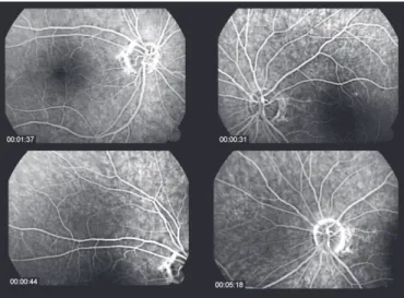
Fluorescent angiography revealed leakage and venous staining suggesting periphlebitis OU, which were compatible with ocular sarcoidosis (Figure 1).
According to the presented case the patient was oriented to maintain the multidisciplinary follow up.
In his follow up visit, 90 days past his first visit, his BCVA was 20/20 OU, as well as unremarkable biomicroscopy and fundoscopy OU. Fluorescent angiography was also normal OU (Figure 2).
The patient was advised to keep regular ophthalmic evaluations every six months and, in the case of any unexpected visual alteration, to come back immediately.
D
ISCUSSIONThe prevalence of sarcoidosis in Brazil was estimated in 10/100,000 persons, which is quite uncommon when compared with other countries that can show a prevalence twice as high, and also with an increased incidence in afro-descendants.
Brazil, due to its mixed population, cannot consider for its own population, epidemiologic studies performed by countries that show homogenous ethnic populations. In this case report, we are righteously describing one example of this situation: a patient with white skin, but not necessarily Caucasian.
On the other hand, it is important to highlight that in Caucasian countries like Norway and Sweden, the incidence is almost three times higher than in Brazil. This fact may find an answer in a poorly organized national public health system, that may underestimate the real Brazilian prevalence and incidence of sarcoidosis.
The ocular sarcoidosis can show low vision, floaters, pain, and photophobia, which are usually brought up by anterior
Figure 1: Leakage and venous staining suggesting periphlebitis Figure 2: Fluorescent angiography after treatment Asymptomatic ocular sarcoidosis
118
Corresponding author:
Luiz Guilherme Azevedo de Freitas
Av. T-2 nº 401 - Setor Bueno
CEP 74210-010 - Goiânia (GO), Brazil
Tel: (62) 3252-5566 - Fax (62) 3252-5500
E-mail: [email protected]
uveitis. The posterior segment inflammation is restricted to one third of the sarcoidosis patients, that present retinal vasculitis and granulomatous periphlebitis(8).
Herein, the patient has not shown any findings in the ante-rior segment, like keratic precipitates, synechiae, or iris nodules, anterior or intermediate uveitis. He has not showed low visual acuity or eye pain as well. The evolution of this ocular sarcoidosis was restricted to the posterior segment, presenting solely as retinal periphlebitis OU, constituting an atypical case.
The diagnosis of ocular sarcoidosis can become tricky because its features may resemble closely those from other cau-ses. It is important to put together a multidisciplinary medical evaluation and a proper laboratory investigation in order to reach the precise diagnosis. Ophthalmologists have an important task in the diagnosis of sarcoidosis, since the eyes are affected in the beginning of the disease in up to 50% of cases(5), and not always
the patient shows eye abnormalities as his first sign.
C
ONCLUSIONOcular sarcoidosis may be underdiagnosed when asymptomatic. It is very well known that patients with extra-ocular sarcoidosis may have concomitantly the extra-ocular form in 50% of cases which may be missed due to the absence of an ocular fundus examination. The ophthalmologic evaluation of a patient with sarcoidosis is, therefore, indispensable.
R
EFERENCES1. Daldon PEC, Arruda LHF. Granulomas não-infecciosos: sarcoidose. An Bras Dermatol. 2007;82(6):559-71.
2. Bethlem NM. Epidemiology of sarcoidosis in Brazil. Sarcoidosis. 1985;2:162.
3. Nunes H, Bouvry D, Soler P, Valeyre D. Sarcoidosis. Orphanet J Rare Dis. 2007;2:46. Review.
4. Stanbury RM, Graham EM, Murray PI. Sarcoidosis. Int Ophthalmol Clin. 1995;35(3):123-37.
5. Karma A, Huhti E, Poukkula A. Course and outcome of ocular sarcoi-dosis. Am J Ophthalmol.1988;106(4):467-72.
6. Chee SP. Retinal vasculitis associated with systemic disease. Ophthalmol Clin North Am. 1998;11(4):657-67.
7. Grutters JC, van den Bosch JM. Corticosteroid treatment in sarcoido-sis. Eur Respir J. 2006;28(3):627-36.
8. Bonfioli AA, Damico FM, Curi AL, Orefice F. Intermediate uveitis. Semin Ophthalmol. 2005;20(3):147-54. Review.
Freitas LGA, Gabriel LAR, Isaac DLC, Freitas CA, Ávila MP