INTRODUCTION
1. Departamento de Microbiologia, Imunologia e Parasitologia, Universidade Federal de Santa Catarina, Florianópolis, SC. 2. Laboratório Experimental de Esquistossomose, Instituto Oswaldo Cruz, Fundação Oswaldo Cruz, Rio de Janeiro, RJ. 3. Departamento de Análises Clínicas, Faculdade de Ciências Farmacêuticas, Universidade de São Paulo, São Paulo, SP.
Address to: Dr Maria Márcia Imenes Ishida. Depto MIP/UFSC. Campus Universitário, Trindade,
88040-900 Florianópolis, SC, Brasil. Phone: 55 48 3721-5209
e-mail: [email protected]
Received in 18/10/2010
Accepted in 07/02/2011
Seroepidemiological study of human cysticercosis with blood samples
collected on ilter paper, in Lages, State of Santa Catarina, Brazil,
2004-2005
Estudo soroepidemiológico da cisticercose humana com amostras de sangue total coletado
em papel iltro, em Lages, Estado de Santa Catarina, 2004-2005
Maria Márcia Imenes Ishida
1, Marília Sirianni dos Santos Almeida
2, Noeli Maria Espíndola
3, Alberto Iha
3,
Diana Ana Pereira
1, Jean Gabriel de Souza
1, heopi Rados Varvakis
1and Adelaide José Vaz
3ABSTACT
Introduction: Human serofrequency of antibodies against Taenia solium antigens was determined and risk factors for cysticercosis transmission were identiied. Methods: Individuals (n=878) from periurban and rural locations of Lages, SC, were interviewed to gather demographic, sanitary and health information. Interviews and blood sample collections by inger prick on Whatman ilter paper were performed from August 2004 to May 2005. Observation determined that 850 samples were suitable for analysis and were tested by ELISA using vesicular luid of Taenia crassiceps
heterologous antigen. To ensure the reliability of the results, 77 samples of the dried blood were matched with sera. he reactive samples were submited to a serum conirmatory immunoblot (IB) test using puriied Taenia crassiceps glycoproteins. Results: he ELISA results for the dried blood and serum samples were statistically consistent. ELISA was positive in 186 (21.9%) out of 850 individuals. A group of 213 individuals were asked to collect vein blood for IB (186 with positive result in ELISA and 27 with inappropriate whole blood samples) and 130 atended the request. he IB was positive in 29 (3.4%) out of 850 individuals. A signiicant correlation (p = 0.0364) was determined among individuals who tested positive in the IB assay who practiced both pig rearing and kitchen gardening. Conclusions: ELISA with dried blood eluted from ilter paper was suitable for cysticercosis population surveys. In Lages, human infection was associated with pig rearing and kitchen gardening. he prevalence index was compatible with other Latin American endemic areas.
Keywords: Cysticercosis. Taenia crassiceps.Immunodiagnosis.Epidemiology.
RESUMO
Introdução: O primeiro levantamento sobre cisticercose humana e identiicação dos fatores de risco associados à transmissão, foram realizados em Lages, SC. Métodos: Oitocentos e setenta e sete voluntários de regiões periurbana e rural foram entrevistados e forneceram informações demográicas e condições sanitárias e de saúde. Amostras de sangue foram coletadas por meio de punção digital em papel iltro entre agosto 2004 e maio 2005. Veriicou-se que 850 amostras estavam adequadas para análise. No ELISA, utilizou-se o antígeno heterólogo liquido vesicular de Taenia crassiceps. Para assegurar a coniabilidade dos resultados de ELISA, foram pareadas 77 amostras de soro e sangue eluido do papel iltro. A conirmação do diagnóstico sorológico foi feita por immunoblot (IB) com glicoproteínas puriicadas de Taenia crassiceps. Resultados: A reatividade de IgG eluída de sangue em papel iltro mostrou-se compatível com a dos soros correspondentes. A triagem por ELISA de 850 indivíduos revelou 186 (21,9%) positivos. De 213 pessoas convidadas a colher soro para IB (186 ELISA positivo e 27 com amostras de sangue total inadequadas), compareceram 130. O IB foi positivo em 29 (3,4%) de 850 amostras. Houve correlação signiicativa entre IB positivo e a prática de criação de suínos e de horta caseira (p = 0,0364). Conclusões: ELISA com sangue total em papel iltro mostrou-se adequado para inquéritos populacionais para cisticercose. A transmissão da cisticercose humana na área estudada mostrou correlação com criação suína domestica e horta caseira. A prevalência obtida foi semelhante à relatada em áreas endêmicas da América Latina.
Palavras-chaves: Cisticercose. Taenia crassiceps.Imunodiagnóstico.Epidemiologia.
Serology is an appropriate screening technique for identif ying possible carriers of specific parasitic infections. Although the first report of an immunodiagnosis method for cysticercosis was presented by the Brazilian researcher Arthur Moses in 19111, Brazil still lacks well deined human
prevalence igures for cysticercosis, although the Ministry of Health made it compulsory to report cases ater 19962.
Human cysticercosis is prevalent in many developing countries and is reemerging in societies where human migration is intense3. Despite
the worldwide distribution of this parasite, the prevalence of neurocysticercosis (NC) is rarely higher than 10% and the number of symptomatic NC cases is even lower4,5.
Difficulties related to acquiring expensive equipment for diagnosis, such as computed tomography (CT) scanners, inhibit current knowledge of the real prevalence of the disease in some Brazilian states6. Immunological assays for
the detection of speciic Taenia solium cysticercus antibodies are a valuable tool when used together with brain imaging exams7-9. However, false-negative
results can be obtained in the enzyme-linked immunosorbent assay (ELISA) with serum or cerebrospinal luid (CSF) from proven NC patients, as well as false-positive results due to cross-reactivity with other parasites8,10. The currently accepted
gold standard antibody-based diagnostic assay was developed by the Centers for Disease Control and Prevention (CDC), Atlanta, USA11,12. he
enzyme-linked immunoelectrotransfer blot (EITB) test, which uses seven puriied glycoproteins as antigens13,
is highly sensitive and speciic for the detection of active multiple lesions but, conversely, the sensitivity for detecting single and calciied lesions is low11,12,14.
All of the components of these diagnosis antigens have been previously characterized, expressed and cloned, eliminating the need for parasite material
Article/Artigo
METHODS
as a source of antigens15,16. Atempts to adapt these antigens to the
ELISA rather than the EITB format, to make it less work intensive and suitable for use in large scale screening assays, have resulted in a considerable reduction in speciicity17.
Some authors have demonstrated that larvae from Taenia solium and Taenia crassiceps (laboratory strain) share antigenic components18,19, such as the 18 and 14kDa fractions from which
have been considered speciic for the immunodiagnosis of NC20.
This paper reports the use of purified 18/14kDa proteins obtained by immunoainity chromatography, which provided good performance and high speciicity for serum samples21.
Studies concerning neurocysticercosis have been conducted on epileptic patients10,22,23. Ishida et al10 reported the irst correlation
between tomography exam results and a serological survey on epileptic neurocysticercosis patients conducted in the State of Santa Catarina, indicating the good features of ELISA and the immunoblot assay in discriminating active lesions from inactive (calciied) lesions.
In ELISA, blood serum is generally used to detect antibodies. Using microquantities of whole blood as specimens is more practical for large scale studies in seroepidemiological surveys with regard to preservation, storage and costs24- 26.
he aims of this study were to assess the frequency of cysticercosis in a population from the State of Santa Catarina by ELISA, using Taenia crassiceps vesicular luid as antigen and dried blood on ilter paper as source of anti-cysticercus antibodies, in association with conirmatory immunoblot serum test, using 18-14kDa fractions from Taenia crassiceps cysticercus and establish an association between infection and sociodemographic and environmental factors.
Study site
his study was conducted in the rural and periurban areas of Lages, a town in the State of Santa Catarina (SC), Brazil (27o48’S;
50o20’W) located around 200km southeast of the State capital
Florianópolis, in southern Brazil. he main economic activity of the region is livestock production on small farms and other types of farming. he annual temperature varies from 7.4 to 35°C with an annual mean of 15oC. he subtropical climate has one rainy season
from May to December and the average annual rainfall is 120mm. he total population of the municipality is 162,000.
This region has been identified as important in terms of cysticercosis23 and, as with many other towns and cities in Brazil, it has
inadequate sanitary and socioeconomic conditions that can facilitate the transmission of Taenia solium through open air defecation, fecal contamination of the environmental, rustic pig rearing methods, poor person hygiene and dietary habits and pork consumption without proper inspection by the authorities.
Study subjects
he population studied was a total of 877 individuals belonging to 580 families from eight locations in the rural area and 29 districts in the periurban area, located around the center of the town. In the periurban areas, the survey was conducted door to door by our staf, always accompanied by a community health worker from the Family Health Program of the Health Ministry. he rural inhabitants were interviewed at community health centers during their visits. All individuals in the study were interviewed using a questionnaire.
Demographic information concerning age, sex, education and occupation were collected, together with any history of teniasis/ cysticercosis and personal hygiene habits. A family questionnaire was applied to each household with questions relating to pig-rearing, the origin of vegetables consumed, sanitation facilities in the home, among other information. he data were collected from August 2004 to May 2005.
Terms of free, informed consent were signed by all participant subjects or from parents or legal guardians of minors or individuals with mental illness. he exam results were sent to the Family Health Program Oice. Individuals identiied as immunoreactive for cysticercosis were advised to inform their local health service in a subsequent visit.
Samples
All 877 (594 females and 283 males) interviewees provided a blood sample collected on Whatman ilter paper-3 (0.5cm x 1cm) by inger prick until each strip was completely soaked with blood on both sides. he Whatman ilter paper samples were air dried at room temperature, labeled, sealed in airtight packets and stored at –20ºC until use. For elution, the specimens were cut out and antibodies were eluted by soaking the samples in 240µl of PBS for 24h at 4ºC. Since 27 samples were determined as inappropriate, these volunteers, along with those who were ELISA positive, were invited to atend to a health center to collect vein blood for a conirmatory immunoblot. Of these, 130 individuals responded to the invitation and blood samples were obtained by vein puncture collected in sterile screw-capped botles. he samples were allowed to clot at room temperature and the serum was then separated, labeled and stored in a deep freezer (–20ºC) until testing (herein referred to as the classical method). A group of 77 individuals were tested for both sources of antibody by ELISA, serum and whole blood, in order to verify the agreement between the results. Control samples: six serum samples from patients with NC conirmed by imaging exams (CT and/or MRI) and serological assays (ELISA and IB), obtained from the Immunology Laboratory of the Department of Clinical Analyses of São Paulo University (Laboratório de Imunologia, Departamento de Analises Clinicas, Universidade de São Paulo) serum collection, were used as a positive control group. Twenty six serum samples from blood donors collected from Hemotherapy Service of the University Hospital of the Federal University of Santa Catarina (Universidade Federal de Santa Catarina, UFSC), Brazil, were used as a negative control group. All samples were examined at theMicrobiology and Parasitology Department of the UFSC, Brazil.
Antigens
Cysts of the Ontario Research Foundation (ORF), Canada, strain of Taenia crassiceps were obtained from the peritoneal cavity of experimental infected Balb/C mice, as described by Vaz et al27.
Vesicular luid of the Taenia crassiceps antigen (Tcra-vf) was produced according to the method described by Espíndola et al19. he 18-and
14-kDa proteins from Taenia crassiceps cysticerci (18/14-Tcra) were puriied by immunoainity chromatography using a sepharose column coupled with monoclonal antibody anti-Taenia crassiceps, as described by Espíndola et al21.
Enzyme-linked immunosorbent assay
ELISA was performed with 850 total blood samples and 77 matched sera, according to previously standardized protocols with Tcra-vf18. Each well of the plates was coated with 10µg/ml of
RESULTS
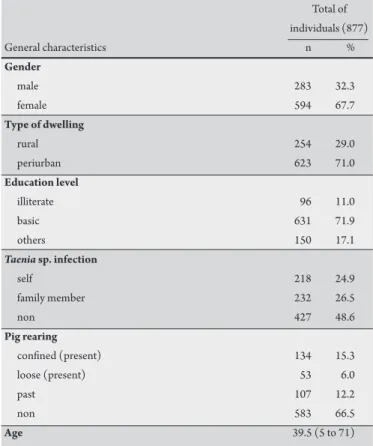
DISCUSSION TABLE 1 - General characteristics of studied population.
Total of individuals (877)
General characteristics n %
Gender
male 283 32.3
female 594 67.7
Type of dwelling
rural 254 29.0
periurban 623 71.0
Education level
illiterate 96 11.0
basic 631 71.9
others 150 17.1
Taenia sp. infection
self 218 24.9
family member 232 26.5
non 427 48.6
Pig rearing
conined (present) 134 15.3
loose (present) 53 6.0
past 107 12.2
non 583 66.5
Age 39.5 (5 to 71)
and 1:2.5, respectively, in a 1% skim milk saline solution containing 0.05% Tween 20®. Conjugate peroxidase-labeled sheep anti-human
immunoglobulin G was used at titers 1:5,000 for both sera and total blood sample and chromogen substrate ortho-phenylenediamine and H2O2 in citrate bufer (pH 5.0). he reaction was read with a plate spectrophotometer at 492nm. he cutof value for the ELISA-Tcra-vf was determined with sera from 6 proven NC patients (positive controls) and from 68 healthy individuals (negative controls) following the same ELISA serum protocol18. Each plaque assay was performed with the
inclusion of one positive and one negative serum sample as controls, from the same group of samples used in the standardization. he results were expressed as the ratio between the optical density (OD) of the sample and the cutof value. Ratio values > 1 were considered as positive. In order to compare the results obtained for the ilter paper-eluted blood with those obtained for the serum samples, a dilution factor for the ilter paper samples was deined based on the volume of blood soaked in the delimited paper area26,28.
Immunoblot assay
The 18/14-Tcra antigen obtained by elution from the immunoainity chromatography with speciic monoclonal antibodies was analyzed by sodium dodecyl sulfate-polyacrylamide gel electrophoresis (SDS-PAGE)29. Then (3µg/mm) was solubilized
with sample bufer (0.01M Tris-HCl, pH 6.8, containing 2% SDS, 5% 2-mercaptoethanol, and 10% glycerol) at 100°C for 5min and separated electrophoretically on 15% polyacrylamide gel. he separated antigen was transferred to a 0.22-μm-pore size membrane of polyvinylidene diluoride (Millipore Corp., Bedford, Mass). he membrane was blocked by treatment with 5% skim milk in PBS for 2h, washed in PBS containing 0.05% Tween 20, and then incubated for 18h at 4°C with serum in 1:50, in 1% skim milk in PBS. Following further washes, the strips were incubated for 1h with goat anti-human immunoglobulin G (IgG)-biotin/avidin peroxidase (Sigma) conjugate (1:3,000). Following additional washes, the antigen-antibody complexes were obtained by incubation with an appropriate substrate: 4-chloro-1-naphthol (Sigma) predissolved in methanol (20% of the volume) and then diluted to 0.05% with Tris-bufered saline (0.01 M Tris, 0.15 M NaCl, pH 7.4) containing 0.06% H2O2 (for the peroxidase conjugate).
Statistical analysis
he Kappa coeicient (k) was calculated to evaluate the agreement between the ELISA results for the serum and whole blood30.
he Chi square test with signiicance set at p < 0.05 and meta-analysis were used in order to verify the correlation between the immunological test results and risk factors31.
The results of the seroepidemiological studies focusing on cysticercosis were sent to the health authorities of the municipality, in order to contribute to the collection of information on endemicity and on risk factors for transmission.
Ethical considerations
Ethical approval for the study was granted by the Research Ethics Commitee of UFSC, Brazil, under protocol no. 211/03.
Data obtained from 877 questionnaires answered orally provided a demographic proile of this population regarding age, sex, residential location, education and information related to the epidemiology of teniasis/cysticercosis in the area, as shown on Table 1.
he results for ELISA using whole blood were in agreement with those using serum samples (k= 0.73) up to one year ater collection. A total of 850 samples of whole blood collected in ilter paper were able to be analyzed by ELISA using Tcra-vf as antigens and positive reactions were obtained in 186 (21.9%) individuals. A total of 213 volunteers were asked to collect blood samples for use in the immunoblot assay (IB) conirmatory assay (186 positive for ELISA and 27 whose initial sample was considered inappropriate). he IB test was performed on 130 samples collected from people who showed up to provide a blood sample and 29 presented a positive reaction. Considering that these individuals are putative holders of cysticerci, analysis of their socioeconomic proiles was conducted in an atempt to relate infection with known factors that predispose human populations to cysticercosis.
he prevalence encountered in the population of Lages (SC) was 3.4%, since the immunoblot conirmatory test was positive in 29 samples out of 850 tested individuals. A signiicant correlation was determined among individuals tested positive by IB who practice both pig rearing and kitchen gardening (p = 0.0364).
he use of ilter paper proved to be an appropriate option for ield work where no facilities to collect vein blood or to obtain serum from whole blood are available. he data obtained were in agreement with results reported by Peralta et al26 and Fleury et al25,
and thus epidemiological studies on cysticercosis can be successfully conducted using this method.
ACKNOWLEDGMENTS
he authors declare that there is no conlict of interest. CONFLICT OF INTEREST
FINANCIAL SUPPORT
REFERENCES encountered in this population is compatible with reports from other
seroepidemiological surveys conducted in Brazil32.
Bragazza et al20 reported a serum prevalence of 2.1% for
cysticercosis in individuals from a rural population in the State of São Paulo, associated with the consumption of untreated water. Prestes-Carneiro et al33 veriied a frequency of 0.6% of anti-Taenia
solium cysticerci, conirmed by the immunoblot method, in a rural setlement in the State of São Paulo. According to the authors, the index was considered low and was a consequence of continued investment in public health care and private institutions to improve the infrastructure services and education.
In this study, pig rearing and kitchen gardening were determined as risk factors associated with Taenia solium infection in the population studied (p = 0.0364). he highest prevalence (65% of the conirmatory IB) was veriied in the areas known as Ranchode Tábuas and Tributo, a rural and periurban area, respectively. Rancho de Tábuas is an area where pigs are reared for domestic consumption and the transmission of Tania solium to pigs requires access to human feces and human consumption of improperly cooked pork. hese conditions are still very common in the developing world.
More than 80% of the population of Lages lives in urban areas, which account for only 10% of the area of the municipality34.
This intense urbanization results in crowded areas around the town center with poor sanitation and precarious housing and is commonly associated with low education and economic levels of the inhabitants. Tributo is a locality with the conditions necessary for the maintenance of teniasis/cysticercosis transmission. Its population lives on the border between the urban and rural areas, maintaining some rural habits, such as domestic rearing of a small number of pigs in the backyard fed with letover food and oten in free contact with domestic sewage.
his study was conducted by visiting houses or at community health centers during the day, when most men are at work and women, mostly housewives, are at home taking care of children. his explains the higher frequency of women in the population sample.
he indings provided data which will be useful to the authorities when planning interventions in routine healthcare practices and, more importantly, in preventive healthcare practices. Moreover, it was suggested that the positive results obtained in this study were registered in the medical records of each participant to improve medical care in cases of the appearance of related symptoms.
In conclusion, the local seroprevalence of cysticercosis veriied among volunteers from rural and periurban areas of Lages, SC, was similar to that reported in others studies in Latin America. Among all the risk factors analyzed, pig rearing and kitchen garden were positively associated with cysticercosis markers.
he authors would like to thank all the health professionals who contributed directly or indirectly to collecting data, in particular to the Health Authorities and Community Health Agents of the town of Lages.
his work was supported by the Fundação de Amparo à Pesquisa no Estado de Santa Catarina, project # 4663/2004
1. Moses A. Dos métodos biolójicos de diagnóstico nas cisticercozes. Mem Inst Oswaldo Cruz 1911; 3:322-326.
2. Fundação Nacional de Saúde. Projeto para controle do complexo teníase/ cisticercose no Brasil. Brasília: Ministério da Saúde; 1996.
3. Fleury A, Morales J, Bobes RJ, Dumas M, Yánez O, Piña J, et al. An epidemiological study of familial neurocysticercosis in an endemic Mexican community. Trans Royal Soc Trop Med Hyg 2006; 100:551-558.
4. Cruz ME, Schantz PM, Cruz I, Espinosa P, Preux PM, Cruz A, et al. Epilepsy and neurocysticercosis in an Andean community. Int J Epidemiol 1999; 28:799-803. 5. Fleury A, Gómez T, Alvarez I, Meza D, Huerta M, Chavaria A, et al. High prevalence of calciied silent neurocysticercosis in a rural village of México. Neuroepidemiology2003; 22:139-145.
6. Agapejev S. Clinical and epidemiological aspects of neurocysticercosis in Brazil: a critical approach. Arq Neuro-Psiquiatr 2003; 61:822-828.
7. Garcia HH, Herrera G, Gilman RH, Tsang VCW, Pilcher JB, Diaz JF, et al. Discrepancies between cerebral computed tomography and western blot in the diagnosis of neurocysticercosis. he Cysticercosis Working Group in Peru (Clinical Studies Coordination Board). Am J Trop Med Hyg 1994; 50:152-157. 8. Sciuto E, Fragoso G, Fleury A, Laclete JP, Sotelo J, Aluja A, et al. Taenia solium
disease in humans and pigs: an ancient parasitosis disease rooted in developing countries and emerging as a major health problem of global dimensions. Microbes Infect 2000; 2:1875-1890.
9. Bueno EC, Scheel CM, Vaz AJ, Machado LR, Livramento JA, Takayanagui OM, et al. Application of synthetic 8-kD and recombinant GP50 antigens in the diagnosis of neurocysticercosis by enzyme-linked immunosorbent assay. Am J Trop Med Hyg 2005;72:278-283.
10. Ishida MM, Peralta RH, Livramento JA, Hoshino-Shimizu S, Peralta JM, Vaz AJ. Serodiagnosis of neurocysticercosis in patients with epileptic seizure using ELISA and immunoblot assay. Rev Inst Med Trop S Paulo 2006; 48:343-346. 11. Bern C, Garcia HH, Evans C, Gonzalez AE, Verastegui M, Tsang VCW, et al.
Magnitude of the disease burden from neurocysticercosis in a developing country. Clin Infect Dis 1999; 29:1203-1209.
12. Wilson M, Bryan RT, Fried JA, Ware DA, Schantz PA, Pilcher JB, et al. Clinical evaluation of the cysticercosis enzyme-linked immunoelectrotransfer blot in patients with neurocysticercosis. J Infect Dis 1991; 164:1007-1009.
13. Tsang VCW, Brand JA, Boyer AE. An enzyme-linked immunoelectrotransfer blot assay and glycoprotein antigens for diagnosing human cysticercosis (Taenia solium). J Infect Dis 1989; 159:50-59.
14. Flisser A, Gyorkos TW. Contribution of immunodiagnostic tests to epidemiological/intervention studies of cysticercosis/taeniosis in México. Parasite Immunol 2007; 29:637-649.
15. Greene RM, Hancock K, Wilkins PP, Tsang VCW. Taenia solium: molecular cloning and serologic evaluation of 14- and 18-kDa related, diagnostic antigens. J Parasitol 2000; 86:1001-1007.
16. Hancock K, Khan A, Williams FB, Yushak ML, Pattabhi S, Noh J, et al. Characterization of the 8-kilodalton Antigens of Taenia solium Metacestodes and Evaluation of heir Use in an Enzyme-Linked Immunosorbent Assay for Serodiagnosis. J Clin Microbiol 2003; 41:2577-2586.
17. Hancock K, Patabhi S, Whitield FW, Yushak ML, Lane WS, Garcia HH, et al. Characterization and cloning of T24, a Taenia solium antigen diagnostic for cysticercosis. Mol Bioch Parasitol 2006; 147:109-117.
18. Bueno EC, Vaz AJ, Machado LR, Livramento JA, Mielle SR. Speciic Taenia crassiceps and Taenia solium antigenic peptides for neurocysticercosis immunodiagnosis using serum samples. J Clin Microbiol 2000;38:146-151. 19. Espíndola NM, Vaz AJ, Pardini AX, Fernandes I. Excretory/secretory antigens
monoclonal antibody for antigen detection in samples of cerebrospinal luid from patients with neurocysticercosis. Ann Trop Med Parasitol 2002; 96:361-368. 20. Bragazza LM, Vaz AJ, Passos ADC, Takayanagui OM, Nakamura PM, Espíndola
NM, et al. Frequency of serum anti-cysticercus antibodies in the population of a rural brazilian community (Cássia dos Coqueiros, SP) determined by ELISA and immunobloting using Taenia crassiceps antigens. Rev Inst Med Trop S Paulo 2002; 44:7-12.
21. Espíndola NM, Iha AH, Fernandes I, Takayanagui OM, Machado LR, Livramento JA. Cysticercosis immunodiagnosis using 18- and 14- kilodalton proteins from Taenia crassiceps cysticercus antigens obtained by immunoainity chromatography. J Clin Microbiol 2005; 4:3178-3184.
22. Trevisol-Bitencourt PC, Silva NC, Figueiredo R. Neurocisticercose em pacientes internados por epilepsia no Hospital Regional de Chapecó, região oeste do Estado de Santa Catarina. Arq Neuropsiquiatr 1998; 56:53-58.
23. Pfuetzenreiter MR, Avila-Pires FD. Clinical manifestations in patients with computerized tomography diagnosis of neurocysticercosis.Arq Neuropsiquiatr 1999; 57:653-658.
24. Jafri HS, Torrico F, Noh JC, Bryan RT, Balderrama F, Pilcher JB, et al. Application of the enzyme-linked immunoelectrotransfer blot to ilter paper blood spots to estimate seroprevalence of cysticercosis in Bolivia. Am J Trop Med Hyg 1998; 58:313-315.
25. Fleury A, Bouteille B, Garcia E, Marquez C, Preux PM, Escobedo F, et al. Neurocysticercosis: validity of ELISA after storage of whole blood and cerebrospinal luid on paper.Trop Med Intern Health 2001; 6:688-693. 26. Peralta RHS, Macedo HW, Vaz AJ, Machado LR, Peralta JM. Detection of
anti-cysticercus antibodies by ELISA using whole blood collected on ilter paper. Trans R Soc Trop Med Hyg 2001; 95:35-36.
27. Vaz AJ, Nunes CM, Piazza RM, Livramento JA, Da Silva MV, Nakamura PM, et al. Immunoblot with cerebrospinal luid from patients with neurocysticercosis using antigen from cysticerci of Taenia solium and Taenia crassiceps. Am J Trop Med Hyg 1997;57:354-357.
28. Evengard B, Linder E, Lundberg P. Standardization of a ilter-paper technique for blood sampling. Ann Trop Med Parasitol 1988; 82:295-303.
29. Lambin P, Rochu D, Fine JM. A new method for determination of molecular weights of protein by electrophoresis across a sodium dodecyl sulfate (SDS)- polyacrylamide gradient gel. Anal Biochem 1976; 74:567-575.
30. Ayres M, Ayres Jr M, Ayres DL Santos AS. Bio Estat 5.0 Aplicações Estatísticas nas Áreas das Ciências Biológicas e Médicas, Belém: Sociedade Civil Mamirauá. Conselho Nacional de Desenvolvimento Cientíico e Tecnológico; 2007. 31. Nist Sematech. e-Handbook of Statistical Methods [Internet]. 2010 – [cited 2010
April 25th]. Available from: htp://www.itl.nist.gov/div898/handbook/.
32. Prestes-Carneiro LE, Freitas SBZ, Zago SCS, Miguel NA, Primo OB, Iha AH, et al. Taeniosis-cysticercosis complex in individuals of a peasants’ setlement (Teodoro Sampaio, Pontal of Paranapanema, SP, Brazil). Mem Inst Oswaldo Cruz2005; 101:1-6.
33. Prestes-Carneiro LE, Souza DHP, Moreno GC, Troiani C, Santarém V, Zago SCS, et al. Toxocariasis/cysticercosis seroprevalence in a long-term rural setlement, São Paulo, Brazil. Parasitology 2009; 136:681-689.
34. City of Lages, State of Santa Catarina. Oicial website [Internet]. Lages (SC): Lages Cityhall; 2009. [cited 2009 Sep 16th]. Available from: htp://www.lages.