UNIVERSIDADE FEDERAL DO CEARÁ
FACULDADE DE FARMÁCIA, ODONTOLOGIA E ENFERMAGEM PROGRAMA DE PÓS-GRADUAÇÃO EM ODONTOLOGIA
MESTRADO EM ODONTOLOGIA
LÍVIA DE OLIVEIRA BARROS
ESTABILIDADE DA INTERFACE DE UNIÃO DE SISTEMAS ADESIVOS CONVENCIONAIS APLICADOS À DENTINA SATURADA COM ALCOÓIS
LÍVIA DE OLIVEIRA BARROS
ESTABILIDADE DA INTERFACE DE UNIÃO DE SISTEMAS ADESIVOS CONVENCIONAIS APLICADOS À DENTINA SATURADA COM ALCOÓIS
Dissertação de Mestrado a ser apresentada ao Programa de Pós-Graduação em Odontologia da Faculdade de
Farmácia, Odontologia e Enfermagem da Universidade Federal do Ceará como requisito parcial para a obtenção do título de Mestre em Odontologia.
Área de Concentração: Clínica Odontológica
Orientador: Prof. Dr. Vicente de Paulo Aragão Saboia
B279e Barros, Lívia de Oliveira
Estabilidade da interface de união de sistemas adesivos convencionais aplicados à dentina saturada com alcoóis / Lívia de Oliveira Barros. – Fortaleza,Ce; 2011.
50 f. : il.
Orientador: Prof. Dr. Vicente de Paulo Aragão Saboia
Dissertação (Mestrado) - Universidade Federal do Ceará. Programa de Pós-Graduação em Odontologia; Fortaleza-Ce, 2011.
1. Adesivos Dentinários 2. Resistência Adesiva 3. Etanol 4. Solvente I. Saboia, Vicente de Paulo Aragão (Orient.) II. Título.
LÍVIA DE OLIVEIRA BARROS
ESTABILIDADE DA INTERFACE DE UNIÃO DE SISTEMAS ADESIVOS CONVENCIONAIS APLICADOS À DENTINA SATURADA COM ALCOÓIS
Dissertação de Mestrado a ser apresentada ao Programa de Pós-Graduação em Odontologia da Faculdade de
Farmácia, Odontologia e Enfermagem da Universidade Federal do Ceará como requisito parcial para a obtenção do título de Mestre em Odontologia. Área de Concentração: Clínica Odontológica.
Aprovada em: ___/___/___
BANCA EXAMINADORA
_____________________________________________ Prof. Dr. Vicente de Paulo Aragão Saboia (Orientador)
Universidade Federal do Ceará - UFC
_____________________________________________ Prof. Dr. Sérgio Lima Santiago
Universidade Federal do Ceará - UFC
DEDICATÓRIA
Dedico este trabalho, principalmente, a DEUS, pelo milagre da vida, por ser Aquele que me fortalece, me guia, me ampara e me ilumina sempre. Agradeço a Ti pelo que sou e pelas coisas e pessoas maravilhosas que tens
me proporcionado. Sem Ti, nada disso seria possível! Muito obrigada por tudo!
Aos meus pais, FERNANDO E MARY, minha base maior, por todo carinho, dedicação, amor, companheirismo, palavras de sabedoria, amizade, ensinamentos e, principalmente, por serem meus exemplos de caráter,
humildade, determinação e força. Obrigada por sempre respeitarem e apoiarem os meus sonhos e ideais.
Sem vocês, eu não conseguiria. Amo vocês! Muito, muito, obrigada!
E ao meu irmão, LUCAS, por ser constante motivo de alegria e tranqüilidade para mim. Admiro a tua generosidade, bom humor e inteligência. Mano, sou muito de ti e tu és muito para mim; e preciso sempre de você na minha vida.
Obrigada por vibrar com as minhas vitórias como se fossem suas. Amo você!
AGRADECIMENTOS ESPECIAIS
Ao meu orientador, prof. DR. VICENTE DE PAULO ARAGÃO SABOIA, pela confiança em mim depositada, pela orientação, pelos conselhos, pelos ensinamentos transmitidos, pela atenção, pela simplicidade e por despertar em mim o gosto pela ciência. Nossa convivência foi essencial para a minha formação profissional. Sinto-me muito orgulhosa por compartilhar de seus
conhecimentos. Minha gratidão e admiração.
À amiga FABIANNI MAGALHÃES APOLÔNIO, pelo companheirismo, paciência, disponibilidade, dedicação e, sobretudo, pelo auxílio imensurável
na execução deste trabalho. Sinta esta conquista como sua também!
Ao prof. DR. SÉRGIO LIMA SANTIAGO, pela atenção, disponibilidade, convivência e por estar sempre presente ao longo dessa minha jornada, como
peça chave na construção da minha personalidade profissional. Você é um exemplo de competência e dedicação para mim.
A todos os professores do Programa de Pós-Graduação em Odontologia da Universidade Federal do Ceará, pela oportunidade que me deram de crescer
profissionalmente e, principalmente, pelos ensinamentos valiosos durante o curso de mestrado.
A todos os professores da Universidade Federal do Ceará, que me transmitiram a base da arte chamada ODONTOLOGIA. Os ensinamentos de
todos vocês foram fundamentais para minha formação.
Ao prof. DR. ILAN SAMPAIO DO VALE, por estar sempre presente ao longo da minha jornada acadêmica, pelo incentivo e pelo exemplo de
À querida MARY ANNE SAMPAIO DE MELO, pelo exemplo de amor a pesquisa e pela presteza em auxiliar a execução deste trabalho.
Aos alunos de Iniciação Científica, DANIEL, DIANA, ÍTALO e NARA no empenho e ajuda indispensável na realização deste trabalho.
Aos colegas do curso de mestrado, CAIO, JANAÍNA, JIOVANNE, JOÃO PAULO, MÁRCIA E YUSKA, pela troca de conhecimentos e experiências,
pelos momentos de alegria e descontração. Torço muito pelo sucesso de todos vocês!
À minha família, meus avós, tios, tias, primos e primas por sempre torcerem por mim de forma tão verdadeira. Vocês são muito importantes para mim!
AGRADECIMENTOS
À Universidade Federal do Ceará, por meio do reitor prof. JESUALDO PEREIRA FARIAS.
À Faculdade de Farmácia, Odontologia e Enfermagem (FFOE/UFC), na pessoa de sua diretora profa. NEIVA FRANCENELY CUNHA VIEIRA.
Ao Curso de Odontologia, na pessoa da sua Coordenadora profa. MARIA ENEIDE LEITÃO DE ALMEIDA.
Ao Programa de Pós-Graduação em Odontologia da Universidade Federal do Ceará, na pessoa de seu coordenador, prof. DR. SÉRGIO LIMA SANTIAGO.
Aos membros da banca examinadora, pela disponibilidade e presteza em avaliar e enriquecer este trabalho.
À funcionária da pós-graduação em Odontologia da Universidade Federal do Ceará, LÚCIA RIBEIRO MARQUES LUSTOSA, pelo auxílio e disponibilidade.
À Academia Cearense de Odontologia e seus funcionários, pelo auxílio na execução deste trabalho.
À CAPES pela concessão da bolsa de estudo.
“Nós
devemos ser a mudança que queremos ver no
mundo.
”
RESUMO
Existe um consenso de que a instabilidade da interface de união resina-dentina está relacionada com a quantidade de monômeros hidrofílicos presentes no sistema adesivo. Dessa forma, interfaces adesivas mais duradouras seriam alcançadas com a utilização de sistemas adesivos mais hidrofóbicos. Uma maneira de unir monômeros hidrófobos à dentina é através da saturação da matriz dentinária desmineralizada por etanol ao invés de água (ethanol-wet bonding technique). O objetivo deste trabalho in vitro foi avaliar o efeito do protocolo de saturação dentinária, utilizando alcoóis, na estabilidade da interface de união produzida por sistemas adesivos convencionais de dois passos. Para isso, foram utilizados 20 terceiros molares humanos, que tiveram a dentina coronária exposta e foram distribuídos aleatoriamente em quatro grupos experimentais (n=5): Adper Single Bond 2 (SB) aplicado sobre dentina saturada com água ou etanol; e XP Bond (XP) aplicado sobre dentina saturada com água ou tert-butanol. O protocolo de saturação da dentina foi realizado através da aplicação de 2 μl de etanol 100% ou tert-butanol 99,5% diretamente sobre a dentina por um período de 60 s. Um platô de resina composta foi confeccionado sobre superfície dentinária após a aplicação do sistema adesivo e os espécimes foram cortados pela técnica non-trimming de obtenção de palitos para o teste de microtração. Os palitos de cada dente foram igualmente divididos em dois subgrupos: imediatamente testado ou envelhecido em solução de NaOCl a 10% por 1 hora. Os espécimes foram tracionados até a ruptura da união a uma velocidade de 1 mm/min e sua força de união mensurada. Os palitos fraturados foram analisados e classificados de acordo com o modo de fratura em mista, adesiva, coesiva em dentina e coesiva em compósito, e os valores expressos em porcentagem. Os valores de resistência de união foram estatisticamente analisados
usando os testes ANOVA a três critérios e Tukey (α=0.05). Discos extras de dentina foram submetidos aos procedimentos adesivos de cada grupo testado e utilizados para investigar as características morfológicas da interface de união através de microscopia óptica após imersão em solução amonical de nitrato de prata. Os resultados do teste de microtração mostraram que a imersão em solução de NaOCl reduziu significativamente a força de união em comparação aos grupos controle (p<0.05) e aumentou a nanoinfiltração das interfaces adesivas de todos os sistemas adesivos testados. A saturação da dentina com etanol 100% reduziu os valores de resistência adesiva para SB quando comparado ao grupo controle, e esta mesma tendência foi observada na infiltração de prata. O uso do tert-butanol 99,5% não afetou os valores de resistência adesiva para o XP, assim como a deposição de nitrato de prata na interface adesiva.
Conclusão: Os protocolos simplificados de saturação da dentina com alcoóis utilizados no presente estudo não foram capazes de melhorar a estabilidade da interface de união para sistemas adesivos convencionais de dois passos.
ABSTRACT
There is a consensus that the resin-dentin bond instability is correlated with increased hydrophilic resin monomer content in dentin adhesive. Thus, more durable bonds could be created when more hydrophobic resins were used. One way to bond hydrophobic monomers to acid-etched dentin is saturating the demineralized dentin with ethanol instead of water (ethanol-wet bonding technique). The objective of this in vitro study was to evaluate the effect of “alcohol wet bonding” technique on the stability of adhesive interface produced by two-step etch-and-rinse adhesives systems. For this, it was used 20 human third molars that had superficial dentin exposed and were randomly divided into four experimental groups (n=5): Adper Single Bond 2 (SB) bonded to acid-etched dentin saturated with water or ethanol; and XP Bond (XP) bonded acid-etched dentin saturated with water or 99.5% tert-butanol. The simplified dentin dehydration protocol was performed using 2 μl 100% ethanol or 99.5% tert-butanol directly applied in dentin for 60 s. Composite build-ups were built on dentin surface and specimens were cut into non-trimming dentin-composite beams to microtensile testing. Beams from each tooth were divided equally in two subgroups: immediately tested and aged by immersion in 10% NaOCl solution for 1 h. Specimens were pulled until failure at crosshead speed of 1 mm/min and bond strength was calculated. Fractured sticks were analyzed and classified according to the failure mode as mixed, adhesive, cohesive in dentin and cohesive in composite; and expressed in percentage. Data from μTBS test were statistically analyzed using three-way ANOVA and Tukey tests (α=0.05). Additional dentin disks were bonded using the same groups tested and the morphological characteristics of adhesive interface were investigated by light microscopy after immersion in ammoniacal silver nitrate solution. μTBS results showed that NaOCl solution reduced significantly bond strength comparing with the control groups (p<0.05) and increased the silver nitrate interfacial deposit for all adhesives tested. SB used in ethanol saturated-dentin showed significant lower bond strength values in comparison with SB control group, and this same tendency was observed in the silver nitrate deposition. The use of tert-butanol did not influence XP bond strengths values and silver nitrate deposits.
Conclusion: The “alcohol wet bonding” simplified technique used in the present
study was not able to improve resin-dentin bond stability for simplified etch-and-rinse adhesive systems.
SUMÁRIO
1. INTRODUÇÃO GERAL...13
2. PROPOSIÇÃO...18
2.1 – Objetivo Geral...19
2.2 – Objetivos Específicos...19
3. CAPÍTULO...20
4. CONCLUSÃO GERAL...41
REFERÊNCIAS...43
1. INTRODUÇÃO GERAL
A odontologia adesiva é caracterizada pelo uso de materiais resinosos que tiveram seu aperfeiçoamento com a técnica de condicionamento ácido proposto por Buonocore em 1955. Desde então vários estudos têm sido desenvolvidos com a finalidade de aperfeiçoar a adesão do material restaurador ao substrato dentário (VAN MEERBEEK et al., 2003). Embora a adesão ao esmalte dentário seja descrita com alto índice de sucesso clínico, a adesão à dentina ainda é considerada deficiente, principalmente a longo prazo, devido às complexas características morfofuncionais desse tecido (PASHLEY et al., 2004).
A durabilidade da interface dentina/resina é de extrema importância para a longevidade das restaurações adesivas, já que a degradação desta interface pode enfraquecer a resistência de união (CARRILHO et al., 2004) e ocasionar problemas clínicos indesejáveis como sensibilidade pós-operatória, infiltração marginal, perda de retenção da restauração e o aparecimento de cáries secundárias (BRESCHI et al., 2008; GOING, 1972). A estabilidade da interface de união é possível a partir da criação de uma camada híbrida compacta e homogênea. Dessa forma, idealmente, o substrato dentinário, previamente desmineralizado, deve ser completamente infiltrado pelo adesivo, evitando áreas de incompleta impregnação da matriz dentinária (BRESCHI et al., 2008).
No conceito atual de adesão à dentina, a água desenvolve função essencial na prevenção do colapso da rede de fibrilas colágenas, expostas pelo condicionamento ácido prévio, permitindo a infiltração dos monômeros resinosos na intimidade do tecido dentinário e a formação da camada híbrida (SHIN et al., 2009). A água é o solvente com melhores características para manter a trama de colágeno expandida em função da sua capacidade de romper pontes de hidrogênio formadas entre os peptídeos das fibrilas quando a dentina é desidratada e manter as fibrilas em seu grau máximo de expansão (PASHLEY et al., 2007).
monômeros hidrófobos presentes no sistema adesivo, insolúveis em água, resultando na formação de uma interface adesiva deficiente. Há ainda que se considerar que a água presente durante a hibridização da dentina é impossível de ser eliminada por completo pelos procedimentos clínicos usuais (YIU et al., 2005), podendo se tornar um fator de comprometimento da polimerização do adesivo. Esse conjunto de fatores favorece a degradação precoce da camada híbrida através de um efeito combinado de hidrólise de seus componentes resinosos e por um processo hidrólítico/enzimático das fibrilas colágenas expostas pelo condicionamento ácido e não totalmente protegidas pela resina (HASHIMOTO et al., 2000; PASHLEY et al., 2004; CARRILHO et al., 2005; LOGUERCIO et al., 2005).
A fim de promover uma melhor infiltração dos monômeros resinosos na dentina, reduzir a sensibilidade do sistema adesivo à intrínseca umidade dentinária (TAY, PASHLEY, 2003) e minimizar a separação de fase na interface adesiva, monômeros hidrofílicos são adicionados a composição dos sistemas adesivos atuais (SPENCER, WANG, 2002). Entretanto, estudos têm demonstrado que quanto maior a hidrofilicidade do sistema adesivo, maior a sorção de água pela interface adesiva (UNEMORI et al., 2003; YIU et al., 2006), mais rápida a deterioração das suas propriedades mecânicas (YIU et al., 2004; ITO et al., 2005) e, consequentemente, menor a longevidade clínica da restauração (YE et al.,2008; YE et al., 2009). Em estudo realizado por Tay et al. (2002), observou-se que sistemas adesivos simplificados, que não possuem a aplicação de uma camada de adesivo hidrofóbico puro, comportaram-se como membranas semipermeáveis após polimerização, permitindo a passagem de água pela interface adesiva. Diante disso, esforços (TAY et al., 2007; SAURO et al., 2010) têm sido concentrados no sentido de desenvolver uma técnica capaz de otimizar a união de adesivos mais hidrófobos à dentina, promovendo assim, a obtenção de restaurações mais estáveis.
monômeros mais hidrófobos se dissolvem com maior facilidade e a difusão é favorecida por toda a extensão da zona desmineralizada (BECKER et al., 2007), minimizando a existência de fibrilas de colágeno expostas e não protegidas na base da camada híbrida.
Essa estratégia envolve a lenta substituição da água, presente no interior da matriz colágena desmineralizada, através de uma desidratação química, utilizando concentrações ascendentes de etanol (SADEK et al., 2010). Teoricamente, com essa técnica, a matriz colágena desmineralizada torna-se menos hidrofílica e mais receptiva aos monômeros hidrofóbos, reduzindo a separação de fase (WANG, SPENCER, 2003) na interface de união. Além disso, a técnica de saturação do substrato dentinário com o etanol permite uma melhor infiltração do agente de união na trama de colágeno, visto que, a maioria dos monômeros hidrófobos são solúveis em etanol (SHIN et al., 2009), tornando o substrato dentinário mais compatível com os monômeros resinosos contidos nos sistemas adesivos.
Outro fator relevante e que participa em certo grau do processo de adesão, é o enrijecimento das fibrilas quando da aplicação de etanol puro ou misturas de HEMA/ etanol, resultando em menor contração da mesma quando o solvente é evaporado (GARCIA et al., 2005; BECKER et al., 2007). Dessa forma, os espaços interfibrilares são mantidos mesmo após a evaporação do solvente, favorecendo a infiltração posterior da resina. Além disso, a dentina submetida à saturação com etanol apresenta um aumento do espaço interfibrilar quando comparada à dentina saturada com água (HOSAKA et al., 2009).Esse fato poderia promover uma maior quantidade de resina ao redor das fibrilas colágenas (HOSAKA et al., 2009) e, consequentemente, maiores valores de resistência adesiva quando comparada a técnica convencional (NISHITANI et al., 2006).
al., 2010), para a obtenção de resultados satisfatórios, o que dificulta a viabilidade clínica dessa técnica. Por esta razão, protocolos de desidratação mais simplificados têm sido utilizados (HOSAKA et al., 2009; SADEK et al., 2010). Entretanto, estudos recentes têm observado, que curtos períodos de saturação podem não ser suficientes para a completa substituição da água, presente no interior da matriz dentinária desmineralizada, pelo etanol. Dessa forma, a presença de água residual na dentina (OSORIO et al., 2010; SADEK et al., 2010) pode resultar na pobre infiltração dos monômeros hidrófobos (SADEK et al., 2010) presentes nos sistemas adesivos. Nesse caso, o uso de um adesivo contendo monômeros hidrofílicos poderia auxiliar na melhor infiltração do adesivo na dentina desmineralizada, diminuir a permeabilidade em comparação a um adesivo hidrofóbico (SAURO et al., 2009; CADENARO et al., 2009) e aumentar a resistência de união imediata à dentina (NISHITANI et al., 2006).
Os monômeros resinosos dos sistemas adesivos são misturados a solventes, tais como: água, etanol, acetona ou por uma combinação destes (VAN MEERBEEK
et al., 2003). XP Bond (Dentsply, Konstanz, Germany) é um sistema adesivo convencional de dois passos, desenvolvido recentemente, que possui o álcool tert-butanol como solvente. Tem sido relatado que esse adesivo tem a capacidade de penetrar a matriz colágena colapsada, resultando em menor sensibilidade técnica e melhor desempenho clínico (BLUNCK, KNITTER, JANH, 2007). Contudo, não existem estudos que relatem o uso desse sistema adesivo associado à técnica de saturação da matriz dentinária com o seu próprio solvente (tert-butanol).
2. PROPOSIÇÃO
O presente trabalho teve como objetivos:
2.1 Objetivo Geral
Avaliar in vitro o efeito do protocolo de saturação dentinária, através do uso de alcoóis (etanol ou tert-butanol), na estabilidade da interface de união de sistemas adesivos convencionais de dois passos.
2.2 Objetivos Específicos
Avaliar a resistência de união à microtração da interface de união de sistemas adesivos convencionais de dois passos quando aplicados na dentina saturada com etanol ou tert-butanol e submetida a envelhecimento artificial;
3. CAPÍTULO
Esta dissertação está baseada no Artigo 46 do Regimento Interno do Programa de Pós-Graduação em Odontologia da Universidade Federal do Ceará, que regulamenta o formato alternativo para dissertações de Mestrado e teses de Doutorado e permite a inserção de artigos científicos de autoria ou co-autoria do candidato. Por se tratar de pesquisas envolvendo seres humanos, ou parte deles, o projeto de pesquisa foi submetido à apreciação do Comitê de Ética em Pesquisa, tendo sido aprovado (Anexo 1). Assim sendo, esta dissertação é composta de um artigo científico que será submetido ao periódico Operative Dentistry, conforme descrito abaixo:
Stability of interface created by simplified etch-and-rinse adhesives to alcohol-saturated acid-etched dentin
Stability of interface created by simplified etch-and-rinse
adhesives to alcohol-saturated acid-etched dentin
LÍVIA DE OLIVEIRA BARROSa, FABIANNI MAGALHÃES APOLONIOa,
ALESSANDRO DOURADO LOGUERCIOb, VICENTE DE PAULO ARAGÃO
SABOIAc.
a MSc, Faculty of Pharmacy, Dentistry and Nursing, Federal University of Ceará,
Fortaleza, Ceará, Brazil.
b PhD, Adjunctive Professor, Department of Restorative Dentistry, School of
Dentistry, State University of Ponta Grossa, Ponta Grossa, Paraná, Brazil.
c PhD, Associate Professor, Department of Restorative Dentistry, Faculty of
Pharmacy, Dentistry and Nursing, Federal University of Ceará, Fortaleza, Ceará, Brazil.
AUTHORS INFORMATIONS Lívia de Oliveira Barros
e-mail: [email protected]
Fabianni Magalhães Apolonio e-mail: [email protected]
Alessandro Dourado Loguercio e-mail: [email protected]
Vicente de Paulo Aragão Saboia (Corresponding author) e-mail: [email protected]
SHORT TITLE: Stability of adhesive interfaces under alcohol-wet bonding technique
ABSTRACT
Purpose: To evaluate the effect of “alcohol wet bonding” technique on durability of the adhesive interface produced by two-step etch-and-rinse adhesives systems.
Materials and Methods: Twenty human third molars had superficial dentin exposed and were randomly divided into four experimental groups: Adper Single Bond 2 (SB) bonded to acid-etched dentin saturated with: water (control) or ethanol; and XP Bond (XP) bonded acid-etched dentin saturated with water (control) or tert-butanol. A simplified dentin dehydration protocol was performed using 100% ethanol or 99.5% tert-butanol directly applied in dentin for 60 s. Composite build-ups were built on dentin surface and specimens were cut into non-trimming dentin-composite beams. Beams from each tooth were divided equally in two subgroups: immediately tested and aged by immersion in 10% NaOCl solution for 1 h. Specimens were pulled until failure at crosshead speed of 1 mm/min and pattern failure was evaluated. Data were statistically analyzed with three-way ANOVA and Tukey tests (α=0.05). Additional dentin disks were bonded using the same groups tested and investigated by light microscopy after immersion in ammoniacal silver nitrate solution.
Results: SB control group showed significant higher bond strength values in comparison with SB used in ethanol saturated-dentin (p<0.05), and this same tendency was observed in the silver nitrate deposition. The use of tert-butanol did not influence XP bond strengths values (p>0.05) and silver nitrate deposits. NaOCl solution significantly reduced bonding of all groups tested (p<0.05) and also increasing the silver nitrate interfacial deposit.
Conclusion: The “alcohol wet bonding” simplified technique used in the present study was not able to improve resin-dentin bond durability for simplified etch-and-rinse adhesive systems.
INTRODUCTION
In the water wet bonding technique, after acid etching, the presence of water in the demineralized dentin is essential to prevent the collapse of the dentin matrix. The space that is occupied by the water surrounding the non-collapsed collagen fibrils can then be infiltrated by the resin monomers to form the hybrid layer.1
However, the presence of residual water in the current wet bonding technique2 is a potential factor for phase separation of the hydrophobic monomers which has limited water solubility.1 Traditionally, hydrophilic resin monomers are used in the contemporary dentin adhesives to enhance their wetting properties and to avoid phase separation when hydrophobic dimethacrylates are added to water.3 Nevertheless, it has been reported that the current generation of dentin adhesives has been criticized as being too hydrophilic4 and absorbing too much water following polymerization,5 which lowers their stiffness when compared with that of more hydrophobic resins.6 This increased water sorption can result in the accelerated degradation and decreased mechanical properties that affect the long-term survival of the interface and associated restoration.7,8 Presumably, if dentin is bonded with more hydrophobic resins, the interface would absorb less water and produce more durable bonds.5
Recent studies have shown that it is possible to bond hydrophobic resin monomers to acid-etched dentine9,10 with a new technique called “ethanol wet
bonding”.10 This technique involves slowly replacing water within the demineralized collagen matrix with ascending concentrations of ethanol, allowing the latter to penetrate the collagen matrix without causing additional shrinkage of the interfibrillar spaces.11 The ethanol saturated dentine is less hydrophilic and prevents phase separation of hydrophobic resin monomers.12 Moreover, because most hydrophobic monomers are also soluble in ethanol, it is possible to use an adhesive with a higher ratio of hydrophobic to hydrophilic monomers.1 When acid etched dentin was saturated with 100% ethanol instead of water, the bond strengths of both hydrophilic and hydrophobic resin increased significantly.5,13
enough for complete replacement of water within the acid-etched intertubular dentine and the dentinal tubules by ethanol. Most likely, residual water present in within the acid-etched dentin11,15 can result in poor wetting by the hydrophobic adhesive.11 In this case, the use of a hydrophilic monomer could help the infiltration of the adhesive on dentin and decreasing the permeability in comparison with hydrophobic adhesive,16,17 as well as, increasing the immediate bond strength to dentin.5 To extent of our knowledgement, there is no studies that evaluated this issue regarding immediately and aging bond strength on commercial simplified adhesive system.
The monomers of adhesive systems are carried by a solvent which is usually water, ethanol, acetone or a combination of those.18 Recently, it was formulated a two-step etch–and-rinse adhesive (XP Bond, Dentsply) which uses the alcohol tert-butanol as the solvent. It is claimed to be less technique sensitive due to an improved ability to diffuse through partially collapsed demineralized dentin.19 Nevertehless, there is no studies using this adhesive system on dentin saturated with its solvent. Thus, more evidence in relation to ethanol wet bonding regarding to water replacement protocol, time application and bonds durability are needed to justify this extra step.
The present study was performed to evaluate the effect of “alcohol wet bonding” technique on the durability of adhesive interface produced by two-step etch-and-rinse adhesives systems on alcohol (ethanol or tert-butanol) saturated dentin, immediately after bonding and after artificial ageing in 10% NaOCl, by microtensile bond strength evaluation and silver nitrate uptake. The null hypothesis tested was there is no difference in adhesive performance and stability between the use of
“alcohol wet bonding” technique and water wet bonding technique.
METHODS & MATERIALS Tooth Preparation
paper mounted in a polisher machine (Aropol 2V – Arotec, São Paulo, SP, Brazil). Each tooth was etched with 35% phosphoric acid gel (3M ESPE, St Paul, MN, USA) for 15 s and rinsed thoroughly with distilled water for 15 s. Excess water was removed from the surface with absorbing paper (Melitta, São Paulo, SP, Brazil) and the dentin was kept visibly moist.
Bonding procedures
Specimens were equally and randomly assigned to 4 groups (n=5) and treated with one of the two adhesives: Adper Single Bond 2 (SB; 3M ESPE, St Paul, MN USA) and XP Bond (XP; Dentsply DeTrey, Konstanz, Germany), containing ethanol or tert-butanol solvents, respectively. In the control groups, SB and XP were applied
according to the manufacturer’s instructions to visibly moist water-saturated
demineralized dentin in which was applied 2 μl of distilled water for 60 s (Table 1). In
the specimens of the experimental groups, the water-moist dentin surfaces were
saturated with 2 μl of 100% ethanol (Vetec, Rio de Janeiro, RJ, Brazil) for SB and
99,5% tert-butanol (Vetec, Rio de Janeiro, RJ, Brazil) for XP. The solution was directed applied in dentin for 60 s. Composite build-ups were constructed with a light-cured resin composite (Filtek Z250, 3M ESPE, St Paul, MN, USA) in four 1mm-thick increments and individually polymerized for 20 s each, using a halogen light-curing unit (Eliopar Freelight 2, 3M ESPE, St Paul, MN, USA) with an output intensity of 650 mW/cm2.
Testing procedures
The bonded specimens were serially sectioned in both x and y directions across the adhesive interface with a diamond blade in an Isomet saw (Isomet, Buehler Ltd., Lake Bluff, USA) under water cooling to obtain 1mm-thick beams, in accordance with the microtensile test non-trimming technique.
After being stored for 24 h in distilled water at 37°C, beams from each tooth were divided equally into two subgroups. One subgroup was tested immediately, while the other was stored in 10% NaOCl for 1 h at room temperature.
Microtensile testing
mm/min. The number of prematurely debonded sticks per group during specimen preparation was also recorded.
The dentin side of the failed bonds was analyzed using a stereoscopic light microscopy (Stemi 2000–C, Carl Zeiss Jena, Jena, Germany) with 50x magnification and classified according to the failure mode as adhesive (A), cohesive in dentin (CD), cohesive in composite (CC) or mixed (M).
Light microscopy
Eight additional teeth (n=1 for subgroup) were processed for interfacial silver nitrate evaluation. Specimens were prepared as previously described in the item bonding procedure and, after that, specimens were then cut into 4 transverse sections, 1mm-thick, to expose the hybrid layer. Bonded specimens were stored for 24 h in distilled water or 1 h in 10% NaOCl solution.21,22 The slices were covered with nail varnish, leaving 1 mm exposed at the interface and immersed in 50 wt% ammoniacal silver nitrate (AgNO3) solution (pH 9.5) according to the protocol described by Tay et al. (2002).20 After immersion in the trace solution, specimens were then thoroughly rinsed in distilled water and were immersed in photodeveloping solution.
The silver nitrate-impregnated specimens were fixed on glass slides using cyanoacrylate glue (Super Bonder flex gel, Henkel Ltd, Düsseldorf, Germany) and flattened with SiC papers on increasing fine grits (600, 800, 1200 and 2400) in a polisher under running water. Images of all interfaces at 400x magnification under light microscopy (Nikon Eclipse E 800, Nikon, Tokyo, Japan) were obtained and silver nitrate representative images were chosen for each tested group.
Statistical analysis
Bond strength data were collected and analyzed using SPSS 17.0 (SPSS, Chicago, IL, USA). As the normality (Kolmogorov-Smirnov test) assumption of the data appeared to be valid, a three-way ANOVA was employed for statistical analysis to examine the effects of adhesive system, storage period and dentin treatment, and
interaction of these three factors on tensile bond strength. Tukey’s test was
employed for multiple comparisons (α=0.05). Silver nitrate evaluation was only
performed qualitatively.
RESULTS
Number of specimens and failure mode, as well as, means and standard deviations of microtensile bond strength, are shown in table 2 and 3, respectively. The number of prematurely debonded sticks per test group affected all groups to a similar extent and all premature failures occurred during the cutting procedure. For this reason, this report was not included in the statistical analysis.
Examination of the fractures surfaces by light microscopy indicated that although the majority of surfaces failed at the adhesive interface (adhesive failure), lower bond strengths were associated with higher percentages of adhesive failures. When the specimens were stored in 10% NaOCl for 1 h, the number of adhesive failures increased for all the groups (Table 2).
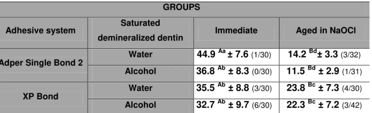
After 24 h, SB showed higher bond strength values on water saturated-dentin (44.9 ± 7.6 MPa) in comparison with 100% ethanol saturated-dentin (36.8 ± 8.3 MPa). On the other side XP did not shown statistical significance among saturated dentin with tert-butanol or with water saturated-dentin (35.5 ± 8.8 and 32.7 ± 9.7 MPa, respectively).
Storage in 10% NaOCl reduced the bond strength in all groups tested. After ageing in 10% NaOCl for 1 h, XP exhibited significantly higher bond strengths values (23.8 ± 7.3 MPa and 22.3 ± 7.2 MPa, respectively for water- and alcohol-based), when compared with the SB groups regardless of the water replacement protocol (14.2 ± 3.3 MPa and 11.5 ± 2.9 MPa, respectively for water- and alcohol-based). Interfacial silver nitrate evaluation
Representative light microscopy images of the adhesive interface produced by SB on water saturated dentin 24 h after bonding, showed no nanoleakage expression (Fig. 1A). However, when SB was bonded to ethanol saturated dentin, minimal silver deposition along the adhesive interface was observed (Fig. 1C). The results of interfacial nanoleakage analysis of the adhesive interface created by XP on water or tert-butanol saturated dentin, 24 h after bonding, showed low levels of silver deposition along the interfaces (Fig. 2A and Fig. 2C). After 1 h of 10% NaOCl ageing, all adhesive interfaces showed extensive and homogeneous deposition of silver throughout the adhesive interface (Fig. 1B, Fig. 1D, Fig. 2B and Fig. 2D).
DISCUSSION
bonding control group. Conversely, the use of 99.5% tert-butanol did not affect bond strength values when compared to the water-wet bonding control group for XP adhesive system. Thus, since there was difference when the alcohol-water replacement protocol was applied, the null hypothesis was partially rejected.
The ethanol-wet bonding protocol is one try to coax hydrophobic monomers to infiltrate in demineralized wet dentin.9,10 The ethanol-wet bonding technique proposed for dentin dehydration14 is analogous to the technique employed by electron microscopists for tissue embedding.23 This chemical dehydration procedure is unlikely to be clinically attractive due its extended time demand, justifying the search of a most simplified dehydration protocols.
Since hydrophobic resins are considerably difficult to apply to dentin, an alternative version of the ethanol wet-bonding technique is to apply hydrophilic adhesives to ethanol-saturated demineralized dentin.24 However, to the simplified ethanol-wet bonding technique, a decreased in the bond strength values for simplified etch-and-rinse adhesive system, like SB, was found, confirming that this technique is extremely technique-sensitive,11,25 even with three absolute ethanol applications.11
When simplified protocol was used, evaporation of water from the water-saturated collagen matrix prior to the rising with absolute ethanol could have resulted in the collapse and shrinkage of collagen fibrils.15 The collapse of demineralized dentin matrices is an active process, involving the rapid, spontaneous development of new hydrogen bonds between adjacent collagen peptides that results in stiffening of the matrix in a collapsed state.10
According to the theory of solubility parameters (δh)26,27 different solvents like ethanol (δh= 20.0), methanol (δh= 24) and water (δh= 40.4), were successful in breaking those interpeptide hydrogen bonds, allowing the matrix to soften to the point that it can expand rapidly with water or methanol, or more slowly with ethanol.10
In the present study we suppose that the amount of water in the SB adhesive system was not able to re-expand the extremely collapsed matrix after the chemical dehydration of dentin by 100% ethanol, resulting in poor monomer infiltration and in a deficient hybrid layer, like observed in the silver nitrate uptake microscopy, as well as observed in the Sadek et al. (2010)11 study.
this, short periods of ethanol saturation could not be enough for complete replacement of water within the acid-etched intertubular dentin and the dentinal tubules by ethanol.11
On the other hand, XP BOND is a recently formulated adhesive based on tert-butanol (2-methyl-2-propanol) as solvent. This solvent consists of a C4-body with an alcohol group surrounded by three methyl groups, making it completely miscible with water and polymerizable resins both.29 Vapor pressure of the different kinds of solvents at 20 °C is given 2,330 Pa for water, 4,133 Pa for tert-butanol and 5,900 Pa for ethanol.30 Once tert-butanol evaporates more slowly than ethanol we suppose that it would result in less shrinkage of the demineralized dentin matrix. It could increase the bond strength as was found in this work.
It has been described that XP adhesive system is capable to penetrate dry and collapsed demineralized dentin to improve the bonding procedure and result in a less technique-sensitive application29 and this was recently confirmed by Orellana et al. (2009).31 In this study, authors compared the microtensile bond strength of three different total etch adhesives (XP Bond/Dentsply, Excite/Ivoclar/Vivadent and Prime & Bond NT/Dentsply) applied on wet and dry dentin. The authors observed that XP Bond adhesive system with tert-butyl alcohol as a solvent, had higher values of microtensile bond strength than the Prime & Bond NT and Excite, which use acetone and ethanol as solvents, respectively, on both moist and dry dentin.31 Authors correlated the results obtained in this study a chemical reaction between collagen and XP Bond adhesive components.
A rapid method of simulating the bond interface degradation in vitro is to expose the resin-dentin interface to a strong, non-specific oxidizing and deproteinizing agent such as sodium hypochlorite (NaOCl).22 The rationale for the use of NaOCl is the expedited NaOCl-induced degradation of suboptimally impregnated collagen fibrils (unprotected by the resin) caused by incomplete impregnation of the demineralized dentin layer, that typically occurs in etch-and-rinse adhesive systems. This is considered to be one of the main reasons for hybrid-layer instability, as shown by fractographic analysis of in vivo-degraded resin-dentin bonds.32
observed increasing of silver nitrate uptake in all light microscopy evaluated after immersion in 10% NaOCl for 1 h.
Although the wet-bonding with ethanol achieved higher bond strengths even with hydrophilic resins than were possible with water-saturated matrices,5 the major problem is the use of hydrophilic monomers. When more hydrophilic adhesive monomers are used, the resultant hybrid layer tends to be more prone to hydrolytic degradation.34,35
CONCLUSION
Within the limitations of this study, it may be concluded that the “alcohol wet bonding” simplified technique used in the present study, it was not able to improve resin-dentin bond durability for simplified etch-and-rinse adhesive systems.
REFERENCES
1. Shin TP, Yao X, Huenergardt R, Walker MP & Wang Y (2009) Morphological and chemical characterization of bonding hydrophobic adhesive to dentin using ethanol wet bonding technique Dental Materials 25(8) 1050-1057.
2. Kanca 3rd J (1992) Improving bond strength through acid etching of dentin and bonding to wet dentin surfaces. Journal of the American Dental Association 123(9) 35-43.
3. Spencer P & Wang Y (2002) Adhesive phase separation at the dentin interface under wet bond conditions Journal of Biomedical Materials Research 62(3) 447-456.
4. Tay FR & Pashley DH (2003) Have dentin adhesives become too hydrophilic? Journal of the Canadian Dental Association 69(11) 726-731.
5. Nishitani Y, Yoshiyama M, Donnelly AM, Agee KA, Sword J, Tay FR & Pashley DH (2006) Effects of resin hydrophilicity on dentin bond strength Journal of Dental Research 85(11) 1016-1021.
7. Yiu CK, King NM, Pashley DH, Suh BI, Carvalho RM, Carrilho MR & Tay FR (2004) Effect of resin hydrophilicity and water storage on resin strength. Biomaterials 25(26) 5789-5796.
8. Ye Q, Park JG, Topp E, Wang Y, Misra A & Spencer P In vitro performance of nano-heterogeneous dentin adhesive (2008) Journal of Dental Research 87(9) 829-833.
9. Tay FR, Pashley DH, Kapur RR, Carrilho MR, Hur YB, Garrett LV & Tay KC (2007) Bonding BisGMA to dentin - a proof of concept for hydrophobic dentin bonding Journal of Dental Research 86(11) 1034-1039.
10. Pashley DH, Tay FR, Carvalho RM, Rueggeberg FA, Agee KA, Carrilho M, Donnelly A & García-Godoy F (2007) From dry bonding to water-wet bonding to ethanol-wet bonding. A review of the interactions between dentin matrix and solvated resins using a macromodel of the hybrid layer American Journal of Dentistry 20(1) 7-20.
11. Sadek FT, Mazzoni A, Breschi L, Tay FR & Braga RR (2010) Six-month evaluation of adhesives interface created by a hydrophobic adhesive to acid-etched ethanol-wet bonded dentine with simplified dehydration protocols Journal of Dentistry 38(4) 276-283.
12. Wang Y & Spencer P (2003) Hybridization efficiency of the adhesive/dentin interface with wet bond Journal of Dental Research 82(2) 141-145.
13. Hosaka K, Nishitani Y, Tagami J, Yoshiyama M, Brackett WW, Agee KA, Tay FR & Pashley DH (2009) Durability of resin-dentin bonds to water vs. ethanol-satured dentin. Journal of Dental Research 88(2) 146-151.
14. Sadek FT, Pashley DH, Nishitani Y, Carrilho MR, Donelly A, Ferrari M & Tay FR (2008) Application of the hydrophobic resin adhesive to acid-etched dentin with an alternative wet bonding technique Journal of Biomedical Materials Research Part A 84(1) 19-29.
15. Osorio E, Toledano M, Aguilera FS, Tay FR & Osorio R (2010) Ethanol wet-bonding technique sensitivity assessed by AFM Journal of Dental Research 89(11) 1264-1269.
16. Sauro S, Watson TF, Mannocci F, Miyake K, Huffman BP, Tay FR & Pashley DH (2009) Two-photon laser confocal microscopy of micropermeability of resin-dentin bonds made with water or ethanol wet bonding. Journal of
17. Cadenaro M, Breschi L, Rueggeberg FA, Agee K, Di Lenarda R, Carrilho M, Tay FR & Pashley DH (2009) Effect of adhesive hydrophilicity and curing time on the permeability of resins bonded to water vs. ethanol-saturated acid-etched dentinDental Materials 25(1) 39-47.
18. Van Meerbeek B, De Munck J, Yoshida Y, Inoue S, Vargas M, Vijay P, Van Landuyt K, Lambrechts P & Vanherle G (2003) Buonocore memorial lecture. Adhesion to enamel and dentin: current status and future challenges Operative Dentistry 28(3) 215-35.
19. Blunck U, Knitter K & Jahn KR (2007) Six-month clinical evaluation of XP Bond in noncarious cervical lesions The Journal of Adhesive Dentistry; 9 (Supplement 2) 265-268.
20. Tay FR, Pashley DH & Yoshiyama M (2002) Two modes of nanoleakage expression in single-step adhesives Journal of Dental Research; 81(7) 472-6. 21. Yamauti M, Hashimoto M, Sano H, Ohno H, Carvalho RM, Kaga M, Tagami J,
Oguchi H & Kubota M (2003) Degradation of resin-dentin bonds using NaOCl storage Dental Materials 19(5) 339-405.
22. Saboia VP, Silva FC, Nato F, Mazzoni A, Cadenaro M, Mazzotti G, Giannini M & Breschi L (2009) Analysis of differential artificial ageing of the adhesive produced by a two-step etch-and-rinse adhesive European Journal of Oral Sciences 117(5) 618-624.
23. Newman GR & Hobot JA (1999) Resins for combined light and electron microscopy: a half century of development The Histochemical Journal 31(8) 495-505.
24. Liu Y, Tjäderhane L, Breschi L, Mazzoni A, Li N, Mao J, Pashley DH & Tay FR (2011) Limitations in bonding to dentin and experimental strategies to prevent bond degradation Journal of Dental Research (in press).
25. Sauro S, Toledano M, Aguilera FS, Mannocci F, Pashley DH, Tay FR, Watson TF & Osorio R (2010) Resin-dentin bonds to EDTA-treated vs. acid-atched dentin using ethanol wet-bonding Dental Materials 26(4) 368-379.
26. Hansen CM (1991) Hansen solubility parameters: a user’s handbook CRC Press: Boca Raton.
28. Pashley EL, Zhang Y, Lockwood PE, Rueggeberg FA & Pashley DH (1998) Effects of HEMA on water evaporation from water-HEMA mixtures Dental Materials 14(1) 6-10.
29. Compendium S (2006) XP BOND universal total-etch adhesive Dentsply DeTrey, Konstanz.
30. Roempps Chemie-Lexikon (1981) Franckh-Fachlexikon, Stuttgart.
31. Orellana N, Ramirez R, Roig M, Giner L, Mercade M, Durán F & Herrera G (2009) Comparative study of the microtensile bond strength of three different total etch adhesives with different solvents to wet and dry dentin. Acta Odontologica Latinoamericana 22(1) 47-56.
32. Hashimoto M, Ohno H, Kaga M, Endo K, Sano H & Oguchi H (2000) In vivo degradation of resin-dentin bonds in humans over 1 to 3 years Journal of Dental Research 79(6) 1385-1391.
33. Yoshida E, Hashimoto M, Hori M, Kaga M, Sano H & Oguchi H (2004) Deproteinizing effects on resin-tooth bond structures Journal of Biomedical Materials Research 68(1) 29-35.
34. Breschi L, Mazzoni A, Ruggeri A, Cadenaro M, Di Lenarda R & De Stefano Dorigo E (2008) Dental aging review: aging and stability of the bonded interface Dental Materials 24(1) 90-101.
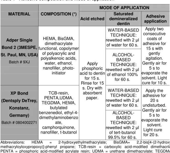
Table 1 - Adhesive composition and mode of application
Abbreviations: HEMA = 2-hydroxyethylmethacrylate; BisGMA=
2,2-bis[4-(2-hydroxy-3-methacryloyloxypropoxy)]-phenyl propane; TCB-resin = carboxylic acid-modified dimethacrilate; PENTA = phosphoric acid-modified acrylate resin; UDMA = urethane dimethacrylate; TEGDMA –
triethyleneglycol dimethacrylate.
* Information as received from manufacturer.
Table 2 – Number of specimens (%) and failure mode according to each experimental
group
GROUPS FAILURE MODE (%)
Adhesive system Saturated demineralized
dentin
Immediate Aged in NaOCl
A M CD CR A M CD CR
Adper Single Bond 2
Water 87.1 3.2 9.7 0.0 94.4 2.8 2.8 0.0
Alcohol 90.0 0.0 3.3 6.7 100 0.0 0.0 0.0
XP Bond Water 90.9 6.0 0 3.1 94.2 0.0 5.8 0.0
Alcohol 94.4 0.0 2.8 2.8 100 0.0 0.0 0.0
A = adhesive failure; M = mixed failure; CD = cohesive failure in dentin and; CR = cohesive failure in resin composite.
MATERIAL COMPOSITION (*)
MODE OF APPLICATION
Acid etched demineralized Saturated dentin
Adhesive application
Adper Single Bond 2 (3MESPE, St. Paul, MN, USA)
Batch # 9XJ
HEMA, BisGMA, dimethacrylate functional, copolymer
of polyacrylic and polyalkenoic acids, water, ethanol, nanofiller, photo-initiator Apply phosphoric acid to dentin
for 15 s. Rinse for 15
s. Dry with absorbent
paper.
WATER-BASED TECHNIQUE:
rewetted with 2 μl
of water for 60 s.
Apply two consecutive
coats of adhesive for
15 s with gently agitation. Gently air for
5 s to evaporate the
solvent. Light cure for 10 s.
ALCOHOL-BASED TECHNIQUE:
rewetted with 2 μl
of ethanol 100% for 60 s.
XP Bond (Dentsply DeTrey,
Konstanz, Germany)
Batch # 0804002271
TCB-resin, PENTA,UDMA, TEGDMA, HEMA, butylated benzenediol, ethyl-4-dimethylaminobenzo ate, camphorquinone, nanofiller, t-butanol WATER-BASED TECHNIQUE:
rewetted with 2 μl
of water for 60 s.
Apply the adhesive for
20 s undisturbed. Gently air for
5 s to evaporate the
solvent. Light cure for 20 s.
ALCOHOL-BASED TECHNIQUE:
rewetted with 2 μl
Table 3 – Mean and standard deviation (MPa) for microtensile bond strength values according to each experimental group, as well as, statistical analysis (*)
GROUPS
Adhesive system Saturated
demineralized dentin Immediate Aged in NaOCl
Adper Single Bond 2 Water 44.9
Aa ± 7.6 (1/30) 14.2 Bd± 3.3 (3/32)
Alcohol 36.8 Ab ± 8.3 (0/30) 11.5 Bd ± 2.9 (1/31)
XP Bond Water 35.5
Ab ± 8.8 (3/30) 23.8 Bc ± 7.3 (4/30)
Alcohol 32.7 Ab ± 9.7 (6/30) 22.3 Bc ± 7.2 (3/42)
A
D
C
C
Fig. 1 – Light microscopy images showing representative nanoleakage interfacial expressions. Pointers: silver deposits; c: composite resin; d: dentin. (A) Adhesive interface produced by SB bonded on water saturated dentin (control group), showing no silver uptake. (B) After 10% NaOCl storage, the adhesive interface showed silver deposits. (C) Adhesive interface created by SB on ethanol saturated dentin, showing minimal silver deposition along the adhesive interface. (D) After aged in 10% NaOCl, the adhesive interface showing extensive and homogeneous silver deposits throughout the adhesive interface (400 x).
C
C
D
D
C
D
C
A
B
C
Fig. 2 – Light microscopy images showing representative nanoleakage interfacial expressions. Pointers: silver deposits; c: composite resin; d: dentin. (A) Adhesive interface produced by XP bonded on water saturated dentin (control group), showing minimal silver uptake. (B) After 10% NaOCl storage, the adhesive interface showed an increase in size of these silver deposits. (C) Adhesive interface created by XP on tert-butanol saturated dentin, showing some silver deposits points. (D) After aged in 10% NaOCl, the adhesive interface showing significant increases in silver deposits throughout the adhesive interface (400 x).
D
C
C
D
4. CONCLUSÃO GERAL
Da avaliação dos resultados obtidos neste trabalho, pode-se concluir que:
REFERÊNCIAS BIBLIOGRÁFICAS
AGEE KA, BECKER TD, JOYCE AP, RUEGGEBERG FA, BORKE JL, WALLER JL, TAY FR, PASHLEY DH. Net expansion of dried demineralized dentin matrix produced by monomer/ alcohol saturation and solvent evaporation. J Biomed Mater Res 2006; 79(2): 349-58.
BECKER TD, AGEE KA, JOYCE AP, RUEGGEBERG FA, BORKE JL, WALLER JL, TAY FR, PADHLEY DH. Infiltration/evaporation-induced shrinkage of demineralized dentin by solvated model adhesives. J Biomed Mater Res B Appl Biomater 2007; 80(1): 156-65.
BLUNCK U, KNITTER K, JAHN KR. Six-month clinical evaluation of XP Bond in noncarious cervical lesions. J Adhes Dent 2007; 9(Suppl 2):265-8.
BRESCHI L, MAZZONI A, RUGGERI A, CADENARO M, DI LENARDA R, DE STEFANO DORIGO E. Dental adhesion review: Aging and stability of the bonded interface. Dent Mater 2008; 24(1): 90-101.
BUONOCORE MG. A simple method of increasing the adhesion of acrylic filling materials to enamel surface. J Dent Res 1955; 34(6): 849-53.
CADENARO M, BRESCHI L, RUEGGEBERG FA, AGEE K, DI LENARDA R, CARRILHO M, TAY FR, PASHLEY DH. Effect of adhesive hydrophilicity and curing time on the permeability of resins bonded to water vs. ethanol-saturated acid-etched dentin. Dent Mater 2009; 25(1):39-47.
CARRILHO MR, TAY FR, PASHLEY DH, TJADERHANE L, CARVALHO RM. Mechanical stability of resin-dentin bond components. Dent Mater 2005; 21(3): 232-41.
GARCIA FCP, OTSUKI M, PASHLEY DH, TAY FR, CARVALHO RM. Effects of solvents on the early stage stiffening rate of demineralized dentin matrix. J Dent 2005; 33(5): 371-7.
GOING RE. Microleakage around dental restoration: a summarizing review. J Am Dent Assoc 1972; 84(6):1349-57.
HASHIMOTO M, OHNO H, KAGA M, ENDO K, SANO H, OGUCHI H. In vivo degradation of resin-dentin bonds in humans over 1 to 3 years. J Dent Res 2000; 79(6):1385-91.
HOSAKA K, NISHITANI Y, TAGAMI J, YOSHIYAMA M, BRACKETT WW, AGEE KA, TAY FR, PASHLEY DH. Durability of resin-dentin bonds to water vs. ethanol-saturated dentin. J Dent Res 2009; 88(2):146-51.
ITO S, HASHIMOTO M, WADGAONKAR B, SVIZERO N, CARVALHO RM, YIU C, RUEGGEBERG FA, FOULGER S, SAITO T, NISHITANI Y, YOSHIYAMA M, TAY FR, PASHLEY DH. Effects of resin hydrophilicity on water sorption and changes in modulus of elasticity. Biomaterials 2005; 26(33):6449-59.
NISHITANI Y, YOSHIYAMA M., DONNELLY AM, AGEE KA, SWORD J, TAY FR, PASHLEY DH. Effects of resin hydrophilicity on dentin bond strength. J Dent Res 2006; 85(11):1016-21.
OSORIO E, TOLEDANO M, AGUILERA FS, TAY FR, OSORIO R. Ethanol wet-bonding technique sensitivity assessed by AFM. J Dent Res 2010; 89(11): 1264-9.
PASHLEY DH, AGEE KA, NAKAJIMA M, TAY FR, CARVALHO RM, TERADA RS, HARMON FJ, LEE WK, RUEGGEBERG FA. Solvent- induced dimensional changes in EDTA- demineralized dentin matrix. J Biomed Mater Res 2001; 56(2): 273- 81.
PASHLEY DH, TAY FR, YIU C, HASHIMOTO M, BRESCHI L, CARVALHO RM, ITO S. Collagen degradation by host-derived enzymes during aging. J Dent Res 2004; 83(3): 216-221.
PASHLEY DH, TAY FR, CARVALHO RM, RUEGGEBERG FA, AGEE KA, CARRILHO M, DONNELLY A, GARCÍA-GODOY F. From dry bonding to water-wet bonding to ethanol-wet bonding. A review of the interactions between dentin matrix and solvated resins using a macromodel of the hybrid layer. Am J Dent 2007; 20(1): 7-20.
SADEK FT, PASHLEY DH, NISHITANI Y,CARRILHO MR, DONELLY A, FERRARI M, TAY FR. Application of the hydrophobic resin adhesive to acid-etched dentin with an alternative wet bonding technique. J Biomed Mater Res A 2008; 84(1):19-29.
SADEK FT, MAZZONI A, BRESCHI L, TAY FR, BRAGA RR. Six-month evaluation of adhesives interface created by a hydrophobic adhesive to acid-etched ethanol-wet bonded dentine with simplified dehydration protocols. J Dent 2010; 38(4):276-83.
resin-dentin bonds made with water or ethanol wet bonding. J Biomed Mater Res B Appl Biomater 2009; 90(1):327-37.
SAURO S, TOLEDANO M, AGUILERA FS, MANNOCCI F, PASHLEY DH, TAY FR, WATSON TF, OSORIO R. Resin-dentin bonds to EDTA-treated vs. acid-etched dentin using ethanol wet-bonding. Dent Mater 2010; 26(4): 368-79.
SHIN TP, YAO X, HUENERGARDT R, WALKER MP, WANG Y. Morphological and chemical characterization of bonding hydrophobic adhesive to dentin using ethanol wet bonding technique. Dent Mater 2009; 25(8):1050-7.
SPENCER P, WANG Y. Adhesive phase separation at the dentin interface under wet bond conditions. J Biomed Mater Res 2002; 62(3): 447-56.
TAY FR, PASHLEY DH, SUH BI, CARVALHO RM, ITTHAGARUN A. Single-step adhesives are permeable membranes. J Dent 2002; 30(7-8): 371-82.
TAY FR, PASHLEY DH. Have dentin adhesives become too hydrophilic? J Can Dent Assoc 2003; 69(11):726-31.
TAY FR, PASHLEY DH, KAPUR RR, CARRILHO MR, HUR YB, GARRETT LV, TAY KC. Bonding BisGMA to dentin - a proof of concept for hydrophobic dentin bonding. J Dent Res 2007; 86(11): 1034-9.
UNEMORI M, MATSUYA Y, MATSUYA S, AKASHI A, AKAMINE A. Water absorption of poly(methyl methacrylate) containing 4-methacryloxyethyl trimellitic anhydride. Biomaterials 2003; 24(8):1381-7.
WANG Y, SPENCER P. Hybridization efficiency of the adhesive/dentin interface with wet bond. J Dent Res 2003; 82(2):141-5.
YE Q, PARK JG, TOPP E, WANG Y, MISRA A, SPENCER P. In vitro performance of nano-heterogeneous dentin adhesive. J Dent Res 2008; 87(9):829-33.
YE Q, WANG Y, SPENCER P. Nanophase separation of polymers exposed to simulated bonding conditions. J Biomed Mater Res B 2009; 88(2):339-48.
YIU CK, KING NM, PASHLEY DH, SUH BI, CARVALHO RM, CARRILHO MR, TAY FR. Effect of resin hydrophilicity and water storage on resin strength. Biomaterials 2004; 25 (26):5789-96.
YIU CK, PASHLEY EL, HIRAISHI N, KING NM, GORACCI C, FERRARI M, CARVALHO RM, PASHLEY DH, TAY FR. Solvent and water retention in dental adhesive blends after evaporation. Biomaterials 2005; 26(34): 6863- 72.