*Correspondence: I. Martins. Laboratory of Toxicants and Drugs Analysis, Faculty of Pharmaceutical Sciences, Federal University of Alfenas. Rua Gabriel Monteiro da Silva, 700 – 37130-000 - Alfenas - MG, Brazil. E-mail: [email protected]
A
rti
Pharmaceutical Sciences vol. 46, n. 4, out./dez., 2010
Single gas chromatography method with nitrogen phosphorus
detector for urinary cotinine determination in passive
and active smokers
Lusiane Malafatti, Patrícia Penido Maia, Matheus Coutinho Gonçalves Martins,
Maria Elisa Pereira Bastos de Siqueira, Isarita Martins*
Laboratory of Toxicants and Drugs Analysis - LATF, Faculty of Pharmaceutical Sciences, Federal University of Alfenas
Nicotine is a major addictive compound in cigarettes and is rapidly and extensively metabolized to several metabolites in humans, including urinary cotinine, considered a biomarker due to its high concentration compared to other metabolites. The aim of this study was to develop a single method for determination of urinary cotinine, in active and passive smokers, by gas chromatography with a nitrogen phosphorus detector (GC-NPD). Urine (5.0 mL) was extracted with 1.0 mL of sodium hydroxide 5 mol L-1, 5.0 mL
of chloroform, and lidocaine used as the internal standard. Injection volume was 1 µL in GC-NPD. Limit of quantiication was 10 ng mL-1. Linearity was evaluated in the ranges 10-1000 ng mL-1 and
500-6000 ng mL-1, with determination coeficients of 0.9986 and 0.9952, respectively. Intra- and inter-assay
standard relative deviations were lower than 14.2 %, while inaccuracy (bias) was less than +11.9%. The eficiency of extraction was greater than 88.5%. Ruggedness was veriied, according to Youden’s test. Means of cotinine concentrations observed were 2,980 ng mL-1 for active smokers and 132 ng mL-1, for
passive smokers. The results revealed that satisfactory chromatographic separation between the analyte and interferents was obtained with a ZB-1 column. This method is reliable, precise, linear and presented ruggedness in the range evaluated. The results suggest that it can be applied in routine analysis for passive and active smokers, since it is able to quantify a wide range of cotinine concentrations in urine.
Uniterms: Gas chromatography/with nitrogen phosphorus detector. Urinary cotinine/determination. Urine/toxicological analysis. Smokers/Toxicology.
A nicotina é uma substância presente no cigarro capaz de causar dependência, sendo biotransformada em vários metabólitos nos seres humanos, dentre eles a cotinina urinária, que é considerada um indicador biológico de exposição à nicotina, devido a suas altas concentrações, comparado a outras matrizes. Assim, o objetivo deste estudo foi desenvolver um único método para determinação de cotinina urinária, em amostras de urina de fumantes ativos e passivos, através de cromatograia em fase gasosa com detector de nitrogênio- fósforo (CG-DNF). Para o preparo de amostras foram utilizados 5 mL de urina, 1 mL de hidróxido de sódio 5 mol L-1, 5 mL de clorofórmio, tendo como padrão interno a lidocaína. Na faixa
de concentrações de 10-1000 ng mL-1 e 500- 6000 ng mL-1, o coeiciente de determinação foi 0,9986
e 0,9952, respectivamente e, o limite de quantiicação foi 10 ng mL-1. A precisão intra- e interensaio
apresentou desvio padrão relativo (%) menor que 14,2% e a inexatidão foi menor que +11,9%, com uma eiciência de extração de 88,5%. O método apresentou robustez, de acordo com o teste de Youden. As concentrações médias de cotinina observadas foram 2980 ng mL-1, para fumantes ativos e 132 ng mL-1,
para fumantes passivos. Os resultados sugerem que o método é coniável, preciso, linear e apresentou robustez, na faixa avaliada, podendo ser aplicado na rotina para análises de amostras de fumantes ativos e passivos, pois é capaz de quantiicar uma ampla faixa de concentrações de cotinina urinária.
INTRODUCTION
It is well known that tobacco smoke, which contains many toxic and tumorigenic compounds, is recognized as a major cause of mortality and morbidity (Richard, Eian, Graham, 2004). Tobacco is the single most preventable cause of death in the world today. By 2030, the death toll will exceed eight million a year (WHO, 2008).
More than 4000 compounds have been identiied in tobacco smoke in which nicotine is the principal alkaloid (Hansen et al., 2001). Nicotine, a major chemical found in all tobacco products, ispresent in both mainstream and sidestream tobacco smoke. In both smokers and non-smokers, nicotine enters the bloodstream when tobacco smoke is inhaled. It is then circulated to various body organs (Benowitz, 1996).
Cotinine is the major proximate metabolite of nico-tine and has been widely used as a biomarker of tobacco smoke exposure (Kuo, Yang, Chiu, 2002) and offers sev-eral advantages over biochemical markers as an objective indicator of nicotine intake or conirmation of non-smoker status. Its concentrations are not inluenced by other sub-stances since it is a speciic nicotine biomarker and con-centrations within a given individual varies by only 15 to 20% over 24 h (Oddoze, Pauli, Pastor, 1998). Cotinine in urine accounted for less than 15% of total systemic dose of nicotine (Cope et al., 1996; Benowitz, 1996).
The half-life of cotinine and nicotine are approxi-mately 19 and 2 h, respectively. Consequently, cotinine, because of its longer half-life, is currently the marker of choice for demonstrating cigarette smoke exposure (Hau-froid, Lison, 1998). Its determination in biological luids has aroused particular interest. These biochemical mark-ers have been used to estimate active smoking behavior, to validate abstinence from smoking and to evaluate the levels and signiicance of environmental tobacco smoke (ETS) exposure (Benowitz, 1996). Urinary samples are more convenient to collect and urinary cotinine is a well-known biomarker of ETS (Jarvis et al., 1987; Wall et al., 1988; Kuo, Yang, Chiu, 2002).
Many methods have been proposed for the determina-tion of cotinine in human urine. These methods use radio-immunoassay (Kuo, Yang, Chiu, 2002); high performance liquid chromatography (Zuccaro et al., 1995; Oddoze, Pauli, Pastor, 1998; Tyrpien et al., 2000; Abou-Qare, Abou-Donia, 2001; Doctor et al., 2004; Kowalski et al., 2007) and gas chromatography with nitrogen phosphorus detection (Kuo, Yang, Chiu, 2002; Moriya, Hashimoto, 2004). Liquid or gas chromatography coupled mass spectrometry is commonly employed to determine cotinine in passive smokers (Ji Jr. et al., 1999; Meger et al., 2002; Man et al., 2006; Chadwick,
Keevil, 2007). Other less sensitive detectors, such as lame ionization detector (FID), have also been used to quantify cotinine in urine of active and passive smokers. However, the sample preparation needed larger urine volume (25 mL) (Vacchino et al., 2006).
Among these methods, the chromatographic tech-niques are more preferred than others because they are potentially more sensitive and more speciic. This fact is due to sample enrichment by extraction prior to analysis and sample cleanup through chromatographic separation during analysis (Song et al., 2005). In this context, many methods have been proposed for extraction of samples containing cotinine, among then, liquid-liquid extraction (LLE) (Shin et al., 2002; Man et al., 2006; Kowalski et al., 2007) and solid phase extraction (SPE) (Oddoze, Pauli, Pastor, 1998; Moyer et al., 2002; Xu, Iba, Weisel, 2004). LLE presents the advantages of being a simple method of sample preparation that can use a great number of solvents, pure and available commercially, which sup-ply a wide range of solubility and selectivity (Queiroz, Collins, Jardim, 2001).
The aim of this study was to develop a single method able to determine urinary cotinine, in active and passive smokers, by GC-NPD with a previous LLE.
MATERIAL AND METHODS
Reagents and standard solutions
Cotinine (COT) with approximately 98% purity (Lot no. 055k4053) and lidocaine (LID) with approximately 98% purity (Lot no. 162008), were purchased from Sigma-Aldrich Inc. (St. Louis, USA). Analytical grade isopropyl alcohol and chloroform were purchased from Vetec Ltda. (Rio de Janeiro, Brazil), methyl alcohol from Isofar Ltda. (Rio de Janeiro, Brazil) and sodium hydroxide from La-bsynth Ltda. (São Paulo, Brazil).
Stock solutions of cotinine and lidocaine (internal standard) were prepared in isopropyl alcohol at 1 mg mL-1
and stored at -20oC, protected from light. These solutions
were used for at least one month. Working solutions were freshly prepared in isopropyl alcohol immediately before analysis. Throughout the study, water was obtained from a Milli-Q system by Millipore (São Paulo, Brasil).
Instrumentation and chromatographic conditions
The GC system consisted of a GC model Clarus 400, equipped with NPD from Perkin Elmer® with
Total-chrom Workstation® Software. Chromatographic analysis
polydimethylsiloxane, 30 m x 0.53 mm i.d.; 5 µm ilm thickness). Nitrogen was used as a gas carrier at a pressure of 4.3 psi. One microliter injection volume using splitless mode was injected manually, at an injector temperature of 260 oC. The oven temperature was programmed from
180 oCincreasing by 30 oC/minute to 250 oC for 0.5 min,
increasing by 15 oC/min to 259 oC for 0.1 min and then
in-creasing by 0.1 oC/min to 260 oC. The detector temperature
was 280 oC. The total run time was 13.4 min.
Urine samples
Cotinine-free urine, sourced from non-smokers not exposed to environmental nicotine, was used as a blank matrix to perform validation parameters. Urine samples from forty-one active smokers, ten passive smokers and eighteen unexposed non-smokers were used for application of this method. This study was approved by the Ethics Committee of Federal University of Alfenas (23087.002423/2008-12). An informed consent term was obtained from each volunteer.
All samples were collected in polyethylene urine containers and density was measured at each sampling time with a refractometer (Atago®) for standardization of
cotinine levels. Samples were then frozen and stored at -20 oC until analysis.
Sample preparation
Before extraction, samples were thawed and equi-librated to room temperature. Urine (5.0 mL) was placed in a 15 mL centrifuge glass tube. Sodium hydroxide (5 mol L-1, 1 mL), lidocaine as the internal standard (500 or
1000 µg mL-1, 0.05 mL) and chloroform (5.0 mL) were added and the mixture was mixed for 15 minutes in a bench top shaker. After centrifugation for 15 minutes at 840 g, the aqueous layer was discarded and 4.5 mL of organic phase was transferred into a conical glass tube. The extract was evaporated in a stream of nitrogen, within a bath at 40 oC. The
residue was reconstituted in 0.05 mL of isopropyl alcohol.
Method validation
Appropriate validation is necessary to ensure the suitability of analytical methods for the purpose (Kowalski
et al., 2007), and the conidence parameters of the present method were assayed according to the Guidance for In-dustry of the FDA for Bioanalytical Method Validation (2001). The parameters evaluated in the present study were: linearity, limit of quantiication, recovery, intra- and inter-assay precision, accuracy and ruggedness.
The linearity was studied in two linear ranges (10-1000 ng mL-1 and 500-6000 ng mL-1)to minimize errors
from a wide range of urinary cotinine concentrations. Urine samples were spiked with COT at concentrations 10; 100; 250; 500; 750; 1000 ng mL-1 and 500; 1000; 2000;
3000; 4000; 6000 ng mL-1, and each treatment was assayed
in six replicates. Concentration of internal standard was adjusted according to the range evaluated (500 µg mL-1, for 10-1000 ng mL-1 of cotinine or 1000 µg mL-1 for
500-6000 ng mL-1, of cotinine).
These samples were analyzed according to the item sample preparation. Calibration curves were constructed by plotting peak area ratios of analyte and its internal standard versus original concentrations, and evaluated by linear least square regression analysis.
The limit of quantiication was estimated after suc-cessive dilutions of cotinine solutions, until obtention of a concentration in which the peak area was ten times the signal-to-noise ratio (S/N= 10), provided by the blank extract (N= background noise).
Recovery was determined by three replicate analyses of samples after the additional spiking of a known mass of the analyte, in the non-contaminated samples, at three levels. The results were compared with those obtained when the analyte was spiked after the clean-up procedure of the sample, at the same levels.
Intra- assay precision was assessed using three replicates of each concentration of linear range, on the same day. Inter-assay precision was evaluated for three replicates analyzed on separate days (n=6). The results were expressed as a percentage of relative standard de-viation (% RSD).
Accuracy was established by spiking urine sam-ples with 30, 500, 1000, 3000 and 6000 ng mL-1 of COT
(n= 3/ concentration). After extraction and chromatogra-phic analysis, results were compared to the theoretical added values.
Ruggedness was evaluated through the Youden approach, which is based on a fractional factorial de-sign according to the Oficial Journal of the European Communities (2002). For this purpose, eight determi-nations were carried out, combining the nominal and with-variation parameters. The variables evaluated are described in Table I. The experiments are described in Table II, and Table III contains the formula used to eva-luate the variation effect.
RESULTS
validated method. Lidocaine was used as the internal stan-dard at concentrations of 500 µg mL-1 in a) non smokers; and at 1000 µg mL-1 in b) and c), passive and active
smokers, respectively. The method revealed satisfactory chromatographic separation between the analyte and inter-ferents, I requiring only a short time for chromatography (less than 14 min) and proved suitable for determining the cotinine in urine in routine analysis.
LOQ is the lowest concentration of an analyte on a calibration curve, and for this method the concentration 10 ng mL-1 resulted in a signal-to-noise ratio of 10, and
TABLE I - Robustness parameters
Parameters Nominal (+) Variation (-)
Sample volume (mL) 5.0 4.0
NaOH concentration (mol L-1) 5.0 4.5
Agitation in vortex Yes no
Time in bench top shaker (min) 15 10
Time in centrifuge (min) 15 10
Detector temperature (oC) 280 275
Gas carrier pressure (psi) 4.3 4.2
TABLE II - Combinations assayed for nominal or variable
parameters
Parameters Combination assayed
1 2 3 4 5 6 7 8
Sample volume (mL) + + + +
-NaOH concentration (mol L-1) + + + +
-Agitation in vortex + + + +
-Time in bench top shaker (min) + + - - - - + +
Time in centrifuge (min) + - + - - + - +
Detector temperature (oC) + - - + + - - +
Gas carrier pressure (psi) + + + +
-Results a b c D e f g h
TABLE III - Variation effects evaluation
Factor Formula for variation effect
Sample volume (a+b+c+d)/4 – (e+f+g+h)/4
NaOH concentration (a+b+e+f)/4 – (c+d+g+h)/4
Agitation in vortex (a+c+e+g)/4 – (b+d+f+h)/4
Time in bench top shaker (a+b+g+h)/4 – (c+d+e+f)/4
Time in centrifuge (a+c+f+h)/4 – (b+d+e+g)/4
Detector temperature (a+d+e+h)/4 – (b+c+f+g)/4
Gas carrier pressure (a+d+f+g)/4 – (b+c+e+h)/4
FIGURE 1 - Chromatograms after LLE of urine: a) non
smokers; b) passive smokers; c) active smokers. (1) Cotinine: rt 5.7 min; k’ 2.0 min; α 1.25 (calculated between cotinine and immediately before peak) (2) Lidocaine (IS): rt 8.2 min; k’ 3.3 min; α 1.1 (calculated between cotinine and immediately after peak). Chromatographic conditions: ZB-1 column, injector
temperature at 260 oC, the oven temperature programmed with
initial temperature of 180 oC, ramp 1: 30 oC/ minute to 250 oC
for 0.5 min; ramp 2: 15 oC/ min to 259 oC for 0.1 min and ramp
3: 0.1 oC/min to 260oC, detector temperature at 280 oC. Nitrogen
TABLE IV - Linearity of method
COTININE
Linear Range (ng mL-1) 10-1000 500-6000
Slope (a) 0.0036 ± 0.0002 0.0012 ± 7.53 x10⁻⁵
Intercept (b) 0.0936 ± 0.061 0.2979 ± 0.203
Determination Coeficient (R2) 0.9986 0.9952
TABLE V - Recovery and accuracy for the analysis of cotinine in urine, by CG-NPD after ELL
COTININE
Urine concentration (ng mL-1) Relative Recovery (mean, %) Accuracy (bias, %)
30 102.2 +11.9
500 99.2 +0.6
1000 88.5 +2.9
3000 104.7 +7.6
6000 108.2 -1.7
Mean 100.6 +4.3
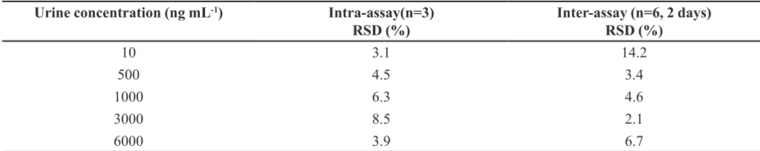
TABLE VI - Intra- and inter-assay precision for the analysis of cotinine in urine, by CG-NPD after ELL.
Urine concentration (ng mL-1) Intra-assay(n=3)
RSD (%) Inter-assay (n=6, 2 days)RSD (%)
10 3.1 14.2
500 4.5 3.4
1000 6.3 4.6
3000 8.5 2.1
6000 3.9 6.7
the % relative standard deviation ( n=5) obtained was 8.99%. Tables IV, V and VI show the results of the method validation.
Ruggedness was assessed using the Youden approa-ch. Eight determinations were made using a combination of the factors with variations (see Table I). Variation in-luence was evaluated by comparing the values obtained by the formulas in Table III, with those values obtained by the proposed method (nominal parameters). Variations of more that two standard deviations from the result obtained using the proposed method (nominal parameters) were considered to indicate that a parameter caused alteration in the method. Ruggedness was demonstrated, since no statistically signiicant difference was observed between the nominal parameters and the values obtained with the variation described in Table II.
The values for cotinine concentrations observed in urine were 2,980 ± 2,160 ng mL-1, for active smokers and
132.00 ± 80 ng mL-1, for passive smokers.
DISCUSSION
Urine is the preferred specimen over plasma and saliva because it is much easier to obtain, particularly in epidemic studies (Hariharan, Vannoord, 1991). The advantages of using urine in the investigations included lower viscosity and ease of handling compared with saliva or blood, as well as constituting a relatively nonintrusive sample collection/ donation method without an occupatio-nal health risk (Tuomi, Johnsson, Reijula, 1999; Hagan, Ramos Jr., Jacob III, 2002). Since urinary cotinine is considered a biomarker of exposure to tobacco, the objec-tive of this study was to develop a single method able to determine urinary cotinine in active and passive smokers. The literature describes this determination but the sample preparation required a high urine volume (25 mL) (Vac-chino et al., 2006). The present study used 5.0 mL urine.
for removing unwanted substances from urine matrix and can also be used to concentrate the analyte (Kowalski et al., 2007).
Due to its pKa (4.5), cotinine is found in greater quantity in non-ionized form in the blood (pH 7.4) and the free base form is poorly soluble in lipids, showing a low rate of distribution to tissues, which partly explains their prolonged existence in the blood. Another factor that contributes to its prolonged half-life is the low rate of re-nal excretion in relation to nicotine (Feyerabend, Russell, 1980). Therefore, because of its longer half-life, cotinine is often the marker of choice to demonstrate exposure to cigarette smoke (Chadwick, Keevil, 2007).
A high pH was used in order to co-extract cotinine and lidocaine, with satisfactory extraction eficiency, since the pKa of cotinine was reported to be < 5.0 (Beckett, Gor-rod, 1972) with basic characteristics. In this study, COT was extracted eficiently in chloroform, after alkalization with sodium hydroxide 5 mol L-1.
Linearity was determined for COT using a pool of blank urine that was spiked with the analyte and the internal standard. Peak area ratios (COT/IS) and analyte concentrations were found to be linear in the range eva-luated. The least-squares linear regression was used to determine the slope and intercept. Man et al. (2006), using gas chromatography and mass spectrometry, obtained a linear range from 0.5 to 5000 ng mL-1.
The present study was proposed for determination of urinary cotinine of active, passive and non-smokers. The limit of quantiication obtained (10 ng mL-1) was suficient
for monitoring passive smoking, because according to Kolonen & Pahukainen (1991), urinary cotinine levels in passive smokers are typically less than 100 ng mL-1,
serving as a cut-off point to verify tobacco-free status. Other authors have suggested a cut-point of 50 ng mL-1 for
urinary cotinine as a means to distinguish smokers from non-smokers (Haufroid, Lison, 1998; Song et al., 2005). Besides, zero cotinine concentration is generally observed in urine of non-smokers not exposed to environmental tobacco smoke.
In the present study with a nitrogen phosphorus detector, the method developed was able to distinguish different groups exposed to tobacco smoke, which can be considered an advantage in routine analysis.
Intra- and inter-assay precision were less than 8.5% and 14.2%, respectively, and this is considered satisfac-tory according to FDA guidelines (2001). This parameter should not exceed 15% of the RSD, except for the LOQ, where it should not exceed 20% of the RSD.
Relative recovery of COT was in the range of 88.5% and 108.2% with an accuracy in the range of -1.7 % and
+11.9%. The guidance of validation for bioanalytical me-thods of the FDA (2001) establishes that recovery of the analyte need not be 100%, where lower values are accep-table provided the recovery offers precision and accuracy. Other studies (Voncken, Schepers, Schafer, 1989; Hagan
et al., 1997; Ji Jr et al., 1999; Shin et al., 2002; Chadwick, Keevil, 2007) using liquid-liquid extraction as the sample preparation technique obtained recoveries of between 81 and 112%.
Ruggedness was evaluated through the Youden ap-proach and allowed the conclusion that small variations did not affect the method under tested conditions, since the results did not differ signiicantly across the conditions evaluated.
Cotinine concentrations observed in this study were 2,980 ± 2,160 ng mL-1, for active smokers and 132.00 ±
80 ng mL-1, for passive smokers, in line with urinary
coti-nine concentrations described in the literature (Voncken, Schepers, Schafer, 1989; Oddoze, Pauli, Pastor, 1998; Moyer et al., 2002; Ji Jr et al., 1999; Kuo, Yang, Chiu, 2002; Man et al., 2006; Chadwick, Keevil, 2007) of 1,560 to 6,680 ng mL-1, for active smokers and approximately
50 ng mL-1, for passive smokers.
CONCLUSION
A fast and simple GC-NPD method was developed and validated for urinary cotinine analysis using a low sample volume. The results revealed that satisfactory chromatographic separation between the analyte and inter-ferents was obtained with a ZB-1 column. This method is reliable, precise, linear and presented ruggedness over the range evaluated. The results suggest that it can be applied in routine analysis to passive and active smokers, since it is able to quantify a wide range of cotinine concentrations in urine.
ACKNOWLEDGEMENTS
The authors gratefully acknowledge inancial sup-port from FAPEMIG, Brazil (process CDS APQ-1827-4.04/07) and CAPES, Brazil.
REFERENCES
BECKETT, A.H.; GORROD, J.W. A possible relationship between pKa1 and lipid solubility and the amounts excreted
in urine of some tobacco alkaloids given to man. J. Pharm.
Pharmacol., v.24, p.115-20, 1972.
BENOWITZ, N.L. Cotinine as biomarker of environmental
tobacco smoke exposure. Epidemiol. Rev, v.18, p.188-204,
1996.
CHADWICK, C.A.; KEEVILL, B. Measurement of cotinine in urine by liquid chromatography tandem mass spectrometry. Ann. Clin. Biochem., v.44, p.455-462, 2007.
COPE, G.; NAYYAR, P.; HOLDER, R.; GIBBONS, J.; BUNCE, R. A simple near-patient test for nicotine and its metabolites
in urine to assess smoking habit. Clin. Chim. Acta, v.256,
p.135-149, 1996.
DOCTOR, P.B.; GOVANI, V.N.; KULKARNI, P.K.; PARIKH, J.R.; SAIYED, H.N. Determination of nicotine and cotinine in tobacco harvester´s urine by solid-phase extraction and
liquid chromatography. J. Chromatogr. B, v.802,
p.323-328, 2004.
EUROPEAN COMMISSION. Corrigendum to Commission Decision 2002/657/EC of 12 August 2002 implementing Council Directive 96/23/EC concerning the performance
of analytical methods and the interpretation of results. Off.
J. Eur. Communities, L, Legis, n. L 221, p. 24-25, 17 Aug. 2002, Session 3.
FEYERABEND, C.; RUSSELL, M.A. Rapid gas liquid chromatographic determination of cotinine in biological
luids. Analyst, v.105, p.998-1001, 1980.
FOOD AND DRUG ADMINISTRATION. U.S. Department
of Health and Human Services. Guidance for industry:
bioanalytical method validation. Rockville: FDA, 2001. p.22.
HAGAN, R.L.; RAMOS JR.; J.M.; JACOB III, P.M. Increasing urinary cotinine concentrations at elevated temperatures: the
role of conjugated metabolites. J. Pharm. Biomed. Anal.,
v.16, p.191-197, 1997.
HANSEN, A.M.; GARDE, A.H.; CHRISTENSEN, J.M.; ELLER, N.; KNUDSEN, L.E.; HEINRICH-RAMM, R. Reference interval and subject variation in excretion of urinary metabolites of nicotine from non-smoking healthy
subjects in Denmark. Clin. Chim. Acta, v.304, p.125-132,
2001.
HARIHARAN, M.; VANNOORD, T. Liquid-chromatography determination of nicotine and cotinine in urine from passive smokers: comparison with gas chromatography a
nitrogen-speciic detector. Clin. Chem., v.37, p.1276-1280, 1991.
HAUFROID, V.; LISON, D. Urinary cotinine as a
tobacco-smoke exposure index: a minireview. Int. Arch. Occup.
Environ. Health, v.71, p.162-168, 1998.
JARVIS, M.J.; TUNSTALL-PEDOE, H.; FEYERABEND, C.; VESEY, C.; SALOOJEE, Y. Comparison of tests used to
distinguish smokers from nonsmokers. Am. J. Publ. Health,
v.77, p.1435-1483, 1987.
JI JR, A.; LAWSON, G.M.; ANDERSON, R.; DALE, L.C.; CROGHAN, I.T.; HURT, R.D. A new gas-chromatography-mass spectrometry method for simultaneous determination
of total and free trans-3-hidroxycotinine and cotinine in
the urine of subjects receiving transdermal nicotine. Clin.
Chem., v.45, p.85-91, 1999.
KOLONEN, S.A.; PAHUKAINEN, E.V.J. Assessment of the automated colorimetric and high- performance liquid chromatographic methods for nicotine intake by urine samples of smokers’ smoking low- and medium- yield
cigarette. Clin. Chim. Acta, v.196, p.159-166, 1991.
KOWALSKI, P.; MARSZATT, M.; OLEDZKA, I.; CZARNOWSKI, W. Comparative determination of CE and HPLC for determination of cotinine in human urine. Chromatographia, v.66, p.357-361, 2007.
KUO, H.W.; YANG, J.S.; CHIU, M.C. Determination of urinary and salivary cotinine using gas and liquid chromatography
and enzyme-linked immunosorbent assay. J. Chromatogr.
B, v.768, p.297.303, 2002.
MAN, C.H.; GAM, L.H.; ISMAIL, R.; AWANG, R. Simple, rapid and sensitive assay method for simultaneous quantiication of urinary nicotine and cotinine using gas
chromatography-mass spectrometry. J. Chromatogr. B,
v.844, p.322-327, 2006.
MEGER,M.; MEGER-KOSSIEN,I.; SCHULER-METZ, A.; JANKET, D.; SCHERER, G. Simultaneous determination of nicotine and eight nicotine metabolites in urine of smokers using liquid chromatography–tandem mass
MOYER, T.P.; CHARLSON, J.R.; ENGER, R.J.; DALE, L.C.; EBBERT, J.O.; SCHROEDER, D.R.; HURT, R.D. Simultaneous analysis of nicotine, nicotine metabolites and tobacco alkaloids in serum or urine by tandem mass spectrometry, with clinically relevant metabolic proiles. Clin. Chem., v.48, p.1460-1471, 2002.
MORIYA, F.; HASHIMOTO, Y. Nicotine and cotinine levels in
blood and urine from forensic autopsy cases. J. Leg. Med.,
v.6, p.164-169, 2004.
ODDOZE, C.; PAULI, A.M.; PASTOR, J. Rapid and sensitive high-performance liquid chromatographic determination of nicotine and cotinine in nonsmoker human and rat urines. J. Chromatogr. B, v.708, p.95-101, 1998.
QUEIROZ, S.C.N; COLLINS, C.H.; JARDIM, I.C.S.F. Métodos de extração e/ou concentração de compostos encontrados em fluídos biológicos para posterior determinação
cromatográica. Quim. Nova, v.24, p.68-76, 2001.
RICHARD, R.B.; EIAN, D.M.; GRAHAM, S. An overview of the effects of tobacco ingredients on smoke chemistry and
toxicity. Food Chem. Toxicol., v.42S, p.S53-S83, 2004.
SHIN, H.S.; KIM, J.G.; SHIN, Y.J.; JEE, S.H. Sensitive and simple method for the determination of nicotine and cotinine in human urine, plasma and saliva by gas
chromatography-mass spectrometry. J. Chromatogr. B,
v.769, p.177-183, 2002.
SONG, L.; DAVIS, W.; ABRAMS, S. M.; HEMIUP, J.; LATIF KAZIM, A.; CUMMINGS, K.M.; MAHONEY, M.C. Sensitive and rapid method for the determination of urinary cotinine in non-smokers: an application for the studies
assessing exposures to second hand smoke. Anal. Chim.
Acta, v.545, p.200-208, 2005.
TUOMI, T.; JOHNSSON, T.; REIJULA, K. Analysis of nicotine, 3-hydroxycotinine, cotinine and caffeine in urine passive
of smokers by HPLC- Tandem Mass Spectrometry. Clin.
Chem., v.45, p.2164-2172, 1999.
TYRPIEN, K.; WIELKOSZYNSKI, T.; JANOSKA, B.; DOBOSZ, C.; BODZEK, D.; STEPLEWSKI, Z. Application of liquid separation techniques to the determination of the
main urinary nicotine metabolites. J. Chromatogr. A, v.870,
p.29-38, 2000.
VACCHINO, M.N.; VELURTAS, S.M.; SALINAS, G.P.; GARCIALOREDO, H.H. Determinación de cotinina y
exposición a tabaco. Acta Bioquim. Clin. Latinoam., v.40,
p.181-185, 2006.
VONCKEN, P.; SCHEPERS, G.; SCHAFER, K.H. Capillary gas
chromatographic determination of trans-3-hydroxicotinine
simultaneously with nicotine and cotinine in urine and blood
samples. J. Chromatogr., v.479, p.410-418, 1989.
XU, X.; IBA, M.M.; WEISEL, C.P. Simultaneous and sensitive measurement of anabasine, nicotine and nicotine metabolites in human urine by liquid
chromatography-Tandem Mass Spectrometry. Clin. Chem., v.50,
p.2323-2330, 2004.
WALL, M.A.; JOHNSON, J.; JACOB, P.; BENOWITZ, N.L. Cotinine in the serum, saliva and urine of nonsmokers,
passive smokers, and active smokers. Am. J. Publ. Health,
v.78, p.699-701, 1988.
WORLD HEALTH ORGANIZATION. WHO report on the
global tobacco epidemic: the mpower package. Geneve, 2008. 342 p.
ZUCCARO, P.; ALTIERI, I.; ROSA, M.; PASSA, A. R.; PICHIN, S.; PACIFICI, R. Solid-phase extraction of nicotine and its metabolites for high-performance liquid
chromatography determination in urine. J. Chromatogr. B,
v.668, p.187-188, 1995.
Received for publication on 29th October 2009.